Pain Management Medications

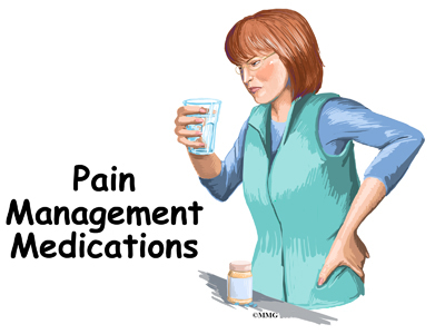
A Patient’s Guide to Pain Management Medications

Introduction

There are several types of medications that can be used to treat pain. No one drug works for everyone. The medications that work best for you will depend on the type of pain you have and your response to them. Your medication regimen will need to be designed just for you.

This guide will help you understand

  • what medications are commonly prescribed
  • why these drugs are prescribed
  • what are the risks and benefits of this treatment

Rationale

If you have lived with pain for a long time, it is common to have other conditions as well. These may include depression, anxiety, and insomnia (trouble sleeping). These conditions can make pain worse. And it’s harder for you to cope with the stress of living with pain. In order to get control over the pain, these conditions usually need to be treated at the same time as the pain.

The goal of using medication, as part of your treatment plan, is to increase your activity level. This can be done by improving pain control with drugs that you can afford while avoiding side effects.

The medications most commonly used in pain management belong to the following groups

  • Opioid (narcotic)
  • Non-opioid (non-narcotic)
  • Acetaminophen (Tylenol®)
  • Non-steroidal antiinflammatory (NSAID)
  • Steroidal anti-inflammatory
  • Antiepileptic (anticonvulsant)
  • Antidepressant and antianxiety agents (anxiolytics)
  • Muscle relaxants (antispasmodics)
  • Sleep aids (hypnotics)
  • Topical agents

Opioid (Narcotic) Pain Medications

Pain management experts prefer the term opioid to describe narcotic medication. The word narcotic is a legal term that is often linked with the illegal use of drugs.

Rationale

Opioid medications are expected to improve your activity level. They do this by decreasing the pain you feel when you are active.

Expected Benefit

Opioids such as morphine (MSContin), fentanyl (Duragesic), and methadone reduce pain by binding to opioid receptors in the brain. Most pain is responsive to opioid medications. However, some types of pain respond better than others. Nociceptive pain refers to skeletal or muscular pain. This type of pain is usually more responsive than neuropathic (nerve) pain.

Some problems such as fibromyalgia with diffuse (widely spread) myofascial (inflamed muscle and fascia) pain may respond at first. But studies show that patients with fibromyalgia do not do better over time on these drugs. Pain caused by headache may be relieved by opioids, but they can cause worse headaches later. These are called rebound headaches.

Potential Risks

There are some possible risks associated with opioid use. Not everyone will have all of these problems. With the right management, you may not have any problems. Risks can include

  • Addiction
  • Physical dependence
  • Tolerance
  • Hyperalgesia
  • Decreased hormone levels
  • Change in mood
  • Sleep apnea
  • Theft
  • Death

Addiction is defined by craving, uncontrolled or compulsive use, and continuing to use the substance despite harm. Addiction is a complicated chronic disease. Simply taking an opioid does not cause addiction. If you have no risk factors, it is rare to develop the disease of addiction.

However, addiction is a possibility if you have the right (or wrong) genetics. Psychological and social stressors are also important factors. Family history must be reviewed before taking an opioid. There are ways to lower the risk of addiction or relapse if you are at risk for this disease. If you have an active addiction, the risk may outweigh the benefit of taking an opioid. Other options for pain control should be explored.

Substance abuse is a term used for unusual or abnormal behavior that does not meet the definition of addiction. Pain experts watch closely for any signs of this kind of behavior. These behaviors include reports of lost or stolen medication or use of medication in ways other than directed. Not taking the medication according to the directions would also fall into this category. Requesting early refills or taking a medication despite serious side effects are red flag behaviors. Taking the drug to deal with stress such as after a heated argument with someone is also a warning sign. These behaviors are often a signal that you may be getting into trouble with your medication. Your treatment plan may need to be altered. The goal is to get the most benefit with the least risk of side effects.

Pseudo-addiction is the term used to describe what appears to be dangerous or unusual behavior but occurs when pain is not being fully treated. Think about this idea. You have constant pain. It lasts all day and night. You are given a pain medication and told to take one every eight hours. The pain medication that you are prescribed is effective and allows you to be active. But it only works for about four hours. Four hours before the next dose is due, you are in great pain again. How would you respond?

Some people take more drug than is prescribed. Then they run out of pills early. Others complain bitterly at every appointment. They demand a refill. To the doctor it seems as if the person is “drug seeking”. Some may go to more than one doctor to get what they think they need to relieve the pain. When these behaviors are caused by under-treatment of pain, an increase in the dose stops the behavior. If these behaviors start up shortly (within days to weeks) after the dose increase, then there may be other reasons for the behavior.

Physical dependence occurs in everyone on opioids. This happens when you take them regularly for more than two to three weeks. Physical dependence does not mean that you are addicted. Physical dependence means that your body has become used to the drug. You feel withdrawal symptoms if the medication is stopped suddenly.

Symptoms of withdrawal can range from mild irritability to sweating, diarrhea, vomiting, and muscle cramps. You may feel like you have a severe case of the flu. You may feel like you are dying. But withdrawal from opioids is not fatal. It can be avoided by slowly decreasing the amount of drug you take. This can be done over several days up to several weeks. The time it takes to get off the medication without having unpleasant symptoms depends on how much medication you are on and how long you have been on it.

Many opioids require a hand signed prescription every time you need it refilled. It is very important to plan ahead for holidays. You don’t want to run out.

Tolerance to a medication occurs when an individual requires more medication to achieve the same amount of pain relief. Tolerance is not the same as addiction. Not everyone develops tolerance to pain relief. If you should have tolerance to your pain medication, there are ways to deal with it. For example, your doctor may increase the amount you are taking. Or you may be switched to a different drug. Sometimes treating other conditions such as depression and anxiety is helpful. Adding a non-opioid drug can help, too. The best approach depends on your situation. You and your healthcare provider will decide together what’s best for you.

Hyperalgesia is an increase in painful sensation. If you become less functional and feel more pain after starting or increasing an opioid, you may be having hyperalgesia. If hyperalgesia develops, decreasing the opioid or stopping it will improve pain control. Other non-opioid medications and non-medication treatments can be useful should you experience hyperalgesia.

Decreased hormone (testosterone and estrogen) levels can develop over time. Let your healthcare provider know if you feel a decrease in energy, increased depression, or new sexual problems. Simple blood tests can be ordered to see if you are having a decrease in hormone levels. Treatment for this problem is possible. It might mean replacing the hormone or stopping the opioid medication.

A change in mood can occur with opioid medications. You may feel worse (depressed mood). Or you may feel better (elevated mood). If you have a mood disorder such as depression or anxiety, an opioid can make you feel better. This can be hard to sort out. You may not be able to tell if you feel better because of the mood elevation or because you are getting pain relief.

But opioids only improve mood for a short period of time such as several weeks to several months. It is not possible for this mood elevation to last very long without an increase in opioid dose on a regular basis. This complicates therapy. And it can be dangerous. If you have a mood disorder, it needs to be treated in order to get good pain relief from an opioid. Mood disorders or worsening of the spinal condition are more often behind dose increases than tolerance to the pain relief. Short-acting opioids such as hydrocodone found in Lortab® and oxycodone found in Percocet® are useful for pain that only lasts a short time (less than three to four hours). These drugs are good choices when pain doesn’t occur very often. The short-acting opioids are linked with mood changes. It is best to use long-acting opioids whenever possible. This will help you avoid the ups and downs of the short-acting medication. First it kicks in and then it wears off within hours.

Sleep apnea can get worse while taking opioids. If you think you have sleep apnea, you should have it checked before starting an opioid medication.

Theft. Opioid medications have value in the illegal drug market. People have been known to open medicine cabinets, go through drawers, and attend open houses just to look for these drugs. Family members have been known to do the same thing. They may go through your medicine cabinets and steal medications to either take for themselves or sell for profit. Be sure to keep your opioid medications safely locked away. This step is for your benefit and for the safety of others. Many healthcare providers will not replace stolen medications.

Death can be caused by taking more of the medication than prescribed. If you take too large of a dose, an opioid can stop your breathing. However, the benefits of the correct dose include the fact that they do not damage organs such as your liver or kidneys. Opioids must be started at a low dose and slowly increased as you become used to them. Methadone is unusual as it builds up in the body over three to seven days. As methadone builds up in the body, it becomes more effective. This means that the full effect of the current dose doesn’t occur for three to seven days. Methadone causes accidental overdoses and death when patients take extra doses. They do this while trying to get more pain relief now. It is extremely important to take opioids only as directed. Never take extra. Talk to your doctor if you aren’t getting the pain relief you need.

Potential Side Effects

The most common side effects from daily use of opioids are constipation, stomach upset, itching, and drowsiness. All of these side effects (except constipation) become milder. They can be treated if needed. They usually go away over five to10 days.

Exercise and eat a healthy diet to prevent constipation. Drink plenty of water and eat fiber-rich foods such as vegetables and fruits. Prunes are an excellent choice. Even with a good diet and exercise, you will most likely need a stimulant laxative while on an opioid.

Avoid bulk laxatives such as Metamucil® because excessive bulk can worsen constipation caused by opioids. A combination of senna (stimulant laxative) with docusate (stool softener) such as is found in Senokot S® (or any generic equivalent) is recommended. Start with two tablets at bedtime and increase as needed. This will help you have a bowel movement at least every other day. Do not take more than eight tablets per day. Tell your healthcare provider if this does not prevent constipation for you. You may need a more aggressive treatment approach.

Non-Opioid (non-narcotic) Pain Medications

Acetaminophen (Tylenol®)

Rationale

Acetaminophen improves your level of activity by decreasing the pain you feel while active.

Expected Benefit

Acetaminophen or Tylenol® inhibits pain-generating substances called prostaglandins. It blocks the pain pathway to the brain. Acetaminophen works well for mild-to-moderate pain by itself. When taken with other medications, it can improve your pain control.

Potential Risks

Liver failure is the main risk linked with acetaminophen. The overuse of acetaminophen is the most common reason for liver failure and the need for a liver transplant. Acetaminophen is in many prescription and non-prescription products. So be sure to read all labels to see if it is in any of your other medications. Never take more than four grams of acetaminophen per day. Four grams is equal to eight extra-strength 500 mg tablets or 12 regular strength 325 mg tablets. Tell your healthcare provider if you have any liver disease and/or a history of alcohol abuse. You may need to limit your use to less than two grams per day. Or you may have to avoid acetaminophen entirely. This depends on your risk factors for liver disease.

Potential Side Effects

Acetaminophen is well-tolerated and unlikely to cause any significant side effects when taken as prescribed.

Nonsteroidal anti-inflammatory Drugs (NSAIDs)

Rationale

NSAIDs can improve your level of activity by decreasing pain and swelling.

Expected Benefit

NSAIDs inhibit the production of inflammatory substances such as prostaglandins. NSAIDs are commonly used for acute painful conditions such as a sprained ankle or a flare-up in back pain. They are also used to decrease the pain and stiffness you might feel with arthritis.

Potential Risks

Let your healthcare provider know if you have any problems with your stomach, intestines, or kidneys. Any problem with bleeding should also be reported. Some NSAIDs have been shown to increase the risk of cardiovascular disease resulting in heart attack or stroke. These medications can be very helpful in the short-term ranging from several days to several weeks. The benefit of using them for months-to-years needs to be carefully weighed against your personal risk factors for cardiovascular disease, kidney disease, and stomach ulceration.

If you have kidney disease, you are at an increased risk for kidney failure. If you are diabetic, you have an increased risk of developing kidney disease and kidney failure.

Bleeding can be increased as a result of taking NSAIDs. Most NSAIDs such as ibuprofen (Advil®), naproxen (Naprosyn®, Aleve®), and piroxicam (Feldene®) can increase bleeding. These drugs should be stopped before surgery. Avoid taking them if you are taking a blood thinner. Celecoxib (Celebrex®) is the only NSAID on the market at this time that does not increase bleeding and is allowed to be taken prior to surgery.

Stomach ulcers can be caused by NSAIDs irritating the stomach or intestinal lining. You may decide to take an NSAID even if you are at risk for ulcers. If this is the case, there are products that can help protect the stomach and intestinal lining such as omeprazole (Prilosec®) and esomeprazole (Nexium®).

Potential Side Effects

Stomach upset is the most common side effect. Taking NSAIDs with food usually helps. If taking the medication with food doesn’t help enough, your healthcare provider may prescribe medication to protect your stomach. While on the NSAID, you may also be given famotidine (Pepcid®), omeprazole (Prilosec®), or esomeprazole (Nexium®).

Steroidal Antiinflammatory Medications

Rationale

Steroids decrease inflammation. They do this by decreasing the body’s immune response to injury or disease.

Expected Benefit

In an acute flare-up of pain, a short course of a steroidal anti-inflammatory may be helpful. Drugs such as methylprednisone (Medrol®) can decrease pain by decreasing swelling and inflammation. The effect takes place around the spinal nerves. With this treatment, you may be able to return to your activities sooner.

Potential Risks

Short-term use, such as several days to several weeks is usually safe. Long-term use (months to years) can cause major problems. Osteoporosis, diabetes, poor wound healing, and increased risk of infection are among the most common problems.

Potential Side Effects

There are some common side effects from taking steroids. These include mood changes (either feeling better or worse), increased appetite, indigestion, increased nervousness, and decreased sleep. If you have diabetes, you will need to check your blood sugar closely.

Antiepileptic Medications (Anticonvulsants)

Rationale

Anti-epileptic Drugs (AEDs) are expected to improve your activity level. They do this by decreasing the pain you feel. They also help you cope with the stresses of living with pain.

Expected Benefit

Anti-epileptic Drugs (AEDs) are used to treat epilepsy, stabilize moods, and manage neuropathic (nerve) pain. Nerves can become pain generators. AEDs quiet the nervous system. If you have lived with pain for very long, you most likely have some nerve pain. Pain described as streaking, lightening, burning, tingling, pins and needles, or pain radiating down legs or arms is most likely neuropathic pain. Many people get good pain relief when given a medication from this group such as gabapentin (Neurontin®) or pregabalin (Lyrica®.)

Potential Risks

Abruptly stopping the medication can bring on seizures. This can be avoided. Slowly decrease the medication before stopping. There are many different medications in this group. Some have specific risks associated with them. Topiramate (Topamax®) is linked with an increased risk of kidney stones. Lamotrigine (Lamictal®) can cause a severe skin rash. Carbamazepine (Tegretol®) has rare reports of blood disorders. Be sure to ask your healthcare provider about the specific risks of any AED you are thinking about taking.

Potential Side Effects

Most AEDs are well-tolerated if they are started at a low dose and slowly increased. Drowsiness, dizziness, tiredness, swelling, and stomach upset are the most common side effects. Tremor, blurred vision, dry mouth, constipation, anxiety, and weight gain are also common.

Antidepressant and Anti-anxiety Medications

Rationale

Antidepressants decrease pain while improving mood and your ability to cope with the pain. Some antidepressants also improve neuropathic pain.

Expected Benefit

Pain is very much affected by mood and mood is very much affected by pain. Both need to be taken care of in order to get good pain relief. Most antidepressants also treat anxiety. In addition to anxiety and depression, certain antidepressants are good at treating neuropathic pain. The antidepressants that are good at decreasing neuropathic pain are the ones that increase both serotonin and norepinephrine activity. These include the older tricyclic antidepressants (TCAs) such as amitriptyline (Elavil®) and nortriptyline (Pamelor®) and the newer serotonin norepinephrine reuptake inhibitors (SNRIs) duloxetine (Cymbalta®) and venlafaxine (Effexor®). The selective serotonin reuptake inhibitors (SSRIs) such as fluoxetine (Prozac), paroxetine (Paxil®), and sertraline (Zoloft®) are good antidepressants and anti-anxiety medications. They do not specifically treat neuropathic pain.

Potential Risks

If you are bipolar, adding an antidepressant without a mood stabilizer can bring on mania. Most of these medications can cause withdrawal symptoms if stopped abruptly. Because of a few reports of liver failure, duloxetine (Cymbalta®) should be avoided in alcoholics and anyone with liver disease.

Benzodiazepines are not antidepressants but are very effective anti-anxiety agents. They have the additional risks of addiction and physical dependence. They are only used when other drugs fail. Examples of benzodiazepines used to treat anxiety are lorazepam (Ativan®) and clonazepam (Klonopin®). Be sure to ask your healthcare provider about the specific risks associated with any antidepressant or anti-anxiety agent that you are planning to take.

Potential Side Effects

The most common side effects are stomach upset, feeling wired, drowsiness, and tiredness. Sexual dysfunction, flat mood, insomnia, headache, and appetite changes can also occur. Some cause carbohydrate craving and weight gain. When first starting one of these medications, some people feel increased anxiety. But these medications are generally well-tolerated if started at a low dose and slowly increased. If you do not respond well to one of these drugs, changing to a different one may give you better results.

Muscle Relaxants (Antispasmodics)

Rationale

Muscle relaxants decrease the pain associated with muscle spasms. This should allow you to be more active such as being able to participate in physical therapy.

Expected Benefit

Surgery, injury, or increasing activity often causes muscle spasms. Pain caused by muscle spasms does not respond well to other pain medications. Use of a muscle relaxant helps keep pain from muscle spasms under control.

Potential Risks

The muscle spasm agents do not usually work well for long periods of time. After an initial injury, they often serve as tranquilizers rather than as effective treatment for muscle spasms. Methocarbamol (Robaxin®) and cyclobenzaprine (Flexeril®) are examples of commonly prescribed muscle relaxants. Physical dependence and addiction are risks with the benzodiazepine group of antispasmodics. Examples of benzodiazepines used to treat muscle spasms are diazepam (Valium®) and lorazepam (Ativan®).

Potential Side Effects

Drowsiness, dizziness, lightheadedness, upset stomach, blurred vision, insomnia, low blood pressure, and dry mouth are all potential side effects associated with muscle relaxants.

Sleeping Aids (Hypnotics)

Rationale

Many patients have trouble sleeping. Sleeping aides help you sleep. Improving sleep should help you cope with pain and the stresses that come with the pain.

Expected Benefit

Lack of sleep makes everything worse, including pain. You may have trouble falling asleep. Or you may wake up during the night unable to fall back asleep. Which sleep aid is best for you depends on your sleep pattern. Other factors such as pain and depression can make a difference. Short-term use of sleeping aids is very helpful for some people. However, learning good sleep habits and treating the pain properly are the preferred long-term solutions.

Potential Risks

The risks depend on the medication chosen to improve sleep. Diphenhydramine (Benadryl®) is in many products such as Tylenol PM®. Diphenhydramine is an antihistamine. Because its main side effect is drowsiness, it is also used for sleep. It causes more problems in elderly patients than in young patients. Urinary retention, low blood pressure resulting in falls, and confusion are risks for the elderly. You should not take products containing diphenhydramine if you are over 65 years old.

Some medications such as zolpidem (Ambien®) come with the risk of withdrawal or rebound insomnia when they are abruptly stopped.

Some medications can cause sleepwalking and amnesia. Or you may forget what you have done during the night. This has been associated with several medications including zolpidem (Ambien®) and triazolam (Halcion®).

Trazodone (Desyrel®) is an antidepressant that causes drowsiness. In low doses, it is used for sleep. It is considered safe for older patients to use. Young males are at risk for priapism. This is a painful erection that does not go away and can require a visit to the emergency room. Occasionally surgery is needed to resolve the problem.

Potential Side Effects

Daytime drowsiness, nightmares, and dry mouth are the most common side effects. Eszopiclone (Lunesta®) can cause such a severe bad after taste that patients refuse to take it. If you experience a bad taste with Lunesta®, try taking it with orange juice.

Topical Agents

Rationale

Topical medications can improve pain control without causing the side effects that result from taking an oral medication.

Expected Benefit

Some medications such as diclofenac (Voltaren®) can be absorbed through the skin allowing it to be mixed into a lotion or a cream and applied directly to the area that hurts. The drug acts locally and doesn’t have to be absorbed into the blood stream. If it doesn’t go throughout the body, side effects such as stomach ulcers can be avoided.

Capsaicin (Zostrix®) is made from hot peppers. Applying it for 4-6 weeks depletes substance P in the painful area. Substance P stimulates nerves to transmit the pain signal to the brain. By eliminating substance P, nerve pain can be drastically reduced. It has also been beneficial for pain caused by osteoarthritis in the joints such as in the knees. Lidocaine can be mixed into a lotion such as in Emla® or absorbed from a patch such as in Lidoderm®. Lidocaine can decrease the pain you feel by numbing the painful area.

Potential Risks

Most of these are considered safe when applied to the skin. The cost can go up if they have to be mixed by a pharmacist. Excessive use can result in too much absorption. When that happens you can get the same side effects as the oral medication. Be sure to follow the instructions as with any medication.

Potential Side Effects

Skin irritation is the most common side effect. Capsaicin works well but has to be used very carefully. Using too much at first can cause a severe burning sensation. You can avoid this by applying a tiny amount to a dime-sized area one to two times per day. Slowly increase the amount you use over several weeks. Do not give up too soon. It takes four to six weeks of daily use for it to work. The pain control is worth the patience it requires. It is doesn’t cost very much. It doesn’t need a prescription. And it delivers good neuropathic pain control without any other side effects.

Nutrition and Your Spine

A Patient’s Guide to Nutrition and Your Spine

Introduction

Nutrition and Your Spine

You are what you eat. Is that simply a funny saying or is there some truth to that old adage? The spine is not something that usually comes to mind when one thinks about nutrition – but it should. Nutrition is important in having a healthy spine. Good nutrition also helps control pain and disability when we are suffering from many different types of spine conditions.

This guide will help you understand

  • what is nutrition
  • how nutrition affects the spine
  • how nutrition affects injury, inflammation, and pain
  • how to use good nutrition to get ready for and recover from spine surgery

How you eat and exercise (or don’t exercise) will make you more or less likely to have problems with your bones, joints, and connective tissue. This guide will help you learn how to use nutrition for healing after an injury. We will describe how you can make simple changes to your diet and other lifestyle habits. These changes can be helpful if you have a painful spine condition. You will learn how to tell if you have given the changes enough time to work for you. You’ll learn to know if it’s time to move on to other health care solutions for your problem.

Nutrition

What is nutrition?

Nutrients are the chemical elements that make up a food. Nutrients are the basics of what you eat that give your body what you need for “running the show”, that is, for metabolism. Certain nutrients such as carbohydrates, fats, and proteins provide energy. Other substances such as water, electrolytes, minerals, and vitamins are needed for metabolic processes.

Nutrition is all of the internal chemical changes that happen as a result of what we eat (or do not eat) each day. Good nutrition means that what we are swallowing is something that adds to our health. Once we have digested it, food has an important job to do in our body.

Good nutrition is needed for tissue growth and repair. We get good nutrition by eating foods and taking supplements that contain all the proper and necessary ingredients. We also get good nutrition by being able to completely digest the things we swallow. Then we must be able to absorb the nutrients into the blood and other body fluids. With the right nutrients given to the cells, metabolism, or the work of the body, can occur in the most efficient and healthy way.

By the definition above, we know that a lot of what we eat is not nutritional. When we eat a purple pill or swallow a blue-colored sports drink, what we are taking in has no job to do in our body. That purple or green coloring is not a chemical your body has any use for.
The same is true for things like the preservatives added to your cereal. These chemicals are put in so that the cereal doesn’t get moldy in the box. The same thing is true for traces of hormones and antibiotics left in our meat and dairy foods. When you eat French fries from a fast-food restaurant, the oil they have been cooked in has changed into a type of fat that can’t be used by your body. In fact, it has become something called a trans-fat. Trans-fats damage the walls of your body’s cells. This will make more work for your body.

Metabolism

What is metabolism?

Metabolism refers to all of the physical and chemical changes that are taking place in your body every moment. Making energy in the body is part of metabolism. All the physical work that occurs inside your cells is part of this process, too. It includes all the work and chemical changes that happen every day in your bones, connective tissues, body fluids, and organs.

Metabolism refers to the work of changing the chemical energy in nutrients into mechanical energy or heat in your cells.

Metabolism involves two basic processes. There is anabolism (building up) and catabolism (disintegration or breaking down). During anabolism the body works to change simple chemicals from what you have eaten into complex parts, like blood, bone, or connective tissue. During catabolism, complex parts are broken into simpler pieces. One catabolic process is the breaking down of an apple you have chewed and swallowed. It is broken down into water, fiber, vitamins, and minerals. The end of catabolism is usually something being passed out of the body. We are healthy when both anabolism and catabolism are in proper balance.

Our bodies have very good ways to know when food we eat is not useful and to get rid of it. But it takes up a lot of good nutrients to sort out what’s good and what’s not. Getting rid of damaging things you eat uses up energy in the cells, too.

Nutraceuticals

If you have aches and pains, if your joints are inflamed, or if you are overweight, your diet may not have enough good nutrition to get all the necessary work done. That is why nutraceuticals have become so important.

Nutraceutical is a new word, invented by Dr. Stephen DeFelice in 1989. It is a combination of the words nutritional and pharmaceutical. Nutraceuticals are dietary supplements that are sometimes also called functional foods. Many people take nutraceuticals to offset an inadequate or unhealthy diet.

Even with a good diet of fruits and vegetables, whole grains, and the right amount and kind of protein, the standard American diet usually does not have enough nutrition for all the work your body needs to get done. We grow our fruits and vegetables with lots of fertilizers. Chemicals from the fertilizers remain on it when it’s part of your meal. The same is true for pesticides and herbicides sprayed on the plants before harvest.

We harvest fruits and vegetables when they are not quite ripe. Then they are trucked thousands of miles to our stores. Food that is not quite ripe when picked means it doesn’t have its full nutritional value. Food harvested too early will not bruise as easily when it is loaded on and off trucks. It will look good when you buy it, but it won’t have all the nutrition you need to get from eating it.

Proper Balance

We face challenges our grandparents never knew. There are extra chemicals in our food and water. Our food supply just is not as nutrient-rich as we need it to be. A proper balance between protein and high fiber, starchy foods is important for good nutritional health.

It is important to understand that nutrients always work together. Nutraceuticals can help when you aren’t able to eat a perfectly balanced diet. The same is true when you can’t eat everything organic. Supplemental vitamins, minerals, amino acids, and fatty acids are the way to help yourself meet all the needs of your body. This includes growth and repair after injury. It’s also true for the metabolic work needed to feel good, be strong, and live well.

Healthy fats are needed to grow and repair normal connective tissue, bones, and body fluids. The fiber and carbohydrates in whole grains and fresh produce are what your body is designed to thrive on. The fats found in olive oil, deep ocean fish (like salmon and sardines), and nuts and seeds are all part of good nutrition.

Organically raised beef, poultry, and wild ocean fish give the best protein. When you can’t eat organically raised meats, you can decrease your exposure to damaging chemical residues. You can do this by removing all visible fat from the meat before you cook it.

Specific problems can be related to not enough (or too much) of a single vitamin or mineral. But the proper function of the human body requires the right amounts of ALL the nutrients. You can think of it like a recipe. Your soup will taste good when all the ingredients are there in the right balance. Having too much of some of the spices, or not enough salt, will make a pot of otherwise really good food taste terrible. The same is true for the nutrient “soup” in your body. You need to have the right amounts in the correct proportions to have all your body systems work at their best. Every vitamin, mineral, amino acid, and fatty acid has hundreds of jobs to do. None of these nutrients can work well if it is not in the right relationship with all of the other nutrients.

Nutrition and the Spine

How does nutrition affect the spine?

Nutrition will determine how strong your teeth, bones, and connective tissue are. We begin to build our skeleton and connective tissue before we are born. Our diet in childhood has a major effect on how strong we are as adults. What you eat during your whole life will decide how able you are to repair bones, cartilage, ligaments, tendons, and muscles.

Everyone has to replace body tissues due to normal every day wear and tear. Some of us also have repair work to do after injuries or surgery. The raw material for repair comes from our diet. Vitamin C, all of the B vitamins, vitamin D, vitamin K and the minerals calcium, magnesium, copper, zinc, boron and manganese are especially important for bone and connective tissue health. Drinking enough water is also essential.

Your spine is your backbone. The bony pieces of the spine are called vertebrae. There are 33 of these bones. Between each vertebra is a disc made of tough cartilage with a fluid center. These discs provide the cushion that allows your backbone to bend and twist. Discs also act like shock absorbers as we walk, run, and jump. Each vertebral segment consists of bone next to bone with a cartilage cushion between. They are tied together with connective tissue, ligaments, and tendons.

Related Document: A Patient’s Guide to Lumbar Spine Anatomy

Degenerative disc disease is an example of damage to connective tissue that is affected by nutrition. Everyone is going to have a certain amount of damage to the spine. This occurs throughout a lifetime. The discs can flatten, and protrude from between the bones. In time, most people will have small tears in the outer layers of these discs. You are more likely to have injuries if you have poor nutrition. And you’re less likely to have good healing.

Poor nutrition means not getting enough vitamins C, A, B6 and E, as well as the minerals zinc and copper. Daily wear and tear plus injuries from work, sports, or accidents can damage your spinal discs. Good nutrition and adequate hydration (getting enough fluids) play a vital role in your body’s ability to repair the damage and recover from the inflammation that causes the pain of back injury.

Connective tissue, like the cartilage between your joints and the ligaments and tendons that hold them together is made mostly of collagen. Collagen is a type of protein and water. Strong collagen fibers require a steady supply of dietary protein. They also need vitamin C along with vitamins A, B6, and E, and the minerals zinc and copper.

Building Bone with Good Nutrition

Joints are made and maintained, repaired, and protected with proper nutrition. Bone is made of minerals like calcium, phosphorus, magnesium, and boron. Bones also contain water and collagen. The upkeep and repair of bone and connective tissue requires the right amounts of vitamins and other nutrients working together.

Another diet and nutrition-related bone disease is called osteoporosis. Osteoporosis means the bones are weakened, brittle, and can break easily. Lifestyle and nutritional factors can lead to the bone loss of osteoporosis. This includes what you eat during bone-building stages in childhood and adolescence. Nutrition throughout the adult years is also important to maintain good bone density. Calcium intake is a major factor for building bone density. You will find yourself with weakened bones if you do not get enough calcium. The same is true if you do not absorb or properly metabolize the calcium you do eat. You can also lose too much calcium through the urine because of dietary choices. Other conditions like chronic mental or emotional stress that cause inflammation of the digestive tract can prevent calcium absorption. A lack of proper acidity in the digestive tract can also make calcium pass through unabsorbed.

Vitamin D is essential for maintaining and regulating the health of bones and teeth along with many other functions in the body. It is a compound that is more of a hormone than a nutrient or vitamin and thus has an important role in regulating immune function and cell growth.

Vitamin D is needed to absorb calcium from the gut. It prevents bone loss and helps rebuild new bone. Vitamin D is needed for the enzymes that strengthen collagen. Collagen is a major component of bone and connective tissue. Vitamin D has also been shown to aid in nerve and muscle (neuromuscular) function.

How can you get enough vitamin D to ensure good body and bone health? Sunlight will produce vitamin D in your skin. Exposure of the skin to sunlight for five to 30 minutes between 10 am and 3 pm (in most geographical locations) twice a week is usually enough to make your own Vitamin D. As you get older, your capacity to produce vitamin D from sunshine slows down.

Diet may aid you but not with natural foods. Except for cod liver oil and some types of fish (salmon, tuna, mackerel, sardines), vitamin D is not found in what we eat. Only foods that have been fortified with vitamin D (e.g., milk, cereal, yogurt, orange juice) have any significant amounts of this vitamin.

A very low fat diet will make it harder for you to absorb vitamin D from your food. Lack of sunshine and limited consumption of foods fortified with Vitamin D may mean you need to take a nutritional Vitamin D supplement. Your physician will help you decide what kind and how much to take for your age, health, and risk factors. Vitamin D supplementation may be based on current levels of vitamin D in your blood.

You may be someone who does not rebuild bone as quickly as you lose it. Nutritional deficiencies can make this problem worse. Caffeine-containing drinks like coffee and colas will cause increased loss of calcium through the urine. Cola drinks with high levels of phosphorus also disrupt calcium metabolism and healthy bones.

Magnesium is as essential as calcium for strong bones. As much as 50 per cent of your magnesium is found in your bones. Magnesium is required to move calcium into bone. Magnesium is also needed to make vitamin D active. A typical American diet contains much less than the recommended daily requirement for magnesium.

Magnesium is lost through the urine. This happens when people are stressed. Studies have shown something as common as loud noise levels will increase the loss of magnesium. Alcohol and many drugs used for heart disease and high blood pressure also cause magnesium loss. Bone repair calls for amounts of magnesium usually much higher than the recommended daily allowance.

L-lysine is an amino acid that you need to activate intestinal absorption of calcium. You will lose too much calcium through your kidneys if you do not have enough lysine. Lysine is an essential element for building the collagen framework. Minerals like calcium and magnesium weave into collagen to create bone.

You may need supplementation with these minerals, vitamins, and amino acids. This applies to you if you work indoors or don’t get out in the sun much. You are also at risk if you are elderly, or are a sedentary person who drinks a lot of coffee or cola drinks. If you do not digest well, or if you eat a very low fat diet, it may be important to use a good quality supplement to protect your bones.

Specific nutritional factors that will increase your risk of pain and inflammation are

  • not enough omega 3 fatty acids from ocean fish
  • not enough fruits and vegetables in the diet
  • not enough vitamin D from sun exposure or fortified foods
  • risk factors for vitamin D deficiency (e.g., older age, dark skin, use of sunscreen, obesity, kidney disease, liver disease, use of some medications, milk intolerant)
  • not enough of the minerals potassium and magnesium
  • not enough protein and high quality fat in the diet to control enzymes that produce inflammation
  • too many sweets and starches in the diet, leading to weight gain and to excess insulin
  • too many free radical ions from rancid and hydrogenated fats, low nutrient refined foods, chemical additives and residues

Nutrition and Inflammation

Inflammation is most often thought of as the redness, warmth, swelling, and pain that occur with an injury. The body responds this way whether it’s a surgical incision or a spider bite. Inflammation is also present in an infection like a strep throat or the achy, hot finger joints of rheumatoid arthritis.

Wound healing and fighting infection are just some of the ways inflammation is activated. Inflammation is happening all the time in more or less obvious ways in your body. Medical science has begun to understand the connection between inflammation and most of the chronic degenerative diseases. Some of these diseases – like cancer, heart disease, or diabetes – develop quietly for many years before causing problems. We aren’t even aware they are present. Pain isn’t always a part of these diseases. People can be pain free even when a lot of tissue damage has occurred. Others conditions, like osteoarthritis, gall bladder disease, or degenerative disc disease, make their presence known. Pain with these conditions can range from mild to unbearable.

These conditions all have one thing in common. The tissues of the body parts involved are inflamed. The process of inflammation depends on a number of different chemical elements. These are called inflammatory mediators. Chemical mediators are released from certain kinds of white blood cells. These white blood cells are part of our immune system. First they travel to a target area. Then they cause a series of reactions that create the tissue changes we refer to as inflammation. At first this process is actually a repair response to some sort of injury or insult to the tissue. Later, the inflammatory process can become chronic and the cause of further injury.

Inflammation and Back Pain

Healthy repair depends on good nutrition. Poor nutrition can lead to damaging inflammation in the joints of your spine. Inflammation causes the loss of the cellular framework that holds bone and connective tissues together. Studies show that some people with osteoarthritis have more rapid damage to their joints. This is because they have more inflammatory chemistry in their bodies.

Back pain may or may not be present in people with bone or cartilage changes in their spine. For instance many people have x-ray images that show they have flat and bulging discs. Or they may have brittle bones. Yet they have no pain. It is inflammation that causes the tissue changes that create the sensation of pain.

Inflammation stimulates the growth of new blood vessels in joint tissues. This growth process also causes new nerves to grow in areas around joint cartilage. Doctors think this new nerve growth may be why back pain goes along with inflammation. The increased tissue activity and the swelling that comes with inflammation can make the new nerves very sensitive. All of the steps in this inflammatory process (new blood vessel growth, new nerve growth) keep each other going in a never-ending cycle. Stopping inflammation will relieve pain and slow down joint damage.

Back Pain and Obesity

Abdominal obesity adds to spine problems in very important ways. Fat around your middle can cause strain on the muscles and ligaments that support your spine. The joints of your spine are especially vulnerable to daily wear and tear from lack of support.

Most of us get fat by eating too much of the kind of starchy, refined foods that call up more insulin. Insulin will signal enzymes in your body. These enzymes increase levels of inflammatory cells. They also increase cholesterol and constrict (close down) blood vessels. All of these actions help increase the levels of pain you feel from all over your body.

Abdominal fat is made of the type of cells most active at making the kind of chemistry that causes damaging inflammation to all of your joints. The more belly fat you have, the more inflammatory chemistry you are making. Some lean people are also at risk.

How does nutrition affect healing after injury or surgery?

Good circulation is needed to build and repair a surgical incision, injured bone, or connective tissue like cartilage and ligaments. Your blood vessels carry all the raw materials needed to maintain proper strength and function of your bone and connective tissues. Your blood vessels also carry away from these body parts all the waste material from normal wear and tear as well as from injuries. A diet that has too much starch and sweets, and not enough protein and healthy fats, will cause blood vessels to constrict. Then there is less blood flow to the areas that are injured and need repair.

All of the chemical reactions that make up the work of growth and repair require good nutrition. Herbs, fruits, and vegetables contain the dietary sources of antiinflammatory chemistry needed for tissue healing. Plant foods have antioxidants that decrease the chemistry that triggers inflammation. This type of plant is called a flavonoid. Flavonoids are plants that have biologic and metabolic properties in the body. They also strengthen the healing process. They do this by knitting collagen fibers into tightly woven connective tissue. The result is well-knitted skin and blood vessels; dense bone; and strong, elastic ligaments and tendons.

What changes can you make to your diet and supplement choices if you have a spine condition?

It can be confusing to try to sort out what supplements to take. It’s not always easy to know what foods to eat or not eat to help with a spine-related problem. Different musculoskeletal conditions will have some different nutritional requirements. The form of each supplement will also be important, in terms of how useful it is for your condition.

For example, powdered nutrients in capsules or liquid forms are much more likely to be fully digested and absorbed. Tablets are often less expensive. But they don’t break down in many people’s digestive tracts.

Osteoporosis is an example of a spine-related condition with a clear link to nutritional status. Most people with osteoporosis will be advised to take at least a calcium/magnesium supplement. The best quality mineral supplements for osteoporosis are powdered and in the citrate form (for example, calcium citrate). Vitamin D should always be included in an osteoporosis formula.

Inflammatory conditions benefit from antioxidant nutrients like vitamin E. It must be natural vitamin E, not synthetic. It should always have mixed tocopherols in order to be most effective. Any inflammatory condition can be improved with the addition of at least five fish meals a week. A good, pure fish oil supplement taken daily can also help.

Here are some changes you can make to improve your spine condition. Most people will notice results in less than two weeks by following these general rules

  • Drink at least eight large glasses of water or herbal tea daily. Avoid fruit juices or other beverages with coloring and preservatives added. This includes soda pop.
  • Eliminate simple sugars. Get rid of sweets and starchy, refined white flour foods from your diet.
  • Avoid packaged foods with added preservatives and colorings
  • Take a high quality multiple vitamin/mineral supplements three times daily with each meal
  • If you have any form of arthritis or any inflammatory condition, take a pure fish oil supplement. Most people are helped by one to three grams of combined omega 3 fatty acids daily. Look for EPA and DHA on the label.
  • Add vitamin D3 to your supplements; make sure you are get 800 IU to 1000 IU daily. You may need more if you have a history of malabsorption (e.g., celiac disease, inflammatory bowel disease, cystic fibrosis, gastric bypass surgery)

Long-term dietary changes can benefit your spine condition. If you are overweight, ask a health professional to help you lose weight, especially abdominal fat. Most people can do this safely by

  • eating fresh, raw, or steamed vegetables every day
  • eating two or three pieces of fresh fruit every day
  • eating five to seven fish meals a week
  • eating three to six ounces of clean, lean beef, poultry, lamb, or game meat daily – eggs are also an excellent source of protein for most people
  • using olive oil on salads and for cooking daily
  • eating fresh nuts and seeds. Almonds, walnuts, and pumpkin seeds give us high quality, healthy fats

If you do not have a regular habit of exercise, invest in instruction with a professional who can teach you how to strengthen your muscles and protect your joints. Certain exercises will be very good for some spine conditions, and possibly harmful for others. An exercise professional is your best choice for guidance to design a safe, effective program just for your needs.

How long does it take to see results from these changes?

Many people who change their diets see results right away. The difference in body pain levels can be noticed in a matter of days. Reducing inflammation by stopping the triggers that sweets and starches create can be felt very quickly. The effects of diet changes are even more when added to the supportive chemistry of antioxidants. Dietary supplements can encourage your healing even more dramatically.

It can take some months of steady supplementation to rebuild your tissues after illness or injury. It depends on how deficient you are in certain nutrients. It may take three to six months for to experience the benefits of a specific supplement program. This time frame may vary based on your condition. Your doctor can advise you about this.

For expert help to start a nutrition plan for your spine health, see a nutritionist or contact a licensed naturopathic physician. Specially trained nutritionists may be available in your area. Many registered dietitians and conventionally trained nutritionists are limited in their ability to give personalized attention to in-hospital patients. You may have to look for an independent practitioner with more advanced training. The ongoing support of a progressive nutritionist can help you start new, healthy habits that will become a permanent part of your daily life.

Naturopathic doctors (NDs) are also available to help patients develop healthier nutritional habits for the spine. Naturopathic physicians practice the art and science of natural health care. They are trained at accredited medical colleges. Partnerships between medical doctors and naturopathic physicians are becoming more common all over the U.S. and Canada.

Chronic Pain and Nutrition

A Patient’s Guide to Chronic Pain and Nutrition

Introduction

Your nutrition has a major role in how you feel pain. What you eat will give your body the chemistry it needs to make an inflammatory response. Inflammation is what your immune system creates when there is some kind of insult or damage to your tissue. Inflammation is not the only cause of pain but it can make your pain feel more intense and last longer.

This guide will help you understand

  • how nutrition affects your pain
  • what nutritional changes you should make
  • types of supplements to consider
  • the role of nutraceuticals

Basic Information

The amount of inflammation that occurs in your body can be affected by what you eat. This includes food and drinks as well as other chemical exposures. Environmental pollution and artificial colorings and preservatives can also cause painful inflammatory responses in your body. Eating foods that leave you low in many micronutrients (vitamins and minerals) can make it more likely that your body will produce pain chemistry.

Fat cells in particular are a source of inflammatory chemistry. And for those who are overweight, chronic low back and hip, knee, or foot pain may be caused by where and how the bones and joints are supporting that weight.

The chemistry that creates pain signals in your body is increased by starchy and sugary foods. It’s can be decreased by protein foods. Controlling inflammation and therefore pain is done best by avoiding carbohydrates you don’t need. This means sweets and many of the grain products. Meals that regularly include lean meat, fish, and eggs are essential for controlling pain chemistry. Portion control is also central in controlling inflammation, and to successful weight loss. Portion control means not eating more food than you are using for fuel on a daily basis.

Research suggests that losing as little as seven to 10 per cent of your current body weight can help. Such a weight loss can change your body chemistry for the better. These changes can help decrease physical pain. Diet and exercise are crucial. Medication, herbs, and nutritional supplements can help but won’t be enough without your efforts to improve your muscle tone and lose extra fat.

Weight Loss

There is a lot of mistaken information about how to lose weight. Ninety per cent of people regain fat once lost. Preventing re-gain is crucial to long-term health. Recent interest in the epidemic of obesity has resulted in new information about how you can successfully lose weight and keep it off long-term. It’s important that you lose fat in a healthy way. The goal is to keep it off the rest of your life.

Changing how you eat, drink, and exercise can be hard but the results are always very rewarding. All the changes you make to reduce your pain by losing weight will help every part of your life. This includes your ability to think clearly, your memory, and your moods. Eating to reduce pain can also help you avoid illnesses of all kinds, including heart disease, diabetes, and cancer. Avoiding foods with artificial colorings, preservatives, and other chemical pollution allows your body to put its energy toward healing.

There is no single diet that will work well for everyone who needs to lose weight. Food choices should be made to allow for your personal preferences. Try to limit foods that are high in fat. Get plenty of lean protein. Increase your intake of vegetables and fresh fruit. Drink plenty of of clear liquids. These are the basic rules of any healthy diet.

For some people using meal replacement products can be useful. However learning to eat the right amount of home-cooked meals will always be part of complete recovery from obesity. Following structured meal plans and regular contact with supportive professionals and friends or family will also make weight loss success more likely.

What nutritional changes should I make?

What To Do

  • Lose excess fat
  • Eat two or more cups of fresh vegetables daily
  • Avoid sweets and reduce starchy foods
  • Increase clean, lean meat, poultry, fish, and eggs
  • Use olive oil and snack on fresh fruit, nuts, and seeds
  • Avoid artificial colorings, preservatives, hormones, antibiotics, and herbicide and pesticide residues in your food
  • Take a good quality multiple vitamin/mineral supplement every day
  • Think about other specific nutraceuticals for your own specific needs

What nutritional supplements should I consider?

There are some nutritional supplements that can help with weight loss. They are safe and effective when used as recommended. None are a substitute for changing eating and exercise habits. Research is showing that quite a few herbs and nutritional supplements are good options for pain control. Comparing these nutraceuticals to non-steroidal anti-inflammatory drugs (NSAIDs) reveals that some of them are as effective at pain relief while having few-to-no side effects. These natural products often cost less than many drugs.

Anti-inflammatory Herbs

Willow bark (Salix spp) is the original source of the salicylic acid used to make aspirin. A study was done of 228 people with low back pain compared willow bark (standardized for 240 mg salicin) with an antiinflammatory drug (Vioxx). (Vioxx is no longer on the market due to dangerous side effects for some people.) However, the study showed these medicines were equally effective. But willow bark was safer and 40 per cent less expensive. The daily dose of willow bark products should not have more than 240 mg of salicin. The best products will include other parts of the whole plant.

Willow bark works in multiple, complex ways to relieve pain. It is an antioxidant. Antioxidants are molecules that slow down or prevent potentially harmful chemical reactions in the body. Willow bark slows the production of at least three pain-causing chemicals. Except for rare allergy, no adverse effects are known. It can be blood thinning so it is not advised for use during pregnancy or with anti-coagulant medication.

Boswellia (Boswellia serrata) is a well-researched plant medicine. It is an effective anti-inflammatory pain reliever. It also reduces fever and helps with muscle relaxation. It works on many tissues including joints, the digestive tract, the colon, and the airways. It reduces swelling in cases of brain cancer. It does not cause the sort of stomach irritation and ulcers that are common side effects of many NSAIDs. Studies show it is very safe even after many weeks of use. There are only rare side effects such as mild nausea, loose stools, or skin rash.

The usual dose is about 150 mg three times daily. The product should contain 37.5–65 per cent boswellic acids.

Ginger (Zingiber officinale) is familiar as a spice and food. It also has a long history of use as an anti-inflammatory and anti-nausea medicine. It reduces the production of three different kinds of chemicals that can lead to inflammation. Ginger has been shown to safely reduce the pain and disability associated with arthritis, muscle aches, and migraine headaches.

Ginger appears to have a protective benefit against stomach ulceration. This is different from over-the-counter or prescribed NSAIDs. Ginger doesn’t cause any major side effects. It has mild effects that may interfere with blood thinners and drugs used to treat gallstones.

The recommended doses for the treatment of body pain has ranged from one gram (one-half teaspoon) of powdered ginger to up to 50 grams per day of fresh or lightly cooked root.

Devil’s Claw (Harpagophytum procumbens) has a long history of use in the treatment of musculoskeletal complaints. Recent research has shown that it is useful for relief of mild to moderate pain. Twelve clinical trials found Devil’s claw to be both safe and effective. It has no more adverse effects than a placebo (sugar pill). These studies showed that people had at least as much pain relief from the plant medicine as they got from commonly used non-steroidal anti-inflammatory drugs (NSAIDs). And they had fewer uncomfortable side effects than with the NSAID. Less than 10 per cent of users reported mild diarrhea or GI upset. Even fewer had some dizziness. Devil’s claw may increase the blood thinning activity of drugs taken for anti-coagulant therapy.

Devil’s Claw usually costs less than NSAIDs. The most useful products will have standard amounts of harpagosides. The effective dose for most people is between 30 mg and 60 mg of harpagoside daily. Treatment should continue for at least four weeks. Many people will still improve eight or more weeks after starting treatment.

Bromelain is a mixture of digestive enzymes from the stem of the pineapple plant. It reduces pain and inflammation caused by surgery, arthritis, trauma, or sports injury. It helps to heal and regenerate the digestive lining of the stomach. The usual dose of bromelain is two or three 2,400 mcg capsules. This is taken two or three times daily on an empty stomach. Studies have shown that for larger people, the best pain relief comes with larger, more frequent doses.

Bromelain reduces edema and inflammation. A study of people with knee pain showed bromelain improved stiffness, physical function, and overall psychological well-being. Other studies show it increases healing as well.

Studies have shown that bromelain can
be as effective and a safe alternative to NSAIDs in the treatment of painful episodes of osteoarthritis of the knee. Results of these studies showed that people with sinusitis who took bromelain had better breathing and decreased mucosal inflammation.

Bromelain can cause an allergic response in some people. These are individuals who are also allergic to pineapple, honeybee venom, or olive tree pollen. No toxic effects have been seen using recommended doses for as long as six months. Not enough research has been done to know if bromelain is safe for pregnant or nursing mothers.

Bromelain may increase the action of anticoagulant drugs. It will also increase the action of certain antibiotics. Bromelain helps the cancer drugs 5-fluorouracil and vincristine to work better.

Curcumin (Curcuma longa) is the yellow-colored chemical of the turmeric root. Curcumin (or tumeric) acts as a powerful antioxidant. Curcumin also has antiinflammatory effects equal to some NSAIDs in acute injuries. It is also potent in chronic inflammation. Curcumin can stop a number of different inflammatory molecules. It assists with wound healing by helping to repair the lining of the colon.

People using up to 8,000 mg of curcumin per day for 3 months found no toxicity from it. Five other research studies showed people could use 1,125-2,500 mg of curcumin per day without problems. Turmeric is not advised if you have certain health problems. These include bile duct blockage, a blood-clotting disorder, a history of stomach ulcers, or gallbladder disease. Turmeric may increase the risk of bleeding or increase the effects of blood thinning drugs. It has been used for hundreds of years in East Indian and Asian cooking. Even so, there isn’t enough research yet to say whether turmeric or curcumin is safe to use by pregnant women or nursing mothers.

Quercetin is one of the most important plant medicines we have studied. It slows down the production of histamine and other inflammatory chemicals from white blood cells. It reduces acute inflammation and the swelling and pain of arthritis. There are currently no reported adverse reactions to quercetin. There is not yet enough safety data available to recommend the use of quercetin during pregnancy or while nursing a child.

Nutrients

Vitamin D (cholecalciferol): New research shows a big increase in the number of people in the United States who don’t have enough Vitamin D. Low levels of Vitamin D are especially likely in people with chronic musculoskeletal pain, limb pain, and low-back pain.

People with too little vitamin D can take an oral (by mouth) pill to supplement their vitamin D. This can have anti-inflammatory benefits. Treatment with vitamin D can safely lead to a big decrease in musculoskeletal pain for many people. It is best to have a yearly blood test to measure your vitamin D levels. Then it can be adjusted as needed with supplements. People taking moderate doses of vitamin D (adult range 4,000 – 10,000 IU per day), should have blood levels of calcium measured every six or so months. Too much blood calcium is the best indicator of vitamin D excess. High doses of vitamin D (up to 100,000 IU per day) have been safely used during pregnancy. Testing of calcium levels in the blood is needed to check for high blood calcium.

How much vitamin D each person requires differs depending on many things. Factors include sun exposure, skin color, food choices, and digestive health. Currently some medical authorities are saying that adult males need about 4,000 IU per day. This is a much higher dose than has been set in the recent past.

Most people would have to take a pill form of Vitamin D to get that much. Otherwise, you would have to have most of your skin exposed to the sun for hours every day. This would be how our ancestors lived. It explains why our bodies need this much vitamin D. In general, a safe dose for most people is 2,000 to 4,000 IU daily. Before and after testing of vitamin D and calcium blood levels is a good idea.

High doses of vitamin D should not be taken by people who are also taking thiazide diuretics. Likewise anyone who has a vitamin D hypersensitivity syndrome should avoid too much vitamin D. This includes people with primary hyperparathyroidism, adrenal insufficiency, hyperthyroidism, hypothyroidism, or granulomatous disease. Granulomatous diseases include sarcoidosis, Crohn’s disease, or tuberculosis.

Niacinamide is a form of vitamin B3. It was first shown to be highly effective in the treatment of osteoarthritis more than 50 years ago. A recent well-designed study found that niacinamide therapy improved joint mobility. It also reduced objective inflammation. This was measured by erythrocyte sedimentation rate (ESR). Therapy with this nutrient decreased the impact of the arthritis on the activities of daily living. It also allowed people to decrease their use of pain medication.

Side effects are rare when daily doses are kept below 3,000 mg per day. However it is a good idea to check liver enzymes after three months of treatment. After that, the liver can be tested once a year. People notice pain relief after two to six weeks of treatment. Many people notice a decrease in anxiety levels as well. This may be due to the binding of niacinamide to brain cells. The result is a calming effect. A dose of niacinamide 750 mg given orally four times per day works better than 1,000 mg three times per day.

MSM (Methylsulfonylmethane) is a fairly popular nutritional supplement. It is used to treat the symptoms of allergies, interstitial cystitis, and joint pain. Research supporting its use is very limited. MSM is fairly inexpensive. It appears safe, especially for short-term use. One clinical trial used 2,600 mg for 30 days with no major adverse effects.

Glucosamine and chondroitin sulfate are the building blocks used to make cartilage. Supplements will help slow the break down of joint cartilage that can occur with arthritis. Clinical trials with glucosamine and chondroitin sulfates have shown positive results for people with arthritis pain of the hands, hips, knees, jaw, and low-back.

Both treatments are safe for many years of use. The rare side effects include allergy and mild gastrointestinal upset. Most people see the change in their joint symptoms after four to six weeks. The effects last for as long as you keep taking it. Using glucosamine and chondroitin sulfate appears to safely reduce the pain and disability from osteoarthritis. At the same time, there is less risk of cardiovascular illness and death. This is in contrast to anti-inflammatory drugs that increase your risk of having serious, even fatal heart disease.

The adult dose of glucosamine sulfate is generally 1,500-2,000 mg per day. It is taken in divided doses. The dose of chondroitin sulfate is around 1,000 mg daily.

Nutraceuticals That Help You Lose Weight

The idea of taking a pill to solve our problems usually sounds good. This seems true no matter how many times we try them and they fail or even cause us harm. Many specific nutrients are very valuable, for many reasons, before, during, and after a weight loss effort. But there’s no pill that can cause weight loss if you do not also change how you eat and exercise.

The following are some safe and effective nutraceuticals. They can help you in your efforts to change your lifestyle habits. These supplements will help you make the most of your weight loss efforts.

Calcium

Studies have shown that people whose diets have the most calcium are least likely to be overweight. More research is needed to understand the links between calcium intake and body fat. For now, these findings remind us how important it is to get enough calcium every day, especially while eating to lose weight.

The dairy industry has turned these observations into an advertising campaign for milk. Milk products are difficult for many people to digest. They add calories and fat to your diet. We know humans don’t actually need dairy products to get enough calcium. We were around for a long time before animals were tamed in order to get at their milk. You probably aren’t making soup and chewing on the bones of the animals you eat. This is what our ancestors did to get calcium. Today, eating lots of dark leafy greens (like broccoli, which is very high in calcium) and taking a supplement is your best bet for having all the calcium you need.

Adult men and women, 50 years of age and younger, should take in about 1,000 mg – 1,200 mg/day of calcium. Older men and postmenopausal women should get 1,200 mg – 1,500 mg/day of calcium. Taking a supplement containing at least 10 mcg (400) IU/day of vitamin D will help to make sure your calcium is absorbed in your gut.

5-Hydroxytryptophan (5-H PT)

This amino acid is a safe and effective support for increasing serotonin. Serotonin is a key brain chemical that helps you feel calm and satisfied. It can help you lose fat by reducing carbohydrate cravings. It helps stop eating binges and therefore reduces calorie intake. It can trigger a feeling of fullness so you have a greater sense of satisfaction from your meals. This product will not help you lose weight if you don’t choose the proper amount of healthy foods and if you don’t exercise regularly. The recommended dose is 100 mg three times daily. Take it 20 minutes before meals for at least four weeks.

A condition known as Serotonin Syndrome can result if your body produces excessively high levels of serotonin. This syndrome can also occur if your body can’t break down serotonin fast enough. Symptoms include confusion, fever, shivering, sweating, diarrhea, and muscle spasms. This syndrome does not occur when 5-HPT is taken alone. There have been some reports of this syndrome when a related amino acid called L-tryptophan was used with prescription drugs that inhibit serotonin activity. These drugs are called monoamine oxidase (MAOs). MAOs break down molecules of serotonin.

You should stop taking an MAO-inhibitor for at least four weeks before beginning therapy with 5-HTP or any other serotonin-active substance. Also, you should not take 5-HTP at the same time as any prescription anti-depressant. For example, don’t take 5-HTP if you are taking Prozac, Zoloft, or Paxil unless supervised by a physician. Do not stop or adjust the doses of any prescription medication without first talking with your physician.

Possible side effects include a mild nausea that doesn’t last. This is more likely to occur at higher doses, in the range of 200 mg to 300 mg. Treatment of depression and insomnia require smaller doses, so this isn’t usually a problem. Even if nausea develops, it disappears for most people in two to 14 days. Consider using ginger tea or capsules to decrease the nausea.

L-Theanine

Theanine, an amino acid found in green tea, helps you relax. When you are more relaxed, you are more in charge of your behaviors. You have better control if you have the food cravings that lead to increased weight. It is safe and fast acting. The adult dose is between 50 to 200 mg of L-theanine. In about 30 to 40 minutes you will notice feeling more alert. You will be in a calm, relaxed state. These feeling usually last from eight to 12 hours.

There are no known safety problems with L-theanine taken in doses up to 600 mg every 6 hours. In Asian countries women have been drinking the teas that theanine is extracted from for hundreds of years with no problems. However, there is no research on L-theanine in capsule form. So we can’t state it is safe for use by pregnant women and nursing mothers.

Green Tea Extract

Green tea has at least two different chemicals that work together to increase the amount of calories you burn in a day. One study showed that people taking green tea extract containing at least 90 mg epigallocatechin-3gallate (EGCG) burned 266 more calories per day than those who didn’t take the green tea extract. The supplement was taken three times daily. Other chemicals in green tea have also been shown to reduce how much fat is digested from a person’s meals.

Green tea is generally thought to be a safe, non-toxic drink. There are no bad side effects. There is caffeine in it. Drinking too much can cause irritability, insomnia, nervousness, and fast heart rate. Green tea extracts in capsules may be caffeine free. Many health care providers advise pregnant women and nursing mothers to avoid caffeine. Some studies suggest large amounts may harm a developing fetus. Some infants may become over stimulated by caffeine in their breast milk.

Summary

Nutrition is another key factor in managing chronic pain. Talk with your healthcare provider about help with a healthy diet. There are many reliable resources available to help you achieve a healthy diet.

Naturopathic doctors (NDs) are available to help patients develop healthier nutritional habits for the spine. Naturopathic physicians practice the art and science of natural health care.

Registered dietitians are another source of nutritional counseling. The ongoing support of a progressive nutritionist can help you start new, healthy habits that will become a permanent part of your daily life.

Complex Regional Pain Syndrome

A Patient’s Guide to Pain Management: Complex Regional Pain Syndrome

Introduction

Complex Regional Pain Syndrome (CRPS) is divided into two categories, CRPS I and CRPS II. CRPS I (caused by an injury to tissues) was previously called Reflex Sympathetic Dystrophy (RSD), Sudeck’s atrophy, and shoulder-hand syndrome. CRPS II (caused by damage to a nerve) was previously called causalgia. The symptoms and treatments of the two types are almost identical. For the purpose of this document we will refer to them jointly as CRPS.

Early recognition of the signs and symptoms of CRPS as well as early treatment are usually effective in preventing it from becoming a chronic condition. When the condition becomes chronic, significant irreversible disability can occur.

This guide will help you understand

  • what parts of the body are involved
  • what causes this condition
  • how doctors diagnose the condition
  • what treatment options are available

Anatomy

What parts of the body are involved?

Complex Regional Pain Syndrome

The sympathetic nervous system consists of ganglia, nerves and plexuses (a braid of nerves) that supply the involuntary muscles. Most of the nerves are motor, but some are sensory.

Sympathetic nerves are responsible for conducting sensation signals to the spinal cord from the body. They also regulate blood vessels and sweat glands. Sympathetic ganglia are collections of these nerves near the spinal cord. They contain approximately 20,000-30,000 nerve cell bodies.

CRPS is felt to occur as the result of stimulation of sensory nerve fibers. Those regions of the body rich in nerve endings such as the fingers, hands, wrist, and ankles are most commonly affected. When a nerve is excited, its endings release chemicals. These chemicals cause vasodilation (opening of the blood vessels). This allows fluid to leak from the blood vessel into the surrounding tissue. The result is inflammation or swelling leading to more stimulation of the sensory nerve fibers. This lowers the pain threshold. This entire process is called neurogenic inflammation. This explains the swelling, redness, and warmth of the skin in the involved area initially. It also explains the increased sensitivity to pain.

As the symptoms go untreated, the affected area can become cool, have hair loss, and have brittle or cracked nails. Muscle atrophy or shrinkage, loss of bone density (calcium), contracture, swelling, and limited range of motion in joints can also occur in the affected limb. These are in part caused by decreased blood supply to the affected tissues as the condition progresses.

Causes

What causes this condition?

CRPS commonly occurs after an injury as minor as having blood drawn, or a sprained ankle. Other times, it may be the result of a more significant injury such as surgery, a fracture, immobilization with casting or splinting, or the result of a stroke.

Risk factors for developing CRPS include immobilization of the affected limb with a cast, splint or sling; smoking; genetics; and psychological factors.

Symptoms

What does the condition feel like?

Complex regional pain syndrome more commonly affects the hand or foot, but may spread further up the affected limb and even into the opposite limb. The common symptoms of CRPS are unrelenting burning or aching pain, skin sensitivity, swelling, discoloration, sweating, and temperature changes. If the condition becomes chronic, dystrophy or deterioration of the bones and muscles in the affected body part may occur.

The course of CRPS is commonly divided into 3 stages.

In the acute or first stage the affected region is painful, tender, swollen, and warm. Also, sweating, discoloration (usually red), and an increase in hair and nail growth is evident.

Complex Regional Pain Syndrome

The second stage is characterized by burning pain, allodynia, hyperalgesia, coolness of the skin, hair loss, changes in skin such as paleness, and brittle or cracked nails. Allodynia is when pain is felt from stimulation which is not usually painful, such as light touch or a breeze. Experiencing pain from the sheet over an affected foot or leg is an example. Hyperalgesia is an increased, prolonged pain response. This is from something that would typically result in only mild discomfort.

The third stage is permanent and is characterized by dystrophy or defective growth of bones or tissue. It consists of muscle atrophy or shrinkage, bone density (calcium) loss, contracture, edema, and limited range of motion of the affected joints. Pain is worsened by any kind of touch and movement of the affected limb.

Sometimes only two stages are used to describe this condition. Temperature of the skin is either warm (stage 1), or cold (stage 2).

Diagnosis

How do doctors diagnose the problem?

The diagnosis of CRPS begins with a thorough history and physical examination. Conditions that may mimic CRPS should be ruled out and the proper treatment provided.

Your doctor will ask questions about possible trauma or a period of immobilization that may have caused your symptoms.

Your physical examination may include a skin examination to evaluate for swelling or a change in skin blood flow. Assessing temperature, color, sweating, hair and nail growth, range of motion, reflexes, and sensation testing may be included.

There is no specific test to make the diagnosis of CRPS. The following symptoms or factors are usually considered when making the diagnosis of CRPS

  • triggering injury or cause for immobilization
  • continuing pain, allodynia, or hyperalgesia that is out of proportion to what would be expected by the injury or immobilization
  • evidence at some time of edema or change in skin blood flow

Other associated symptoms may include

  • atrophy of the hair, nails, skin, and muscles
  • alteration in hair growth
  • loss of joint mobility
  • impairment of motor function, including weakness, tremor

Your doctor may also ask for one or more of the following tests.

Diagnostic Sympathetic Nerve Block

A local anesthetic is injected into the stellate ganglia in the neck when treating the upper extremity. The stellate ganglia are a group of interconnected nerve cell bodies. Injecting anesthetic into the low back is used to treat the lower extremity. The injected anesthetic should numb the affected limb. Pain relief and improved temperature of the extremity is a positive diagnostic test.

Measurement of Blood Flow

Thermal imaging may be used which gives an infrared mapping of the skin temperature. A device called a laser doppler flow meter can also be used to measure skin blood flow.

X-rays

X-rays show problems with bone demineralization or loss of calcium from the bone. These changes can develop as the condition progresses.

Bone Scan

A bone scan is a special test where radioactive tracers are injected into your blood stream. The tracers then show up on special X-rays of the affected limb. In the CRPS affected limb, the uptake will be greater around the joints. Early on in the condition, the bone scan may be normal.

MRI

Your doctor may request that you have a magnetic resonance imaging (MRI) of your spine or other part of your body. This test can show nerve compression either at the spine or of one of the larger nerves in your body that could be causing the pain. If nerve compression is found, then it is treated.

Laboratory Tests

Your doctor may request that you have blood drawn. Doctors use blood tests to identify other conditions such as arthritis or infection.

Electromyogram

Your doctor may request that you undergo an electromyogram (EMG) to help determine the source of your pain. If compression of nerves is found, then referral to a surgeon to release the pressure may be necessary.

An EMG is a special test using needles and electricity that is used to determine if there are problems with any of the nerves going to the limb. It may also be used to assess atrophy of the muscles of the affected limb when CRPS is present.

Treatment

What treatment options are available?

Early intervention is important for long-term positive outcomes. Most of the time CRPS will go away if the condition is recognized and treated early.

Nonsurgical

Treatment may involve a multidisciplinary approach. Nerve blocks, drug therapy, physical therapy, behavioral therapy, and psychologic support may be included.

Anesthetic Interventions

Sympathetic ganglion blocks used to diagnose the condition are also used to help treat CRPS. This treatment option is usually considered in the early stages. It can be performed several times. The stellate ganglia, located near the base of the neck, are injected with numbing medication in order to make the diagnosis of CRPS and to help treat the condition. This is where nerve blocks are done when CRPS involves the arm or hand.

The lumbar ganglia are injected to diagnose and treat symptoms of CRPS in the leg or foot. You may see a physical therapist during this period of time when the pain is decreased. Exercises and activities will focus on restoring motion, strength, and function.

A regional sympathetic block involving the entire limb may be considered. An IV is used to administer a local anesthetic in the affected limb. A tourniquet is used to keep the medication from spreading to the body.

Medication Therapy

Medications used for CRPS include anti-inflammatories. Sometimes steroids, very potent anti-inflammatories are used. Antidepressants can be beneficial as they help block the nerve pain whether you are depressed or not. Medications that prevent or stop seizures are often used. Other medications that dilate (open) blood vessels may be helpful. Topical creams can be used that contain an anesthetic such as lidocaine or other medications. Additionally, medications to prevent or halt the loss of calcium from the bones in the affected area may be considered. For the best results, two or more medications may be needed.

Behavioral Therapy

Psychological support may be useful. Decreased self-esteem from decreased activity tolerance can occur. Anxiety and depression as a result of pain is also common. Relaxation and biofeedback are commonly used.

Surgery

Complex Regional Pain Syndrome

In cases of chronic CRPS that has failed all other therapies, spinal cord stimulation is used with some success. It involves the implanting of wires placed near the spinal cord that are attached to an electrical device. This helps to ‘short-circuit’ the pain pathway to the brain.

If sympathetic blocks were temporarily beneficial but painful symptoms have become chronic and can’t be changed or reduced, then a more permanent solution may be needed. Sympathetic ganglia can be permanently treated. Destruction of the sympathetic ganglion either surgically (radiofrequency ablation), or chemically may be considered.

Complex Regional Pain Syndrome

In the extreme and rare case, amputation of the affected limb may be a surgical option.

Related Document: A Patient’s Guide to Spinal Cord Stimulators

Related Document: A Patient’s Guide to Radiofrequency Ablation

Rehabilitation

What should I expect as I recover?

Nonsurgical Rehabilitation

Physical therapy should be started as early as possible. Since pain from CRPS is made worse with movement, there is a tendency to protect the limb by holding it stiff. This lack of motion can lead to muscle atrophy (shrinkage) and stiff or frozen joints. Early movement of the joints involved is important. Physical therapy may also include alternating hot and cold soaks and massage to the affected region to help control inflammation. Weight bearing activities to prevent bone density (calcium) loss are also important. The therapist may start you on a desensitization program. You will begin by applying various textures and touch to the painful area. For example, you may start by rubbing the affected area with a soft hairbrush or washcloth. This can help desensitize the area and decrease pain. More stimulation is gradually added by rubbing or tapping with various textures and pressures.

Although it may be quite painful, physical therapy is very important. The goal is to prevent the condition from progressing into chronic pain, disability, and deformity.

Immobilization of the limb is discouraged unless necessary in the case of fracture. You may be asked to wear a compression stocking or glove to assist with management of swelling.

After Surgery

You will likely be asked to resume or continue physical therapy following surgical interventions. Limited activity is allowed the first few days following placement of a spinal cord stimulator or destruction of the sympathetic ganglion. Maintaining range of motion and weight bearing activities is encouraged. These exercises are crucial to preventing permanent disability due to joint deformity, contracture, and bone density loss.

Medication Approach to Chronic Pain

A Patient’s Guide to: Medication Approach to Chronic Pain

Introduction

Medication Approach to Chronic Pain

Pain is the most common reason for visiting a doctor. Treatment for pain consists of non-drug therapy and drug therapy. Examples of the non-drug therapies are physical therapy, relaxation exercises, injections, and massage. Most patients with chronic pain will need to use both non-drug and drug therapies to get the best pain relief possible.

This guide will help you understand

  • how healthcare providers choose medications
  • how pain affects the decision
  • what is polypharmacy
  • what are the possible medication side effects

Medication Approach to Chronic Pain

Analgesic Ladder

The World Health Organization (WHO) developed the analgesic ladder. It is designed to help healthcare providers manage cancer pain with medications in a systematic way.

Step 1 of the Analgesic Ladder

The WHO recommends a non-opioid (non-narcotic) medication as the first step. This can be given with an adjuvant medication. Adjuvants are medications that can give additive pain control when used with the primary pain medication. Common adjuvants include certain antidepressants, antiepileptics, and topical medications.

Non-opioid pain medications include acetaminophen (Tylenol®) and the non-steroidal antiinflammatory drugs (NSAIDs). NSAIDs include drugs such as aspirin, ibuprofen (Advil®, Motrin®) naproxen (Aleve®), Naprosyn®), piroxicam (Feldene®), meloxicam (Mobic®), celecoxib (Celebrex®), and many others.

Step 2 of the Analgesic Ladder

If pain is not controlled with a Step 1 medication, then one should proceed to Step 2. This would be adding or changing the medication to include a weak opioid. Weak opioids include the drugs such as hydrocodone or oxycodone with acetaminophen, ibuprofen, or aspirin. Common brand names are Lortab®, Vicodin®, Vicoprofen®, Bancap HC, Percocet®, and Percodan®.

Step 3 of the Analgesic Ladder

If the weak opioid is not enough, then a strong opioid should be tried. Examples of strong opioids are morphine (Kadian®, MS Contin®, Avinza®), oxycodone (OxyContin®), fentanyl (Duragesic® patches), oxymorphone (Opana®), and methadone (Dolophine®.)

Problems with the WHO Analgesic Ladder

The WHO analgesic ladder has been a helpful guide to slowly step patients up as they need stronger medications. There has been some debate over where some medications fit on the ladder.

Tramadol (Ultram®)

Tramadol is a synthetic analogue of the opioid codeine. The Drug Enforcement Agency (DEA) did not classify it as a controlled substance. This means that some see it as a Step 1 non-opioid drug. Others view it as a Step 2 opioid drug. It can be helpful for mild or moderate pain. It is one of the few medications that show benefit in patients with fibromyalgia.

The term weak opioid is a confusing term. Combining a low dose of an opioid with acetaminophen, ibuprofen, or aspirin, improves efficacy (gives the desired effect). Adding hydrocodone and oxycodone to other substances make them weak opioids. This is because there are dose limits to these products. The dose of the combined product is not limited by the opioid. It is limited by the non-opioid component. Exceeding the daily-recommended dose greatly increases the risk of dangerous side effects from the non-opioid component. Hydrocodone and oxycodone are low dose strong opioids without the added acetaminophen, ibuprofen, or aspirin.

At first propoxyphene (Darvon®, Darvocet®, Balacet®) was called a Step 2 weak opioid on the analgesic ladder. However, it is no longer recommended for use in chronic pain. The potential side effects of this drug outweigh the benefits. It gives little pain relief and can cause serious heart and lung problems when taken for long periods of time.

Just because medications are listed as Step 1 or Step 2 does not mean that they are safer medications than Step 3 medications. All medications have risks. The risks, or side effects, need to be balanced with their possible benefit. In chronic pain, the benefit you hope to get is pain control.

Side Effects of Non-opioid (Step 1) Medications

Acetaminophen

Excessive doses of acetaminophen can cause liver failure. Accidental overdose is the most common reason for liver transplants. In healthy individuals the daily dose limit is 4,000 mg per day (8 extra-strength 500 mg tablets or 12 regular strength 325 mg tablets.) The dose limit for patients with a history of liver problems or a history of alcoholism (or heavy drinking) is 2,000 mg per day. This includes acetaminophen from all sources. You must read the labels very carefully. Acetaminophen is often included in other medications such as cold, flu, and sinus preparations.

Ibuprofen and Aspirin (NSAIDs)

Ibuprofen and aspirin belong to the NSAID (non-steroidal antiinflammatory) group. The recommended dose limit for ibuprofen is 3,200 mg daily in healthy adults. This is about 16 of the over-the-counter 200 mg tablets per day. For aspirin, the daily recommend dose is 4,000 mg per day. The problem with all NSAIDs, not just ibuprofen and aspirin, is that major side effects can occur at doses lower than the recommended daily limit. The most concerning side effects are stomach ulcers, kidney failure, and making congestive heart failure worse. The side effects are related not only to the dose but also to the length of time you take the medication. Long-term use of any NSAID can increase the risk of heart attacks and strokes.

Side Effects with Opioids (Step 2 & 3) Medications

Although opioids do not cause liver or kidney damage, they have their own unique set of possible problems.

Common Side Effects

The most common side effects of all opioids are constipation, nausea, vomiting, and drowsiness. Rash, itch and mood changes can also occur. None of these side effects are allergies. Except for constipation, all of these side effects are expected to go away over five to seven days. This works best if the opioid is started at a low dose and your body is allowed to adjust to it. Most patients need to take a daily laxative to prevent constipation. You must do so for as long as you are on an opiate.

Sleep Disorders

Inadequate sleep increases the intensity of pain and decreases your ability to cope with pain. Pain can cause sleep disorders. Anxiety, depression, and fibromyalgia are also linked with sleep disorders. And opioid pain medications can cause sleep disorders. The most common disorders associated with opioids are sleep apnea and altered sleep patterns.

Sleep apnea is a condition in which you stop breathing during the night. The symptoms are loud snoring, gasping, and snorting while sleeping. Daytime drowsiness, fatigue, and/or falling asleep easily such as when driving a car or reading can occur. Opioids can make sleep apnea worse even to the point of causing death. If you think you may have sleep apnea, be sure to discuss it with your healthcare provider. If you have sleep apnea, you will need to treat it before starting an opioid medication.

Opioids can change sleep patterns. Even though you could be getting the same amount of sleep, it may not be restful sleep. While tiredness the first few days on an opioid is common, it should go away quickly. If you find that you are still tired, it is likely that the medication is altering your normal sleep pattern. Changing the medication should improve sleep. It can also improve pain management and your ability to cope with the pain.

Mood Changes

Opioids can cause mood changes. Some patients feel dysphoric (unhappy or sad) or in a worse mood. The biggest problem with opioids is that they can cause euphoria (intense happiness) or improve mood. Some patients describe this as getting energy from the medication. This would be great if this energy high didn’t go away after one to three weeks. Many patients think that euphoria is the same thing as pain relief. Once the euphoria wears off, it’s easy to think the opioid isn’t working any more. This is what causes people to start increasing doses. It takes more drugs over time to get the same change in mood.

Addiction

Addiction is defined by craving, uncontrolled, or compulsive use of a drug. and using it even though it causes harm. Addiction is a complex chronic disease. Simply taking an opioid does not cause addiction. If you have no risk factors, it is rare to develop the disease of addiction. However, if you have the right (or wrong) genetics and psychological and social stressors, addiction can occur.

There is no test for addiction. Addiction reveals itself by aberrant (abnormal) behaviors over time. Your healthcare provider looks for any of the following as a sign of a developing problem

  • reporting that prescriptions have been lost or stolen
  • asking for early refills
  • not following medication directions
  • taking extra medication without being told to
  • increasing the dose without approval
  • obtaining medications from multiple healthcare providers
  • wanting to continue a medication despite major side effects
  • wanting to continue a medication despite worse function
  • using a drug for a reason other than it was prescribed (such as using a pain medication to calm down after a heated argument)

Physical Dependence

Physical dependence occurs when your body becomes used to a medication. Stopping the medication suddenly causes withdrawal symptoms. Withdrawal can occur with many medications including antidepressants and blood pressure medications. Physical dependence is not addiction. Physical dependence can occur in anyone on a regular dose of an opioid for more than one or two weeks. Withdrawal from an opioid can be mild or severe. Symptoms can range from mild irritability to sweating, diarrhea, vomiting and muscle cramps. You may feel like you have a severe case of the flu. Withdrawal may be miserable but it is not life threatening. It can always be avoided. Slowly decrease the medication over several days to several weeks. Let your body readjust to the lower dose.

Pseudo-addiction

Pseudo-addiction is the term used to describe what looks like dangerous or aberrant behavior but occurs when pain is not adequately treated. For example: you are prescribed a pain medication and told that you can only take two tablets per day. You have constant pain all day and night. The pain medication that you are prescribed works and allows you to be active but it only relieves pain for four hours. You are only allowed two tablets per day. How would you respond? Some people take more medication than is prescribed. They run out early. Others complain bitterly at every appointment. They demand more medication, and appear to be drug seeking. Some may go to more than one physician to get what they think they need to relieve the pain. Under-treatment of pain can cause this type of behavior. An increase in the dose stops the behavior.

Therapeutic Trial

It would be wonderful if your healthcare provider could pick the right drug at the right dose at your first appointment. This doesn’t happen very often. Everyone responds differently to different drugs. Pain will respond to some drugs in some people and not in other people. In some people, the pain responds, but they get side effects. Others don’t have any side effects. It really becomes a process of trial and error. It can take several visits and perhaps several trials of different drugs for you to get enough pain relief with side effects you can handle. It is not uncommon to feel like a guinea pig. Don’t give up. Keep telling your healthcare provider how the medication is working for you.

Polypharmacy

It would also be wonderful if one drug worked well. However, most patients with chronic pain will not get enough pain relief from a single medication. Using two or more medications that complement each other is called polypharmacy. The drugs have different mechanisms of action and can give better pain relief. As the WHO Analgesic Ladder suggests, pain specialists often combine opioid medications with other adjuvant medications. The goal is order to treat chronic pain adequately. The most common adjuvants are the following

  • Antidepressants: The two groups that are used in pain management are the serotonin norepinephrine reuptake inhibitors (SNRIs) and the tricyclic antidepressants (TCAs). The SNRIs are duloxetine (Cymbalta®) and venlafaxine (Effexor®). The commonly used TCAs include amitriptyline (Elavil®), nortriptyline (Pamelor®), and desipramine (Norpramin®).
  • Antiepileptics (anticonvulsants): These drugs are used to treat epilepsy and stabilize mood. They also work well treating nerve pain. Some of the antiepileptics commonly used in pain management are gabapentin (Neurontin®), pregabalin (Lyrica®), carbamazepine (Tegretol®), topiramate (Topamax®), levetiracetam (Keppra®), and lamotrigine (Lamictal®).
  • Topicals: Capsaicin ointment and lidocaine patches are some of the medications that can be effective when applied to the skin.

Choosing the Right Medication

How do doctors choose the right medication?

There are many things to consider when choosing the right drug(s) for patients. Patient safety and ability to tolerate the drug come first. Efficacy (how well the medication works for the condition being treated) is also important. Affordability and ease of use (such as how many pills need to be taken daily) are important factors, too.

It may not be possible to use only one medication. But it is still best to treat with the fewest medications possible. Most chronic pain patients also suffer from depression, anxiety, and sleep disorders. It may be possible to pick a medication that not only helps with pain but could also help with the other problems.

Classifications of Pain

Pain can be defined by the underlying mechanism. It can be classified as either nociceptive pain or neuropathic pain. Nociceptive means unpleasant pain. It occurs when the nervous system is working as it should. Pain is a signal that something is wrong. Pain is caused by a stimulus such as injury, infection, or inflammation. The pain signal is sent to your spinal cord and then to your brain. Your brain then interprets the pain and acts on it. Your brain can release substances such as your own natural opioid-like endorphins to calm the initial pain signal down and help you deal with it. The pain intensity is usually related to the degree of injury and amount of actual tissue damage. Nociceptive pain generally responds to opioid medications, NSAIDs, and acetaminophen.

Neuropathic pain is caused by a nervous system that isn’t working right. Think of it as irritable nerve cells that react for no reason. The pain can arise from the central nervous system (spinal cord or brain) or from the peripheral nervous system such as in the legs, arms, skin, and so on. Insults such as trauma, inflammation, or diseases such as shingles, diabetes, and HIV can cause it. Neuropathic pain can be constant or episodic(comes and goes). It is often described as burning, streaking, lightening, tingling, or pins and needles.

Neuropathic pain serves no known purpose. It is not related to the degree of injury or disease. Neuropathic pain is meaningless outside the amount of suffering it causes. It is usually more difficult to treat than nociceptive pain. Most often more than one drug is needed to control neuropathic pain. Opioids aren’t as effective for this type of pain as they are for nociceptive pain. The drugs that are considered first-line choices are the antiepileptics and the antidepressants listed in the previous section.

Controversies About Opiate Treatment of Nonmalignant Chronic Pain

Acute pain lasts a short time. This is the kind of pain you may have after breaking a leg or after surgery. Acute pain goes away as the injury heals. Chronic pain is pain that lasts longer than expected after an injury. Cancer pain can be both acute and chronic. With both acute and chronic cancer pain, there is an expectation that the pain will go away sooner or later. As the injury heals or the cancer is cured (or the patient dies) there is an expectation of an end to the pain. Chronic nonmalignant (or non-cancer) pain is different. The pain can result from an acute injury that lasts beyond the healing or simply appears without a known reason. A few examples are diabetic neuropathy, post-herpetic neuralgia (shingles), low back pain, and fibromyalgia. Patients can have these painful syndromes for decades.

It is acceptable to treat severe acute and cancer pain with opioid medications. However there is debate about treating chronic nonmalignant pain (CNP) with opioids. All medications carry risks. The hoped for benefit of a medication must outweigh the possible risk. Opioids have great risk for abuse and addiction, especially when used for long periods of time. Because of these risks, opioids are only used in CNP after other appropriate medications and non-medication therapies have failed. Most experts consider opioids as the treatment of last resort for CNP.

Risk-Benefit Analysis of Opiates and Sedatives

The biggest risks of taking opiates and sedatives are addiction and death. The overall risk of substance abuse in the general population is around 10%. Your specific risk depends on your own risk factors. If you have no history of addiction and no significant history of mental health problems, then you have a low risk of addiction. If you have a history of addiction or substance abuse, then you are at greater risk for addiction. But if you are in remission and have good support systems, you may still do well on opioids. You are at high risk for abusing an opioid or sedative medication if you are actively abusing alcohol and other substances. Untreated mental health issues and poor support systems add to your risk.

There are ways to decrease the risk of addiction or substance abuse. Diagnosing and successfully treating any mood disorder reduces the risk. The most common mood disorders include depression and anxiety. Avoiding short acting opioids reduces the risk of addiction or abuse. Long acting opioids are safer as they are less likely to feed into the changes in mood that fuel substance abuse. The long acting opioid, methadone is considered the safest of the opioids. It causes little to no mood alteration. Long-acting morphine is usually the second choice.

Opioids cause death by stopping the body’s drive to breathe. This can happen as an intentional overdose such as in suicide. It can also be the result of an accidental overdose when an addict tries to get high. Combining an opioid with another substance such as alcohol also decreases the desire to breathe, or makes sleep apnea worse. Death can be avoided. Don’t take extra medication without approval from your healthcare provider. Let your healthcare provider know about all other medications and drugs you take including alcohol. Finding out if you have sleep apnea and treating it is also important in preventing death.

The benefit of these medications is better function and improved quality of life. If, after weighing your specific risks, you and your healthcare provider decide that the possible benefit is greater than the potential risk, then a medication trial is indicated.

Careful monitoring of your response to a medication trial is essential. Risky behavior or serious side effects can be caught early. Preventing and solving problems before there is a bad outcome is important.

Hyperalgesia

Hyperalgesia is described as an extreme sensitivity to what is normally not painful such as a simple touch. This comes from nerves sending pain messages of increasing frequency and intensity to your brain. It can be from the same area that was previously hurt. Or it can expand beyond the initial painful area. Opioids can cause hyperalgesia. If you feel more pain after starting or increasing an opioid, you may be experiencing hyperalgesia. If hyperalgesia develops, stopping the opioid will improve pain control. Other treatment options can then be explored.

Detoxification

Detoxification (detox) is letting your body get back to its natural state without any pain medications in it. There are several reasons you might want to detox. One reason is hyperalgesia. Another reason is if you are on several medications and you aren’t sure if they are helping or not. Or you might be having a side effect and you aren’t sure which drug is causing it. By slowly going off one drug at a time, you can find out if the medication was helping or not. Likewise, you’ll see if it was causing any side effects. If the pain is worse when you’re off the drug, it can be restarted. It is surprising how many patients feel better after stopping many of their drugs.

Some experts believe that your body stops making its own natural painkillers (endorphins) when it is given artificial ones. Stopping the medications for one or two months lets your body rest and reset its own natural response.

Opiate rotation

Opiate rotation is changing from one opioid to another. Opiate rotation is used when tolerance to the pain relief develops after several dose increases. If this happens to you, be sure to explore all the possible reasons for the medications no longer working as well. One reason for medications not working as well is if the there is a new injury or the underlying disease is getting worse. An example is a new compression fracture in someone with low back pain and osteoporosis. Once that is ruled out, keep in mind anything that makes you feel worse such as worsening depression, increasing anxiety, or stresses at home will make pain feel more intense. The best treatment is to treat the underlying problem.

Once those issues are sorted out, it may be a good idea to switch from one opiate to another. Changing to a different pain medication can result in better pain control at a lower comparative dose and help keep opioid creep (when the dose of the opioid keeps slowly but relentlessly increasing over time) under control.

Opiate Holiday

Similar to detoxification, giving you a holiday from opiates for four to eight weeks can be a good idea. It helps reset your body’s natural ability to cope with pain. And it can help you decide if you even still need the medication. A drug holiday can also reduce tolerance and allow you to restart the medication at a lower dose.

Some surgeons are reluctant to operate on patients taking high doses of opiates. Patients on high doses cope less well with surgical pain and recover more slowly after surgery. Tapering off the medications three weeks before surgery can be a good idea for two reasons. First, you may respond better to lower doses of the pain medication. And second, the surgery may give you significant relief so that you no longer need such a high dose anymore.

Summary

Chronic pain management is a complex process. Most chronic pain patients have both neuropathic and nociceptive pain, along with depression, anxiety, and sleep disorders. Chronic pain can also cause social stresses such as the loss of a job or contribute to the failure of a marriage. When constant pain is combined with these types of stresses, the overall suffering becomes much larger than just the physical pain. It can be a vicious circle as these stresses make the physical pain seem stronger.

The risks associated with the medications used to treat pain also increase the complexity of the problem. However, with careful drug selection and close monitoring, it is possible to decrease pain, increase function, improve quality of life, and decrease the suffering associated with chronic pain.

Spinal Tumors

A Patient’s Guide to Spinal Tumors

Introduction

Spinal Tumors

A tumor is an abnormal growth of tissue. There are several types of tumors that can develop in or near the spine. There are many types of spinal tumors. They can involve the spinal cord, nerve roots, and/or the vertebrae (bones of the spine) and pelvis.

There are two classifications of spine tumors. A spinal tumor can be primary, meaning it comes from cells within or near the spine. Primary tumors of the spine are rare. More commonly a spinal tumor that is found is a secondary spinal tumor. This means that the tumor traveled there from somewhere else in the body.

Tumors can be benign (non-cancerous) or malignant (cancerous).

This guide will give you a general overview of spinal tumors and help you understand

  • what parts of the spine are involved
  • what causes spinal tumors
  • how doctors diagnose the condition
  • what treatment options are available

Anatomy

What parts of the spine are involved?

Spinal Tumors

The cervical spine is formed by the first seven vertebrae. The cervical spine starts at the bottom edge of the skull. It ends where it joins the top of the thoracic spine. The thoracic spine is where the chest begins and is made up of twelve vertebrae. This region is different than the other areas of the spine because it has ribs attached to the vertebrae. It ends where it joins with the lumbar spine. The lumbar spine is made up of five vertebrae in the lower back. It joins with the sacrum or pelvis at the bottom.

Spinal Tumors

Each vertebra is formed by a round block of bone, called a vertebral body. A bony ring attaches to the back of the vertebral body. When they are stacked on top of one another, the rings form a hollow tube called a neural arch. This forms a canal where the spinal cord is located. The spinal cord is protected by the bone. The spinal cord begins at the base of the brain, just below the medulla or brain stem. It ends in the lumbar spine at about the first or second lumbar vertebrae where it is called the conus medullaris. Here it splits into many fibers.

Spinal Tumors

This is called the cauda equina because it looks like a horse’s tail.

The spinal cord is a tube of nerve cells that is hollow in the middle. It carries sensory and motor messages to and from the body and the brain. It is surrounded by layers of tissue and fluid called the cerebral spinal fluid. It is housed in the vertebral or spinal column which is made up of 24 bones, called vertebrae. Vertebrae are stacked on top on one another to form the spinal column. The spinal column is the body’s main upright support.

Spinal Tumors

There are three layers of tissue that surround the spinal cord. The thin, delicate lining of the spinal cord is the pia mater. The next layer is the arachnoid membrane. It was named that because it looks like a spider web. The outermost layer that is thicker and tougher is called the dura mater. These layers are continuous with the layers covering the brain. These layers may give rise to tumors that can spread to the spinal cord.

Related Document: A Patient’s Guide to Lumbar Spine Anatomy

Causes

What causes this problem?

A primary spinal tumor means it comes from cells within or near the spine. They can involve the spinal cord, nerve roots, and/or the vertebrae (bones of the spine) and pelvis.They can be benign (non-cancerous) or malignant (cancerous). In general, benign tumors do not invade other tissues. Malignant tumors may invade other tissues and organs in the body. Although primary spinal tumors often contain a number of abnormal genes their cause remains unknown. In some cases the tumors run in families.

Tumors in the spine become a problem when they compress the spinal cord or nerves. This can lead to serious complications such as paralysis and loss of bladder and bowel control. Others can destroy the vertebral bone that supports the spinal cord making it unstable.

Nerve roots on either side of each spinal segment are made up of bundles of motor and sensory nerves that come from the spinal cord. Nerves are covered by protective cells called Scwhann cells. A form of spinal tumor involving the Schwann cells is called a Schwannoma.

A secondary spinal tumor is more common. This means that the tumor traveled there from cancer somewhere else in the body. These secondary or metastasized tumors are always cancerous. These cancer cells travel and cause tumors that usually involve the vertebrae or bony portion of the spine. They may come from melanoma (skin cancer), cancer in the lung, breast, prostate, kidney, or thyroid gland for example.

Symptoms

What does a spinal tumor feel like?

Sometimes the tumors are found before they cause any symptoms. The symptoms will vary depending on where the tumor is located and what tissues are involved.

At times, pain, bowel or bladder problems, sexual dysfunction, change in sensation, or muscle weakness of the arms or legs may alert you to the problem. This often means that the tumor is compressing the spinal cord or nerve root. Tumors that originate in the spinal cord itself may not cause pain. Pain that awakens you in the night or development of scoliosis (abnormal spine curve) may be a sign that the bone of the vertebra is being destroyed by a spinal tumor.

Symptoms may have a gradual or rapid onset. Symptoms usually worsen without treatment.

Diagnosis

How will my doctor diagnose the problem?

Diagnosis begins with a thorough history and physical exam. Your doctor will ask questions about your symptoms and how your problem is affecting your daily activities. This will include questions about pain, feelings of numbness or weakness, change in bowel or bladder function, and whether you have noticed any changes in the way you walk. Your doctor will want to know if have been diagnosed with any type of cancer in the past.

During the physical exam, sensation of the skin, muscle strength, and reflexes are tested. Your doctor may ask you to walk to see if there are any changes in your walking pattern.

In addition to having lab work done, there are several procedures that can help your doctor diagnose a spinal tumor.

Magnetic resonance imaging (MRI) allows your doctor to look at slices of the area in question. The MRI machine uses magnetic waves, not X-rays to show the soft tissues of the body. This includes the spinal cord and nerves. The test may require the use of dye that is given intravenously.

Computed tomography (CT) scan may be ordered when an MRI is unavailable. Dye will be introduced into the spinal canal fluid so that the spinal cord and nerve root anatomy are identified better. When dye is injected for this purpose, the technique is called a myelogram.

X-rays of the spine may be ordered when there is the likelihood of metastasized tumor from cancer elsewhere. It can show only changes in the bones. Many primary spinal tumors do not affect the bone when they are in the early stage.

Bone scans can be used to detect fracture, bone infection, or cancer. A radioactive tracer, technetium (Tc), is injected into your vein. Where there is an increase in metabolic activity, such as is the case with cancer, the Tc will be more concentrated. Spinal tumors may be malignant, or metastasized from cancer somewhere else in the body. It is helpful to use a scan to see if there are other areas in the body where the cancer may be.

A biopsy helps determine the type of the tumor. A biopsy may require surgery. Sometimes it can be performed using a needle while using CT for guidance. A piece of the tumor is removed and looked at under a microscope.

Types of Spinal Tumors

Malignant Spinal Tumors

Osteosarcoma occurs most often in children, adolescents, and young adults. Males are more likely to be affected than females. Treatment usually consists of removal of the tumor when possible as well as chemotherapy. Radiation therapy is not effective. Older persons with Paget’s disease or who have had radiation therapy may develop this type of tumor.

Spinal Tumors

Ewing’s Sarcoma occurs in the bone of the spine or surrounding tissues. It usually affects adolescents and those in early adulthood. When it involves the spine, it may cause paralysis and loss of bladder or bowel function (incontinence), numbness or tingling. Removing the tumor is preferred if possible, in combination with chemotherapy and sometimes radiation. If the tumor has traveled to the bone marrow, stem cell transplant is a more aggressive form of treatment.

Chondrosarcoma usually occurs in adults and is most common in flat bones such as the pelvis. They usually are noticed because they cause pain and swelling. Surgery is performed to remove the tumor and any tumors that have spread, usually to the lung. In most cases, chemotherapy is not effective.

Astrocytomas are tumors that involve nerve cells within the spinal cord and brain. They most commonly occur in children and adolescents. Neurological symptoms such as weakness and/or sensory changes may be the cause for seeking treatment. They tend to spread throughout the spinal cord and brain. Astrocytomas in the spine can usually be removed surgically. However, they are difficult to completely remove. Radiation therapy may be necessary following surgery to slow the spread of the tumor.

Spinal Tumors

Cancer in the bone marrow is called multiple myeloma. Bone tissue is destroyed by excessive growth of plasma cells in the bone marrow. When X-rayed it appears that holes have been taken out of the bone. These are called osteolytic lesions. Plasma cells are part of the immune system and in multiple myeloma they grow uncontrolled forming tumors in the bone marrow. Any part of the skeletal system (bones) can be affected including the spine. For multiple myeloma, treatment may include chemotherapy and other medications, radiation, and stem cell transplantation.

Benign Spinal Tumors

Spinal Tumors

Osteoid osteoma is the most common of the benign tumors involving the bone of the spine. It is usually found during adolescence. It may be discovered because of scoliosis or curvature of the spine. It may cause pain that does not ease up, and is worse at night. Anti-inflammatory medications are used for treatment. Sometimes removal of the tumor by surgery is necessary. A newer, less invasive treatment is called radio-frequency ablation. These tumors rarely recur (come back).

Related Document: A Patient’s Guide to Radiofrequency Ablation

Spinal Tumors

Osteoblastomas are larger versions of osteoid osteomas. They tend to be found in people under the age of 30. They may cause scoliosis or curvature of the spine. Osteoblastomas tend to be more aggressive and require surgery to remove the tumor. There is a 10% chance that the tumor may recur.

Giant cell tumors are very rare and tend to affect the vertebral body of a spinal segment. They can be aggressive and sometimes spread to other parts of the body, usually the lung. Treatment involves surgery to remove the tumor. Also, radiation therapy may be used.

Enchondromas are tumors involving cartilage. They may grow into the spinal canal or press on the spinal nerve roots. When they cause paralysis, bowel or bladder incontinence, or other neurological symptoms they are surgically removed. They rarely can become chondrosarcomas, which are malignant tumors that can spread to other parts of the body.

Hemangiomas are tumors involving blood vessels that affect the vertebral body of a spinal segment. They are most commonly found in the thoracic or lumbar portion of the spine. They occur more frequently during mid-life. They are found more often in women than men. They can be a source of pain but often do not cause pain. They may be large enough to cause collapse of the vertebral body which could affect the spinal cord or nerve roots.

Meningioma is the most common spinal cord tumor. They tend to occur in older adults and are more common in women. They are usually treated with surgical removal. Rarely, meningiomas recur or spread to other parts of the body.

Ependymoma is a tumor involving the cells lining the canal in the center of the spinal cord. These tumors have the greatest chance for surgical cure.

Schwannoma is a tumor that involves the covering or sheath of peripheral nerve fibers. It may cause weakness, paralysis or sensory changes such as numbness. It can be removed surgically.

Chordomas are tumors that are typically found at the lower end of the spine. Because they can aggressively grow, they can cause compression of the spinal cord or nerve roots causing neurological problems. Treatment involves surgery to remove the tumor. Unfortunately they come back, but do not spread to other areas of the body.

Plasmacytoma is a single tumor involving the bone of a spinal segment. It can have characteristic punched out holes in the bone on X-ray. It can cause a compression fracture of the vertebral body. This can cause neurological symptoms. Spinal surgery to decrease pain and improve function may be necessary. Plasmacytoma is felt to be an isolated form of multiple myeloma, which will then develop lesions in other bones in the body. Multiple myeloma is a malignant process and is the most common primary bone cancer.

Treatment

What treatment options are available?

The type of treatment will depend on symptoms, general health, the results of imaging studies, and a biopsy if done. Many times a combination of surgical and non-surgical treatments are required. If your treatment involves radiation or chemotherapy, you will be asked to see an oncologist (a doctor that specializes in cancer).

Early diagnosis and treatment of a spinal tumor is important. If undiagnosed, the damage caused by the tumor can become permanent.

Nonsurgical Treatment

Sometimes watching the tumor for growth is all that is necessary if significant symptoms are not present. The tumor is monitored with repeat imaging such as MRI. Not all spinal tumors can be removed by surgery due to their location.

A back brace or corset may help to stabilize the spine. This should also reduce pain. It may need to be custom made to fit you.

Medications such as corticosteroids may be prescribed. They are used to decrease the swelling in the spinal cord that can cause compression. They may be taken temporarily or long-term to relieve symptoms. Analgesics such as narcotics may also be used. Medications that diminish the blood supply to the tumor have shown some success. Bisphosphonates are medications more commonly used for osteoporosis. They may be used to treat tumors that can destroy bone in the spine.

Embolization is a term used when the blood supply to a tumor is interrupted. This is done by burning the blood vessel(s). Several types of spinal tumors are treated using this method.

Spinal Tumors

Chemotherapy is used if the primary tumor is a malignant or cancerous tumor. Chemotherapy is also recommended when the tumor is a secondary tumor that has traveled from another area in the body and invaded spinal tissue.

Radiation therapy may also be used to treat tumors that cannot be removed by surgery.

Stem cell transplant may be used for aggressive cancers such as Ewing’s sarcoma.

Surgery

Spinal Tumors

Surgery may be the only treatment for a primary spinal tumor that is non-cancerous. As much of the tumor is removed as possible without causing neurological problems. A decompression refers to removing bone around the spinal cord or spinal nerves in order to take pressure off these structures. Neurological damage during surgery has been improved with the use of newer techniques. These techniques include ultrasonic aspirators, and microsurgery. Ultrasonic aspirators use sound waves to destroy the tumor. It also sucks up the pieces of tumor. Microsurgery uses a microscope for a better view of the operation site. This helps to minimize any damage to surrounding healthy tissue.

If the spine needs to be stabilized or fused as a result of removing a tumor in bone, metal hardware may be required. Metal plates, cages, rods, or screws may be used. The bone may be additionally supported by bone graft or bone cement.

Rehabilitation

What should I expect after treatment?

Nonsurgical Rehabilitation

Your doctor will require you to have periodic follow-up. New lab tests and imaging studies may be required. You may need pain management on a regular basis.

Physical therapy can help with balance, strength, walking, and coordination. Occupational therapy helps with activities of daily living such as dressing, bathing and assistance with mobility. You may need instruction in the use of a brace. Instruction in proper ways to lift and move may be necessary.

After Surgery

The amount of time you will be hospitalized depends on the type of surgery required.

After surgery, activity such as sitting and walking will be allowed as well as activities that do not require stretching of the spine or straining. Lifting is limited during the initial recovery period. You may be required to use a brace or corset after surgery to help with stability.

Radiation is often used following surgery to kill remaining tumor cells. It usually begins one to two weeks following surgery. Radiation lasts only 15-20 minutes per day for two to six weeks.

You can expect recovery and improvement in symptoms between two weeks and several months following surgery. You may need pain management during your recovery, or possibly on a more long-term basis.

Physical and occupational therapy may be beneficial to help restore any lost strength, coordination, or other skills.

Your surgeon will require you to have periodic follow-up. New lab tests and imaging studies may be required. Sometimes the tumor will grow back and need to be removed again.

Nutrition and Surgery

A Patient’s Guide to Nutrition and Surgery

Introduction

Surgery always means a certain amount of risk to your well being. Surgery is a deliberate, skillful injury to your body. It may take you several weeks to months to heal. Infections and blood loss are two possible complications that your surgeon will want to help you avoid.

You can do your part to make sure you heal well without problems. You can do this by choosing the most nutritious diet. Using supportive supplements for some weeks before having an operation is also helpful. Surgery is a big event. It makes sense to give yourself extra nutrition so you can replace any blood loss. Better nutrition can also help your incisions heal. Your diet and nutritional supplements will provide the raw materials your immune system needs to protect you against infection. These same nutritional elements are what you will use to repair your skin, nerves, blood vessels, muscle and bone. Getting good nutrition will help you make the best of your surgery.

This guide will help you understand

  • basic vitamin and mineral information
  • what specific vitamins and minerals do for you
  • what relation vitamins have to surgery

Information

Some vitamins are water-soluble. This means they dissolve in liquid or fluids. Since your body is mostly made up of fluid, you can quickly and easily absorb water-soluble vitamins. They also leave the body quickly through fluid loss such as urination. They must be taken two or three times every day. This way you’ll keep enough in your system to meet the demands of the day. Each vitamin in the water-soluble B-vitamin group must be taken together in order for each one to work properly.

Other vitamins are fat-soluble. They are absorbed by fat cells. These can be taken just once a day or even just a few times a week. Fat-soluble vitamins are used up fast when you are under high-stress. This is true when you are in pain, when you are fighting infection, or when you are healing an injury.

Our appetites change when we are stressed. We can’t always eat as well as we would like. Taking nutritional supplements when you are extra stressed makes good sense.

Vitamins and Minerals

What do vitamins and minerals do for you?

Vitamin B12

Vitamin B12 is the largest and most complex of all the vitamins. You need vitamin B12 so your body can create energy from your dietary fats and proteins. B12 is needed for you to make hemoglobin. Hemoglobin carries oxygen in the red blood cells. Vitamin B12 is needed for your nerves to function properly and for your moods to stay even. It even helps your memory and brain function.

Vitamin B12 deficiency affects about 15 percent of the people over the age of 60. There are several reasons people are low in B12. Most of these are related to changes in the stomach lining. These changes occur with age, from drinking alcohol, or from infection with bacteria that live in stomach ulcers. Certain drugs such as acid blockers used for gastric reflux or Glucophage used for Type 2 diabetes can also affect the stomach lining.

Folate

The terms folic acid and folate can both be used to refer to this B-complex vitamin.

Folate plays a vital role the work and the growth of all your body cells. Not having enough folate causes a number of different problems in the cells. Some of your immune system white blood cells will be affected. A shortage of folate for the rapidly dividing cells of the bone marrow will result in fewer but larger red blood cells. This causes a type of anemia called megaloblastic or macrocytic anemia. The large, immature red blood cells that result do not carry oxygen normally. If you do not take in enough folate you will experience fatigue, weakness, and shortness of breath. Megaloblastic anemia from folate deficiency is the same as megaloblastic anemia resulting from vitamin B12 deficiency. Never take a folate supplement without making sure you have enough Vitamin B12.

Vitamin B6

Vitamin B6 is needed for the proper function of about 100 essential chemical reactions in the human body.

Vitamin B6 is needed to make heme, a component of hemoglobin. Hemoglobin is found in red blood cells. It is vital to the their ability to transport oxygen throughout your body.

People who are low in vitamin B6 have impaired immune function. This is especially true for the elderly. Sleep, pain, mood, memory, and clear thinking are also affected by a shortage of Vitamin B6. The stress of hospitalization for surgery causes many people to lose sleep. Increased pain and mood changes are also common. You may find your appetite is changed when you are in the hospital. And you may not eat as well as you should. Taking a supplement that contains all of the B vitamin complex, including Vitamin B6, will help decrease the effects of your stress.

Vitamin B1 (thiamine or thiamin)

Vitamin B1 plays a critical role in making energy from food. It is needed for your heart, digestive system, and nervous system. Your muscles especially need vitamin B1 to work properly.

Thiamine deficiency is caused by not eating enough thiamine-rich foods. This occurs most often in low-income groups. Their diets are often high in carbohydrates. Alcoholism is linked with low intake of thiamine and other nutrients. Chronic alcohol use is the main cause of thiamine deficiency in higher income groups.

If you drink large amounts of tea and coffee (even decaf), you may end up with not enough thiamine. This is because of the action of certain enzymes in these drinks. Vitamin C and other antioxidants can protect your thiamine levels by preventing it from changing into an inactive form.

There are no known toxic effects from thiamine in food or from long-term oral supplementation (up to 200 mg/day).

Vitamin B2 (riboflavin)

Vitamin B2 is essential for changing the carbohydrates, fats, and proteins in your food into energy in your cells. It also helps manage drugs and environmental pollution in your liver.

If you do not have enough riboflavin you may not be able to absorb the iron you need to make hemoglobin in your red blood cells. Studies show that increasing your riboflavin intake will increase your red blood cell hemoglobin levels. Riboflavin improves your ability to prevent or recover from iron-deficiency anemia.

No toxic or adverse effects of high riboflavin intake in humans are known. High dose riboflavin therapy makes your urine a bright yellow color. This is a harmless side effect.

Vitamin B5 (pantothenate)

Pantothenic acid is essential to all forms of life. It is another of the vitamins you need to create energy from your food. Vitamin B5 is key to making the special fats that cover your nerves. This covering or lining is called a nerve sheath. It’s needed to pass messages along your nerves to all your tissues. Vitamin B5 helps regulate the production of cholesterol and your major hormones. B5 is needed to make hemoglobin to carry oxygen in your red blood cells. Your liver uses it to break down many drugs and environmental toxins.

Vitamin B5 will speed up wound healing. It can also increase the strength of scar tissue. Pantetheine is a form of vitamin B5 that can help lower your cholesterol and triglycerides if these are too high.

Pantothenic acid is not known to be toxic in humans. Diarrhea can occur with very high intakes (10 to 20 grams/day) of calcium D-pantothenate. Pantethine is generally well tolerated in doses up to 1,200 mg/day.

Vitamin B3 (niacin)

Vitamin B3 is also called nicotinamide or nicotinic acid. It is required for the proper function of more than 50 enzymes. Without it, your body would not be able to release energy or make fats from carbohydrates. Vitamin B3 is also used to make sex hormones and other important communicating molecules. People with high levels of vitamin B3 have less throat and mouth cancers. It can also help with cholesterol health. And it’s important for insulin-dependent diabetes. A severe lack of niacin will cause death from the disease called pellagra.

The usual advice is that you take a daily multivitamin/mineral supplement that will give you at least 20 mg of niacin daily. Higher doses of different forms of vitamin B3 can cause problems. This is true for people with liver disease, diabetes, or gout. It’s also true for anyone with active peptic ulcer disease, cardiac arrhythmias, inflammatory bowel disease, migraine headaches, and alcoholism.

Biotin

Biotin is one of the B-complex vitamins. It is needed for four important enzyme reactions in your body. These enzymes cause cell growth and immune system protection against bacterial and fungal infections. If you do not have enough biotin it can lead to depression, a sense of tiredness, and weakness. You may also notice a rash on your face and numbness and tingling of your hands or feet.

Pregnancy can cause a woman’s biotin levels to drop too low. This may cause birth defects. Not enough biotin can also cause problems with your blood sugar. This is especially true if you have diabetes. If you have brittle fingernails or notice increased hair loss, you may be low in biotin.

Biotin is not toxic even in higher doses. Biotin in capsule form is safe to take in doses up to 200 mg/day in people born with problems absorbing biotin. People with normal ability to digest biotin can take doses of up to 5 mg/day without problems.

Vitamin C (ascorbic acid)

Vitamin C is also known as ascorbic acid. It’s a water-soluble vitamin. Unlike most mammals, humans do not have the ability to make their own vitamin C. You have to get the vitamin C you need through what you eat and in the supplements you take.

Vitamin C is required to make collagen. Collagen is an important part of your blood vessels, tendons, ligaments, and bone. Vitamin C also plays an important role in making up your brain chemistry. Chemical balance is essential for how well you can think. Your moods may even be affected by changes in brain chemistry. Vitamin C is also needed to carry fat into cells for use as energy.

Vitamin C works well as an antioxidant. Even in small amounts vitamin C will protect essential molecules in your body. This includes proteins, lipids (fats), carbohydrates, and the genetic material of your cells. Vitamin C protects your cells from damage caused when you are exposed to toxins or pollutants. Powerful chemicals from smoking or medications can also damage cells.

Your ability to heal after surgery will depend on having proper blood flow to the surgical site. Vitamin C will help you heal from the trauma that is part of your surgery. Vitamin C allows blood vessels to be relaxed and open. You will deliver the most amount of blood to your injured tissues if you have enough vitamin C. Blood flow to injured areas is central to how well we heal. After surgery your circulation will be slowed by inactivity. If you have atherosclerosis or hardening of the arteries, your blood vessels are less able to relax. This adds to the challenge of recovering after an operation.

Adding vitamin C will improve the dilation(opening) of your blood vessels. This is important if you have certain health problems. This includes anyone with atherosclerosis, angina pectoris, congestive heart failure, high cholesterol, or high blood pressure. Improved blood vessel dilation can occur with a dose of 500 mg or more of vitamin C daily.

Vitamin A

Vitamin A is required for recovery from surgery. It is commonly known as the anti-infective vitamin. It is central to normal functioning of your immune system. Vitamin A is also needed to maintain the integrity and function of your skin and mucosal cells. Mucosal cells are the cells that line body cavities such as your mouth, intestines, and stomach. Vitamin A is important in making white blood cells. These are critical in the immune response that protects you against infection and promotes healing of your injuries.

All kinds of blood cells depend on vitamin A. Blood cells come from parent cells called stem cells. Stem cells need vitamin A to mature into normal red blood cells, white blood cells, and platelets. All of these types of blood cells are needed by your immune system to respond to surgery.

When you enter a hospital or clinic for surgery, many things will stress you. You will also be exposed to bacteria and viruses that your system is not used to. You are at much higher risk for infection at this time. Infection will quickly use up your vitamin A stores. In this way, infection starts a vicious cycle, because not enough vitamin A is related to increased severity and likelihood of death from infectious disease. It is important to go into surgery with a good supply of vitamin A in your tissues. You will need to continue taking enough vitamin A to keep those levels high.

Vitamin A is also needed to move the iron you have stored in your tissues to the developing red blood cells. This is important for making hemoglobin, the oxygen carrier in red blood cells.

Losing a large amount of blood in surgery can cause iron deficiency anemia. If you do not have enough vitamin A, you are more likely to develop iron deficiency anemia. If you supplement with a combination of vitamin A and iron you will be less likely to have anemia than if you take either iron or vitamin A alone. The usual recommended dose of vitamin A is 2,500 IU (International Unit, a measure used for vitamins) of vitamin A. A safe alternative is up to 5,000 IU of vitamin A when at least 50 percent comes from beta-carotene.

Vitamin E

The main function of vitamin E in humans is as an antioxidant. Oxidation damages tissues when free radical ions (unpaired electron in an oxygen atom) are formed in your body. Free radical ions occur during normal function. They also develop when you are exposed to harmful factors like cigarette smoke or environmental pollutants. Other foreign chemicals such as food additives can also cause free radicals. Free radical ions will destroy your cell walls. Vitamin E, in the form of mixed tocopherols, is best suited to stop the damage caused by free radicals.

Vitamin E has been shown to improve immune system functions that decline as people age. It helps increase blood flow. It does this by preventing blood clots and relaxing blood vessel walls.

Some surgeons might worry that vitamin E will cause too much bleeding during surgery. The Food and Nutrition Board of the Institute of Medicine established a tolerable upper intake level (UL) for vitamin E supplements. The Food and Nutrition Board reviewed the research that’s been done. They published a statement that 1,000 mg/day of vitamin E would be the highest safe dose. The scientists at the Institute of Medicine have found that doses of vitamin E as high as 1,000 mg daily are unlikely to result in bleeding in almost all adults. 1,000 mg of vitamin E equals about 1,500 IU. Most supplements contain between 200 IU and 800 IU.

Some doctors advise patients to stop taking supplements with vitamin E before surgery. This will decrease the risk of excess blood loss. Other doctors want their patients to take vitamin E supplements for optimal recovery after surgery. They are comfortable with doses that are well within safe ranges. The usual recommendation is for not more than 400 IU daily.

Beta Carotene

Beta Carotene is converted to vitamin A in the liver as your body needs it. It is a powerful protector against infection. It is an antioxidant that supports your immune system. It also protects your vision. Beta-carotene protects the body from the irritating effects of smoke and other environmental pollutants. It promotes tissue healing. It may be helpful in preventing mouth and stomach ulcers.

Recently, studies showed that synthetic (man made) beta-carotene caused an increase in lung cancer in smokers. When natural beta-carotene was used, no increase in cancer was seen. Natural beta-carotene has been shown to have antioxidant activity that isn’t found in the synthetic form. The suggested dose of beta-carotene is 5 to 6 milligrams (10,000 IU) up to 15 mg (25,000 IU) per day.

Vitamin D

Vitamin D is a fat-soluble vitamin needed for normal calcium metabolism. You make vitamin D in your skin when you are in the sun without clothing or sunscreen shielding you. You can also get vitamin D from some foods.

Cells that are dividing rapidly are said to be proliferating. This is important for growth and wound healing. Vitamin D makes sure cell growth occurs properly.

Vitamin D regulates your immune system function during times of stress. Adequate vitamin D levels are important for decreasing the risk of high blood pressure.

If you do not have enough vitamin D you will not absorb enough calcium. Then your body will steal calcium from your bones. This will increase your risk of osteoporosis and other health problems.

Vitamin D deficiency causes muscle weakness and pain. Taking 800 IU/day of vitamin D and 1,200 mg/day of calcium for three months will increase muscle strength. It can also decrease your risk of falling.

People with dark skin make less vitamin D on exposure to sunlight than those with light skin. The risk of vitamin D deficiency is high in dark-skinned people who live far from the equator. In the U.S., 42 percent of African American women between 15 and 49 years of age were vitamin D deficient compared to 4 percent of white women. The elderly are more likely to stay indoors or use sunscreen. This means they are less likely to make vitamin D in the skin.

Obesity increases the risk of vitamin D deficiency. Once vitamin D is made in the skin or ingested, it is deposited in body fat stores. Storage makes it less available especially to people with large amounts of body fat.

Osteoporosis has many different causes and not enough vitamin D is one of them. Without enough vitamin D, you will not absorb enough calcium. Decreased vitamin D may lead to bone fractures. In order for vitamin D supplementation to be effective in preserving bone health, it has to be taken along with 1,000 to 1,200 mg/day of calcium.

Medical researchers are discovering that vitamin D is more important, and less toxic than once thought. The importance of vitamin D is causing changes in what we consider a safe and healthy intake. Because sunlight exposure is so different for each one of us, it is impossible to decide on a Recommended Daily Allowance (RDA). Therefore, experts have decided to talk about adequate intake levels. They start by assuming that no vitamin D is being made in the skin. This is based on the idea that people simply do not get outside enough. They don’t get enough sunlight. When they do go outside, they tend to cover up with sunscreen. And they wear clothing that covers most of their body.

Multivitamin supplements for children generally provide 200 IU (5 mcg) of vitamin D. Multivitamin supplements for adults usually have 400 IU (10 mcg) of vitamin D. Single ingredient vitamin D supplements may provide 400-1,000 IU of vitamin D, but 400 IU is the most commonly available dose. A number of calcium supplements may also provide vitamin D.

Medical experts have set a vitamin D dose recommendation of 400 to 800 IU/day for children and adults (based on age). For example, infants up to one year should take 600 IUs of VitaminD daily. Children one year old and adults up to age 70 are advised to take 600 IUs. Adults over age 70 should bump the recommended daily allowance up to 800 IUs.

Research published since 1997 suggests that vitamin D toxicity is very unlikely in healthy people at intake levels lower than 10,000 IU/day. Some adults are advised to take higher dosages of vitamin D (up to 2000 IUs per day). You may need more if you have a history of malabsorption (e.g., celiac disease, inflammatory bowel disease, cystic fibrosis, gastric bypass surgery).

Monitoring for signs of vitamin D toxicity is recommended for anyone taking more than 4000 IUs per day. Signs of toxicity include gastrointestinal distress (e.g., nausea, vomiting, poor appetite), weakness, weight loss, increased urination, and heart palpitations. Vitamin D toxicity doesn’t result from sun exposure. Certain medical conditions can increase the risk of too much blood calcium in response to vitamin D. Check with your doctor to be sure you do not have a condition that increases your risk for too much blood calcium. This is important before you take more than 800 IU of vitamin D for a long time.

Iron

Iron is required for a number of vital functions, including growth, reproduction, healing, and immune function. You need the right amount of iron for hundreds of proteins and enzymes.

Heme is an iron-containing compound found in many biologically important molecules. Hemoglobin and myoglobin are heme-containing proteins. Hemoglobin has the vital role of carrying oxygen from your lungs to the rest of your body. Myoglobin is the molecule that supplies oxygen to your working muscles. These are needed for you to move and store your oxygen.

Cytochromes are heme-containing compounds that are needed for cellular energy production and therefore, to life. Cytochrome P450 is a family of enzymes that help make many important biological molecules. Cytochrome P450 helps your body use the drugs you need to take. It helps break down the pollutants you cannot avoid.

Copper

You must have enough copper in your system for normal iron metabolism and red blood cell formation. Anemia is a sign of copper deficiency. Copper is required for you to be able to move iron to your bone marrow for red blood cell formation.

Vitamin A deficiency will make iron deficiency anemia worse. Taking a combination of vitamin A and iron will protect you from anemia better than either iron or vitamin A alone.

A recent study in an elderly population found that high iron stores were much more common than iron deficiency. Do not take nutritional supplements containing iron unless you know for sure you are low in iron. This is important if you are older than 60. If you are in a low normal range, and you’re having surgery in which a lot of blood loss is likely, a brief period of supplementation prior to surgery is worth considering. The usual dose for supplementing iron is between 18 mg and 45 mg daily.

Calcium

Calcium is the most common mineral in the human body. About 99 percent of the calcium in your body is found in your bones and teeth. The other one percent is found in your blood and soft tissue. Calcium levels in your blood and body fluids must be kept within a very narrow range for normal physiologic functioning. The functions of calcium are so vital to survival that the body will steal calcium from your bones. It does this to keep blood calcium levels normal when your calcium intake is too low. Although this complex system allows for rapid and tight control of blood calcium levels, it does so by stealing from your skeleton.

Calcium plays many important roles. It is vital in controlling the constriction (closing) and relaxation (opening) of your blood vessels. It also aids proper nerve impulses, muscle contraction, and release of your hormones.

Calcium is necessary for optimal activity of many of your proteins and enzymes. The binding of calcium ions is required to cause your blood to clot when you are injured. Calcium is a key factor for good recovery from any surgery involving your bones.

Only about 30 percent of the calcium in your food is actually absorbed in your digestive tract. You lose a certain amount of calcium in your urine every day. This depends on how much caffeine you drink. Too much or not enough protein in your diet will affect your calcium absorption and the strength of your bones. Taking extra calcium makes sense when recovering from bone surgery as well as to prevent bone loss.

After adult height has been reached, the skeleton continues to build bone up to age 30. Adult men and women should consume a least 1,000 mg/day of calcium. This will make sure you make the best skeleton you can have, and limits how much bone you lose later in life.

To minimize bone loss, older men and postmenopausal women should consume a total of 1,200 mg/day of calcium. Taking a multivitamin/multimineral supplement containing at least 10 mcg (400) IU/day of vitamin D will help to make sure you absorb enough calcium.

Magnesium

Magnesium plays important roles in the structure and the function of the human body. Over 60 percent of all the magnesium in your body is found in your bones. About 27 percent is found in muscle, while six to seven percent is found in other cells. Magnesium is required by many other nutrients, like vitamin D and calcium, to function properly.

Your system requires magnesium to turn fats and carbohydrates into energy in your cells. Magnesium is needed to create nerve impulses, muscle contractions, and the normal rhythm of your heart.

Proper wound healing after surgery requires the right amounts of calcium and magnesium in the fluid around the cells involved in the injury.

Several studies have found that elderly people tend to have low dietary intakes of magnesium. Intestinal magnesium absorption tends to decrease in older adults, too. And the amount of magnesium lost through urine tends to increase in older people. If you are older than 50 years and are extra stressed by injury, disease, or surgery, supplementing magnesium intake is important to be sure you have the nutrition you’ll need to recover fully.

If you are having surgery that involves your bones, magnesium is very important for the best outcome. The usual recommended dose of magnesium is 400 to 600 mg daily.

Boron

Boron is a trace mineral that is found in highest amounts in fruits, vegetables, nuts, and legumes (peas, beans, and lentils). It is required for mineral metabolism, brain function, and performance. It also helps maintain testosterone and estrogen hormone levels. Boron is important for the prevention of osteoporosis. It does this by affecting mineral metabolism and hormones needed for bone formation.

Research indicates boron improves the uptake of calcium and magnesium into bone. A lack of adequate boron is linked with arthritic changes in joints. Taking boron can decrease joint swelling and pain. It can also increase the levels of helpful sex hormones in post-menopausal women and older men.

The best dose of boron for prevention of osteoporosis and proper tissue function appears to be between 3-6 mg/day

Strontium Citrate

Strontium citrate is recommended for enhancing bone building when you have been diagnosed with fracture-prone brittle bones. Strontium will slow bone weakening and increase new bone formation. You are likely to see major increases in bone mineral density and enjoy reduced risk of fracture. Strontium is a unique, safe, and effective natural aid for osteoporosis resulting from multiple causes.

Strontium is absorbed best when taken away from food and calcium supplements. It is not advised for pregnant women or for those with kidney disease. The usual dose recommended is about 225 mg of elemental strontium three times daily.

Zinc

Zinc is an essential trace element for all forms of life. Zinc deficiency has recently been recognized by a number of experts as an important public health issue. A diet very high in grains like wheat can cause zinc deficiency.

Nearly 100 different enzymes depend on zinc to complete chemical reactions. Almost all of your tissues use enzymes that require zinc.

You must have enough zinc to keep your immune system healthy. If you don’t have enough zinc, you are more likely to become ill from a number of different infectious bacteria or viruses.

If you are taking iron pills, you may not absorb zinc as well as you should. This may occur if you are taking iron before surgery for your red blood cells. If this applies to you, you should take some extra zinc.

Copper assists in the formation of hemoglobin and red blood cells by increasing iron absorption. It is needed for protein metabolism, cell reproduction, and to make healthy nerve cells. Zinc and copper need to be in proper amounts to each other for each to work well. The recommended ratio of zinc to copper for best function is approximately 8:1, or zinc 30 mg and copper 4 mg.

Manganese

Manganese is a mineral element that is nutritionally essential especially for bone and joint strength.

It is part of the main antioxidant enzymes that work in your cells. Not enough manganese will cause abnormal skeletal development. Manganese is needed for the formation of healthy cartilage and bone.

It is also needed for healing after surgery. Wound healing is a complex process that requires increased production of collagen. Manganese is required for collagen formation in human skin cells.

If you are taking calcium, magnesium, or iron pills, you may not absorb manganese as well as you should. In certain situations, you may need to take some extra manganese. For instance, if you are supplementing with iron before surgery to increase your red blood cells, you may want to boost your manganese intake as well.

Women with osteoporosis have been found to have decreased blood levels of manganese. This improves when they take manganese supplements. A study in healthy postmenopausal women found that a supplement containing manganese (5 mg/day), copper (2.5 mg/day), and zinc (15 mg/day) in combination with a calcium supplement (1,000 mg/day) was more effective in preventing spinal bone loss than the calcium supplement alone.

Two recent studies have found that supplements with glucosamine, chondroitin sulfate, and manganese together will help to relieve pain due to mild or moderate osteoarthritis of the knee.

The usual recommended dose of manganese for adult women and men is between 5 and 10 mg daily.

Selenium

Selenium is a trace element that is essential in small amounts to the work of many other nutrients. This includes vitamin C, vitamin E, copper, zinc, and iron. It aids wound healing by regulating cell growth.

Your thyroid gland needs selenium for normal function. Thyroid hormone tells your body how fast to function. If your cells work too slowly, your healing from wounds and your daily tissue repair will not go well.

People who are low in selenium may be more likely to become ill when stressed by difficult events like surgery or exposure to bacteria and viruses. Taking selenium can improve the immune response even in people who have no symptoms of selenium deficiency.

Studies have shown over and over that people who live in areas with low soil selenium and low selenium in their food will have more death from cancer, especially cancer in men.

The usual recommendation for selenium supplementation is 400 mcg/day.

Chromium (polynicotinate)

Chromium (polynicotinate) is a nutritionally essential mineral. It acts to support your blood sugar and insulin functions. It also helps with your fat and protein metabolism. Normal insulin function is required to provide cellular energy and to prevent diabetes.

If you have kidney or liver disease, you may have problems with side effects from too much chromium. You should limit supplemental chromium intake to not more than 50 mcg daily.

Lysine

Lysine is an essential amino acid. It improves how well you absorb calcium and limits how much you lose through your kidneys. Lysine also contributes to strengthening bone.

There have been some reported cases in animal studies that high dose lysine may cause gallstones and increased cholesterol levels. Doses up to 1000 mg three times daily are commonly used for people who need extra lysine.

Nutraceuticals (Dietary Supplements or Vitamins)

A Patient’s Guide to Nutraceuticals (Dietary Supplements)

Introduction

Nutraceutical is a new word, invented by Dr. Stephen DeFelice in 1989. It is two words put together: nutritional and pharmaceutical. Nutraceuticals are dietary supplements that are also called functional foods.

According to the U.S. Food and Drug Administration (FDA), a nutraceutical is any substance that is a food or a part of a food that has medical or health benefits. Nutraceuticals help prevent and treat disease. These products can be single nutrients like Vitamin C. Or they can be dietary supplements such as a multiple vitamin/mineral combined together. A nutraceutical can also be a genetically engineered designer food. You can get nutraceuticals from herbal products. Nutraceuticals come in processed foods like cereals with iron added. Even sports drinks with electrolytes have nutraceuticals in them. The definition of nutraceutical also includes special diets. One example is a diet used to treat chronic inflammation.

This guide will help you understand

  • what a nutraceutical is
  • why nutraceuticals help
  • how effective are nutraceuticals

What is a nutraceutical (dietary supplement)?

Nutraceuticals are most often thought of as a chemical product taken from foods. They have been shown to have health benefits. They also provide protection against chronic disease. There are some nutraceuticals other than vitamins and minerals that are good for spine and joint problems. These include glucosamine sulfate, chondroitin sulfate, fish oils, and the herb Boswellia.

The U.S. Congress defined the term dietary supplement in 1994. It�s part of the Dietary Supplement Health and Education Act (DSHEA). It says that a dietary supplement is a product taken by mouth that contains any chemical ingredient meant to add to what a person gets in their usual diet. The dietary ingredients in these products may include: vitamins, minerals, and herbs or other plant materials. They also include amino acids and substances such as enzymes or organ tissues. Dietary supplements can be extracts or concentrates. They may be found in many forms such as tablets, capsules, soft gels, gel caps, liquids, or powders. They can also be in other forms, such as a snack bar. If they are offered as a snack food, then information on the label must say the product is not food and not the only item of a meal or diet.

How do nutraceuticals and dietary supplements work?

Supplemental nutraceuticals work by giving you extra nutrition. Whenever your diet is not able to meet all the nutrients needed by your body, supplemental nutrients may be helpful. Illness, injury, or extra hard work can increase the amount of nutrients your body needs.

Nutrients are the chemical elements that make up a food. Nutrients are the basic elements of what you eat that give your body what is needed for running the show. That process is what we call metabolism. Certain nutrients such as carbohydrates, fats, and proteins give us energy. Other nutrients like water, electrolytes, minerals, and vitamins are needed for healthy metabolism.

Metabolism is the work your body does to change the food you eat into the tissues and organs of your body. Metabolic processes help the body make the hormones and other chemical messengers. These products of metabolism signal your organs to work properly. Metabolism also refers to the way your cells change the chemical energy in nutrients into mechanical energy or heat. Metabolism is the work of your cells, fluids, tissues, and organs of your body.

You can think of nutrients and metabolism as a kind of bucket brigade. You have a health problem. For this example, we’ll say it’s like having a house on fire. The goal is to put out the fire (heal the health problem). You have a line of people, passing buckets of water to toss on the fire. These are like the nutrients that do the work of metabolism. If there are not enough people in the line to stretch between the water source and the fire, then the process will not be complete. In our example, this means the fire will burn up the house. What if you have some, but not quite enough of the proper elements of nutrition? You may not heal a wound, or repair an injury as well or as soon as you could. If you had all the nutrients you needed for the job, then wound healing and tissue repair would go faster.

Good health and proper function requires a good diet. It must contain all the elements you need for metabolism needed to do the job. A person who is climbing a mountain will need to metabolize more than a person sitting at a desk all day. A person who is healing from surgery needs extra nutrition to meet the demands of tissue repair.

Why are nutraceuticals prescribed?

Nutraceuticals are prescribed because a healthy diet is hard to find. Many people like to think that they eat a healthy diet. They think they have everything their bodies need for good health. Studies have shown that many people can describe a healthy diet. But when they write down what they actually eat, it’s not a nutritionally complete diet.

Other studies have shown that the standard American diet has more than enough calories. But the food does not have the right amounts of vitamins and minerals. It doesn’t have enough fiber, carbohydrate, or protein. All of these things are needed to really be healthy.

Supplemental nutrition is needed because most people do not eat an ideal diet. Over time, your body will begin to show the effects of a less-than-perfect diet. Feeling tired or having colds too often are some of the signals of nutritional deficiencies in your body tissues. Regular joint stiffness and body aches can also be signs of decreased nutritional health. When you are faced with extra challenges like injury or surgery, nutraceuticals are a quick and reliable way to flood your system with chemical elements. For a short time, you will get all you need to restore, repair, and return to excellent health.

When should I consider taking nutraceuticals?

You should use nutraceuticals when your diet does not give you all the nutrition needed for your situation. Different life events need varied nutritional support. For example, most people know that a pregnant woman needs to eat more. She needs to eat a special diet because she is in a special situation. Pregnancy is a state that requires better nutrition for a better outcome. We know that if a woman does not have enough folic acid, she is much more likely to have a baby with certain birth defects.

Too much of something can be as bad as not enough. A woman who eats too much sugar when she is pregnant can develop a kind of diabetes that will also make her baby sick at birth. This is called gestational diabetes. It increases the child’s risk of being overweight. The child is also at greater risk of diabetes as an adult.

In the same way, many other situations require better nutrition for a better end result. If you are injured, you need more nutrition to give your body the raw materials it needs to rebuild your damaged tissues. If you are sick with an infection, you need extra dietary chemistry to help your immune system. Good nutrition helps the immune system fight the virus or bacteria that is making you sick. And if you are having an operation, you will have an injury that needs to be repaired.

Inflammation is part of chronic conditions like arthritis and degenerative disc disease. The inflammatory process can result in destructive, painful diseases. Inflammation is caused in part by poor nutrition. Correcting your nutritional intake can relieve it. Osteoporosis is a condition in which bone is weakened because of nutritional deficiency. Taking supplemental minerals and vitamins can help improve the strength of brittle bones.

Once a disease is started, it is very hard for most people to eat enough of the right foods and to digest them properly to help. It takes a special effort to get the extra nutrition needed to get over an illness. Dietary supplements are used like medicine in this case. They are used to get the needed doses of nutrition in the right amounts required for healing damaged tissue.

Surgery requires good nutrition for the best possible recovery. In a hospital there are many extra challenges to the immune system. There are unusual bacteria and viruses your body must protect itself from. There is the stress of being away from home. Having pain and surgical wounds to heal are also stressors.

Spine surgery requires a lot of blood building. Your nutrition is the source of all the chemistry you need to make new red blood cells. You will have an excited immune system with a lot of work to do to protect you and help you recover. You will need an extra supply of nutritional elements to help you replace the blood you lost during surgery.

How do we know that nutraceuticals work?

At this point, there have been years of research on this topic. Medical researchers have published many thousands of articles. They tell us how individual nutrients and herbal medicines work in the bodies of animals and humans.

This research is carried out in the same way that drugs are studied. There are strengths and weaknesses in using this method to figure out how any substance will work in the human body. We can study how a single chemical will affect a human cell. That information helps us make a reasonable decision about whether it will be useful for that cell’s function or not.

We can also study how having too much or not enough of a certain substance affects large groups of people. This includes vitamins or minerals or a certain dietary item like fat. We know that groups of people who do not have enough of the mineral selenium in their diet get sick more often with everything from infections to cancer. These studies do not tell us if a particular person is sick because they do not have enough selenium. The study only suggests that if that person is part of the group of people studied, there is a good chance they are low in selenium. This puts them at risk of being sick more often. Common sense tells us that selenium is an important nutrient for protection against disease for all people.

Studies have shown that many people in the U.S. do not get enough Vitamin D. More than 90 per cent of people with chronic muscular or skeletal pain, limb pain, and low-back pain are lacking in Vitamin D. Research makes it clear that taking supplements with vitamin D will reduce inflammation. It will also safely treat musculoskeletal pain for many people.

In the same way, research has shown that people with the brittle weak bones of osteoporosis can be helped by supplements. These are used to rebuild and restore the strength of their bones. Minerals and vitamins containing calcium, magnesium, Vitamin D, and L-lysine are used.

Glucosamine sulfate and chondroitin sulfate are two more nutritional elements. They can be used to build connective tissue like the cartilage that cushions your joints. Studies have shown positive results for people with osteoarthritis of the hands, hips, knees, jaw, and lower back. They get relief from painful symptoms when they take regular doses of chondroitin and glucosamine sulfate.

What are the right nutraceutical supplements for me to take?

No drug or herb is a single, simple chemical item. And no single item, like Vitamin E for instance, can be swallowed by two different people and be digested and absorbed in exactly the same way. There is actually no way we can know exactly what a single chemical, drug, vitamin, mineral, or herb is going to do in any one person’s body. It all depends on how a person digests things. How well their liver and kidneys work makes a difference. And they must have all the other chemical elements that the pill they are taking needs in order to work once inside the body.

Research has given medical specialists a general idea about what amounts of different nutrients will be helpful to most people. It makes sense to take recommended amounts of nutraceuticals. This is especially true in situations where you know you will need to fight infection or heal a wound. In fact nutraceuticals can help your body face any extra stress on your system.

Eating right and adding nutraceuticals will make sure you have all the raw materials you need. This combination will help you repair everyday wear and tear. It gives your body a much better chance to deal with damage from disease, accidental injuries, or surgery.

A healthy diet and the right nutraceuticals will:

  • help you control chemical irritants inflaming your tissues
  • reduce nerve pain stimulation
  • decrease tightening and stiffening of muscles, connective tissue, and joints
  • make sure you have all the raw materials you need to heal a wound or prevent loss of bone or tissue function

Scoliosis

A Patient’s Guide to Scoliosis

Introduction

Scoliosis is a deformity in the spine that causes an abnormal C-shaped (one curve) or S-shaped curvature (two curves). The spine is not straight but curves to one or both sides. There are three types of scoliosis depending on when it develops. Infantile occurs from birth to three years of age. Juvenile scoliosis develops between four and nine years of age. Adolescent presents between 10 years and when growth is complete. Adults can have residuals of childhood scoliosis.

This guide will help you understand

  • what parts of the spine are involved
  • what causes the condition
  • what treatment options are available

Anatomy

What parts of the spine are involved?

The human spine is made up of 24 spinal bones, called vertebrae. Vertebrae are stacked on top of one another to create the spinal column. The spinal column is the body’s main upright support.

When viewed from the side, the spine forms three curves. The neck, called the cervical spine, curves slightly inward. The thoracic spine curves outward. The low back, also called the lumbar spine, curves slightly inward. When viewed from the back, the vertebrae form a straight column keeping the head centered over the body.

Each vertebra is made of the same parts. The main section of each vertebra is formed by a round block of bone, called the vertebral body. Each vertebra increases slightly in size from the neck down. The increased size helps balance and support the larger muscles that connect to the lower parts of the spine.

Causes

How does this problem develop?

A specific cause of scoliosis is unknown or idiopathic. Idiopathic scoliosis is the most common type and affects about two to three per cent of the population. It tends to run in families and is more common in girls than in boys. Most often it develops in middle or late childhood during a rapid growth spurt.

The condition can also be congenital (present at birth) or it may develop as a result of another neurological condition such as cerebral palsy, spina bifida, or spinal muscular atrophy.

Any part of the spine can be affected by scoliosis including the cervical, thoracic, or lumbar vertebrae. Most often the thoracic and lumbar spine are affected. The vertebrae curve to one side and may rotate, which makes the waist, hips, or shoulders appear uneven.

At first, a C-shaped curve may develop causing the shoulders and hips to tilt down on one side. In an effort to keep the head in the middle, the spine may compensate by curving the lower part of the spine in the other direction, forming an S-curve.

Scoliosis

The most common curve in infantile idiopathic scoliosis is in the low thoracic region. Single curves are almost always in the thoracic region with variable amounts of vertebral rotation. As the vertebrae rotate, the attached ribs shift causing a noticeable rib bump or hump on one side of the spine. Most of the curves (85 per cent) are to the right when they occur after the age of two years. The curve doesn’t always attempt to correct but when it does, a double (S-shaped) curve develops.

Scoliosis

Infantile idiopathic scoliosis with the thoracic curve to the left occurs most often in boys observed before one year of age. This type of scoliosis tends to resolve on its own without treatment. It does not get worse with puberty during growth spurts.

Juvenile idiopathic scoliosis develops in boys at an earlier age than in girls. Boys also mature skeletally at a later age. This means there is a greater risk of curve progression in boys with this type of scoliosis compared to girls.

The typical curve patterns of juvenile and adolescent idiopathic scoliosis are similar with right thoracic and double major curves present most often.

Symptoms

What does this condition feel like?

Scoliosis is a painless condition. You may not feel any change in the spine but instead notice that your clothes don’t fit quite right. As the spine starts to curve, the body adjusts to keep the head in the middle over the pelvis.

Scoliosis

As a result, the shoulders and hips may be uneven, causing one shirtsleeve or pant leg to seem shorter than the other. Often there is rotation of the vertebrae causing an uneven waist so that a pair of pants or skirt twists to one side.

The most common signs of scoliosis are:

  • uneven shoulders
  • uneven hips
  • uneven breasts (girls) or nipples (boys)
  • prominent or winging shoulder blades
  • leaning to one side
  • bump or rib hump on one side of the spine, most noticeable when bending forward at
    the waist

The presence of one or more of these signs suggests a need for a medical exam by your pediatrician, primary care physician, or orthopedic surgeon. Severe scoliosis can cause pressure on the heart, lungs, liver, and other internal organs. Early diagnosis and treatment are important to prevent problems with breathing and cardiovascular function.

Diagnosis

How do doctors identify the problem?

Many children are examined during a school screening program by the school nurse or physical therapist. The Adams forward bend test is used to look for prominence of the ribs or changes in the spine. From a standing position, the child slowly bends forward at the waist as if diving into a pool. Anyone with signs of scoliosis is referred to his or her family doctor.

Your doctor will examine the spine and look for any possible causes of scoliosis.

X-rays may be taken to look for any tilt or rotation of the vertebrae causing a curvature. X-rays are not immediately ordered for everyone in order to avoid needless exposure of growing children to radiation. Signs of asymmetry (unevenness) and other changes observed with scoliosis during the exam usually result in x-rays being taken. An MRI may be ordered if the physician suspects an infection, tumor, or problems in the nervous system.

When an x-ray is warranted, your doctor will use a technique called the Cobb method to measure the location and degree of each curve. Curves must be more than 10-degrees to be considered scoliotic.

X-rays are also used to identify skeletal maturation or bone growth. The Risser sign is applied to the x-rays of the pelvic bone to judge whether or not the child has stopped growing. The amount of spinal curvature is compared with the stage of growth activity to help guide treatment.

Treatment

What treatment options are available?

There are several ways to treat scoliosis in children: do nothing (observation), exercise, bracing, and surgery. Studies show that 90 per cent of infantile scoliosis resolve or go away on their own. In such cases, we say the child will “grow out of it.”

The optimal treatment depends on the degree or severity of the scoliosis. Examination and x-rays taken over a period of time will help show if the scoliosis is staying the same or progressing (getting worse). Curves 45 degrees or less are more likely to be treated conservatively with exercises or bracing. Curves that are changing rapidly or greater than 45 degrees may require surgery.

Nonsurgical Treatment

Selecting treatment options for the child with scoliosis involves several factors; the age of the child, the degree of the spinal curve, the skeletal maturity of the spine, and the preferences of the patient and family.

Treatment may be nothing more than observation especially if the curve is 30-degrees or less in a child who is no longer growing. If the curve is progressing and the child is growing rapidly, or if the curve is 30-degrees or more, the child is referred to an orthopedic surgeon for exercise and/or bracing.

Exercise

For many years,exercise was not considered effective in stopping or changing spinal curvatures from scoliosis. But recently, researchers have taken a closer look at exercise. They found that in previous studies, most of the children didn’t do the exercises. Or if they did, they only did them occasionally. Unless the exercise program was designed to prepare for a sports activity, compliance was very low.

Improved technology and the ability to assess muscle function have changed the picture. We now know that there is asymmetry in muscle function for everyone with scoliosis. More specifically, there is an uneven strength in trunk rotation.

The former exercise programs of stretching and general strengthening may have been the wrong approach. Studies using exercise equipment that can measure muscle function show that progressive resistive exercises (PREs) are effective for curves less than 45 degrees.

Bracing

The best bracing results occur in children with slowly progressing curvatures that are detected and treated early. A major thoracic curve before the age of five is more likely to result in other complications. Bracing is less likely to stop curves larger than 40-degrees. Bracing is more effective for single curves in the thoracic spine than for double curves. Most braces are worn over a tightly fitting cotton T-shirt but under the clothes. While you wear the brace, physical activity is restricted.

Two braces used most often for this condition are the Milwaukee brace and the Boston brace. The Boston brace is also called a thoracolumbosacral-orthosis or TLSO. Studies show that bracing controls the curve and prevents progression. In most cases, bracing does not correct the curve; it just keeps it from getting worse.

Scoliosis

The Milwaukee Brace is a corrective brace used with children and adolescents who have scoliosis. It has a pelvic girdle, two posterior uprights, one anterior upright, and a ring around the base of the skull that also supports the lower jaw. Originally made of leather and metal, it has been revised now and is constructed out of rigid plastic with metal uprights. Pads with straps attached to the frame are used to apply corrective forces.

The Boston Brace used for the treatment of scoliosis is very similar to the Milwaukee orthosis. The Boston brace does not extend up as high into the thoracic spine. It does not have the vertical bar in front of the chest or the cervical ring. The Boston brace works best for children who have immature spines or moderate scoliotic curves in the lower thoracic and upper lumbar spines.

Surgery

If bracing doesn’t stop the progression of scoliosis, then surgery may be needed. Metal rods or screws are used to help straighten and hold the spine in a corrected position. The vertebra are fused (joined together) to help correct the curvature. Only a small number of people with scoliosis require surgical intervention. New surgical techniques are designed to give maximum correction with a minimum of incisions and scarring.

Scoliosis

Surgery for idiopathic scoliosis is generally suggested when the curve is 50 degrees or more and bracing fails. Surgery is recommended with two goals in mind; 1) prevent progression of the spine deformity, and 2) to lessen the existing spine deformity. The surgical procedure most often used to correct idiopathic adolescent scoliosis is a posterior (through the back) fusion with instrumentation (rods, hooks, screws and wires) and bone grafting. Sometimes if the curve is severe, additional surgery may be required through the front of the body.

Scoliosis surgery is one of the most complicated orthopedic surgical procedures performed on children. The operation will take several hours.

Rehabilitation

What should I expect during and after treatment?

Nonsurgical Rehabilitation

Exercise

Exercises to improve trunk rotation strength must be done on the correct equipment. It is important to use equipment that can isolate and strengthen torso rotation. The exercise will equalize strength from one side to the other. You will probably start at one-fourth your body weight. Resistance can be increased five per cent when you can do 20 repetitions in each direction. A physical therapist will help you set up this program. It should be done twice a week until the curve stabilizes. Some adults find this exercise program helpful in controlling pain.

Bracing

While in your brace, you won’t be able to participate in sports that require flexibility such as gymnastics or tumbling. Physical contact sports such as football, hockey, or soccer are also prohibited while wearing the brace. Some children are allowed to remove the brace for two to three hours each day and participate in sports with their physician’s approval during that time.

Bracing is generally used for at least two years or until there is no sign of further change. Your surgeon will follow you at regular intervals. Follow-up visits and repeated x-rays are needed more often for the child who has a rapidly progressing curve or who is in a growth spurt.

After Surgery

Post-operative patients are usually discharged from hospital within five to seven days. They are able to progress quickly, returning to routine daily activities, including returning to school. Your surgeon will discuss your activity restrictions.

Clubfoot

A Patient’s Guide to Clubfoot

Introduction

Clubfoot is a congenital condition that affects newborn infants. The medical term for clubfoot is Congenital Talipes Equinovarus. This condition has been described in medical literature since the ancient Egyptians. Congenital means that the condition is present at birth and occurred during fetal development. The condition is not rare and the incidence varies widely among different races. In the caucasian population, about one in a thousand infants are born with a clubfoot. In Japan, the numbers are one in two thousand and in some races in the South Pacific it can be as high as seven infants in one thousand who are born with a clubfoot. The condition affects both feet in about half of the infants born with clubfoot. Clubfoot affects twice as many males as females.

This guide will help you understand

  • what part of the foot is involved
  • what causes the condition
  • what treatment options are available

Anatomy

Clubfoot

What part of the foot is affected?

The tarsal bones are the seven bones that make up the heel and the midfoot. The metatarsals and the phalanges are connected to the tarsals and form the forefoot. Clubfoot primarily affects three bones: the calcaneus, talus and navicular. Other bones can be involved as the deformity can affect the growth of the entire foot to some degree.

The clubfoot is unmistakable. The foot is turned under and towards the other foot. The medical terminology for this position is equinus and varus. Equinus means that the toes are pointed down and the ankle flexed forward (sort of like the position of the foot when a ballet dancer is on her toes). Varus means tilted inward. The ankle is in varus when you try to put the soles of your feet together.

Clubfoot

This twisted position of the foot causes other problems. The ligaments between the bones are contracted, or shortened. The joints between the tarsal bones do not move as they should. The bones themselves are deformed. This results in a very tight stiff foot that cannot be placed flat on the ground for walking. To walk, the child must walk on the outside edge of the foot rather than on the sole of the foot.

One interesting finding is that the calf muscles on the leg with the clubfoot are smaller than normal. If the clubfoot only affects one foot, the calf muscles on this leg will always remain smaller than the opposite side.

Related Document: A Patient’s Guide to Foot Anatomy

Causes

How does this problem develop?

During the nine months of pregnancy, the fetus undergoes remarkable changes. In the skeleton, these changes include the separation of each individual bone in the body from one mass of bone material. In some cases, this process is flawed. A clubfoot occurs when this type of failure of separation occurs in the tarsal bones of the foot.

Until recently, most experts believed that the deformity was due to the foot being stuck in the wrong position in the womb. As development progressed, the foot could not grow normally because it was turned under and held in that position. Today, most information suggests that clubfoot is hereditary, meaning that it runs in families. It is not clear what genetic defect causes the problem. It is not known yet whether the defect affects the development of the muscles, blood vessels, or bones of the foot.

Clubfoot

The foot is an incredibly complex structure. To grow and develop correctly, all of the bones of the foot must move normally in relationship to each other. If the movement between two bones is abnormal, or non-existent, that changes how the bones grow after birth. If untreated, over time this leads to more deformity in the foot.

Symptoms

What does this problem feel like?

Clubfoot

The primary problem of a clubfoot is that the foot can not be placed flat on the ground so that the child can walk on the sole of the foot. The condition is not painful to the child. In developing countries where there is sometimes no treatment for conditions such as this, adults with clubfoot walk on the side of their foot. They do not walk normally. The foot is extremely deformed with calluses where the foot contacts the ground. Shoe wear is very difficult to fit and usually must be custom made. Normal shoes will not fit.

Eventually the abnormality can lead to wear and tear arthritis in the abnormal joints, pain, and decreased ability to walk.

Diagnosis

How do doctors identify the problem?

The history and physical examination make the diagnosis of clubfoot. The appearance alone is usually enough to make the diagnosis. A complete examination of the newborn is critical, since there are other genetic conditions that are associated with clubfeet. Your pediatrician will perform a complete evaluation to make sure there are no other congenital conditions to be concerned with.

A clubfoot can be diagnosed before birth using ultrasound. Many women have routine ultrasound test to assess the status of the pregnancy. When a clubfoot is found, there is no treatment currently available before birth. Because clubfoot is associated with other serious congenital and genetic abnormalities, the obstetrician may recommend amniocentesis to look for genetic problems in the fetus. An amniocentesis is a test where a needle is inserted into the uterus and a small amount of fluid removed. This
fluid is sent to the lab for analysis. If evidence for serious genetic or congenital anomalies are found, then the option of terminating the pregnancy exists.

X-rays are helpful in determining the severity of the condition. This information may become important later in trying to decide what treatment is best to recommend. Usually, no other imaging studies are needed.

Treatment

What treatment options are available?

Treatment for clubfoot usually starts at birth. Treatment in the majority of infants will require both non-surgical treatment and surgery.

Nonsurgical Treatment

The most commonly used treatment in the newborn and infant is manipulation and casting. This is started as soon as possible. The foot is manipulated to stretch and loosen the tight structures. The foot is then placed in a cast to hold it in a corrected position. This is repeated every one or two weeks until the deformity is corrected or surgery is performed.

Clubfoot

As any parent knows, the newborn grows rapidly after birth. The technique of manipulation and casting the foot is used to guide the growth of the foot towards the normal alignment. Without this guidance, the foot will remain deformed and may actually get worse. The greatest chance for correction of deformity occurs early in life when there is so much growth occurring.

There have been many different techniques proposed for the way the foot is manipulated and the way the casts are applied. Treatment of the infant with clubfoot is definitely one of the arts of medicine. Successful treatment requires patience and attention to detail.

The success of treatment of clubfoot by manipulation and casting alone varies greatly. The majority of infants will eventually require surgery. But, the manipulation and casting begins the process of guiding the foot towards a more normal form. In the infant that eventually needs surgery, the manipulation and casting are still required to obtain as much correction as possible prior to the surgery.

Surgery

When it is clear that manipulation and casting alone will not result in success, surgery will be recommended by your surgeon. The main question is when to perform the surgery. The earlier the surgery is performed, the more growth remains in the foot. The more growth remaining, the more the deformity can be corrected. But, a smaller foot is much harder to operate on effectively. The surgery is much harder and the risk of damage to the nerves, blood vessels, and bones is much higher.

Most surgeons recommend waiting until the foot is about eight cm (three inches) long. This usually occurs when the infant is about nine months old. Most surgeons agree that it is ideal to have the surgery over and healed before the infant starts to try and walk. Surgery performed at nine months usually will accomplish this as well.

Clubfoot

The surgical procedure is tedious and complex, but the goals are always the same. Your surgeon will find and cut all the ligaments that are too tight. Ligaments are the connective tissues that connect bones to bones. When they are cut, they eventually heal back with scar tissue. In the growing infant, this scar tissue will grow back to form new ligaments that are not so tight.

Clubfoot

Once the ligaments have been loosened, your surgeon can align the bones of the foot as normal as possible. Metals pins are commonly used to hold the bones in the proper alignment. These metals pins stick out through the skin and are removed three to six weeks after the surgery is completed.

Rehabilitation

What should be expected from treatment?

Nonsurgical Treatment

All treatment, both surgical or non-surgical, is designed to give the child a foot that can be placed flat on the floor. The foot will never be normal, but treatment can provide a very functional foot that can be used for walking without pain. If surgery is required, the goal is to have the foot healed by the time the child is starting to walk.

After Surgery

After surgery for clubfoot, a large bandage is applied to the foot. Some type of cast or brace may also be used. The child will probably need to wear some type of brace for several months – and maybe even years after the surgery – but ideally, the treatment should not interfere with the normal developmental milestones. Once the surgery is over, mother nature takes over. Weightbearing will help guide the growth in the foot towards a more functional orientation where the sole of the foot can be placed flat on the floor.

Complications

What can go wrong?

As with any treatment, complications can result from both conservative and surgical treatment of clubfoot. Failure of manipulation and casting to result in a successful outcome is not a complication. The majority of patients will not be treated successfully with non-surgical treatment alone.

Several complications are possible both during and after surgery. Wound problems may occur after surgery due to abnormal swelling or pressure from the cast. When the foot is markedly deformed, correction of the deformity may stretch the skin so tight that the blood supply is compromised. This may result in a small section of the skin actually dying. This normally heals with time and only rarely does this require a skin graft.

Infection can occur following any type of surgery. A wound infection can occur after clubfoot surgery. This may require additional surgery to drain the infection and antibiotics to treat the infection.

The infant foot is very small. The structures are very difficult to see even using magnifying glasses. Blood vessels and nerves may be damaged or cut during the operation. The bones of the infant foot are mostly made of cartilage. This material can be damaged, resulting in deformities of these bones. This damage usually corrects itself with growth.

Up to half of all patients undergoing clubfoot surgery will require at least one additional surgical procedure later in life.

Radiofrequency Ablation

A Patient’s Guide to Pain Management: Radiofrequency Ablation

Introduction

Radiofrequency Ablation

Radiofrequency ablation is a treatment where radio waves are used to create heat and destroy a part of a nerve. This technique is most commonly used to treat pain that is originating in the facet joints of the spine. The procedure is used to destroy the end of the small nerves that provide sensation to the facet joints. Once the nerve is destroyed, you should no longer feel the pain from the worn out, painful facet joints. This procedure is also sometimes called a RFA, rhizotomy, or a neurotomy.

This guide will help you understand

  • What parts of the spine are involved
  • What is the surgeon is trying to achieve
  • What happens during the procedure
  • What are the possible complications

Anatomy

What parts make up the spine?

Radiofrequency Ablation

The spine is made up of 24 bones called vertebrae that stack on top of one another. Each vertebrae has a ring of bone that encases and protects the spinal cord. When the vertebra are stacked one on top of the other, these rings of bone create a hollow tube called the spinal canal. The spinal cord runs through the spinal canal from the brain to the lower spine. Between the vertebrae are intervertebral discs. The intervertebral discs cushion the spine and make it flexible. There are also two facet joints between each vertebrae of the spine. The facet joints are located on the back of the spinal column in the lumbar and thoracic spine. In the neck, or cervical spine, they are located on the each side of the vertebra.

A facet joint is a small, bony knob that extends out from the vertebral body. Where these knobs meet, they form a joint that connects the two vertebrae. The surface of the facet joints are covered by articular cartilage. Articular cartilage is a smooth, rubbery material that covers the surface of all synovial joints. It allows the bone ends to move against each other smoothly without friction. Each joint is surrounded by a joint capsule. The joint capsule is made up of the ligaments and connective tissues that help hold the joint together. The joint capsule forms a water tight sac that contains the joint fluid. The facet joints allow freedom of movement as you bend forward and back.

Like all joints, the facet joints can wear out – or degenerate. This condition is sometimes called degenerative arthritis or osteoarthritis. When this occurs in the facet joints it can cause neck and back pain. When the facet joints of the cervical spine are affected the pain can radiate into the upper back and shoulder area. When the facet joints of the lumbar spine are affected, the pain may radiate into the buttock and back of the thigh.

Radiofrequency Ablation

Small nerves called the medial branch nerves provide sensation to each facet joint. These nerves carry the pain signals from the facet joint to the spinal cord. The signals eventually reach the brain where you feel the sensation of pain.

Rationale

What do surgeons hope to achieve with this procedure?

There are several structures in the spine that can be a source of pain. One of the most common sources is the facet joint. As these joints degenerate and develop osteoarthritis they become painful. The pain from facet joint arthritis may come and go depending on activity.

The goal of radiofrequency ablation is to destroy the small nerves that carry the pain signal from the joint to the spinal cord. The goal is to reduce your pain, allow you to do more activity, and enable you to reduce your pain medications.

Radiofrequency Ablation

Radiofrequency Ablation

Radiofrequency ablation is usually done only after you have had a successful facet joint injection. Your doctor will perform a facet joint injection to try and determine if the facet joints are the cause of your back pain. The injection may only reduce your pain temporarily, maybe only for a few hours. Once your doctor is sure that it is the facet joint causing your pain, radiofrequency ablation is done to reduce your pain for a longer period of time. Radiofrequency ablation is not a permanent fix, but it lasts much longer than a facet joint block.

Preparation

How should I prepare for the surgery?

To prepare for the procedure your doctor may tell you to remain “NPO” for a certain amount of time before the procedure. This means that you should not eat or drink anything for the specified amount of time before your procedure. This means no water, no coffee, no tea – not anything. You may receive special instructions to take your usual medications with a small amount of water. Check with your doctor if you are unsure what to do.

You may be instructed to discontinue certain medications that affect the clotting of your blood several days before the injection. This reduces the risk of excessive bleeding during and after the injection. These medications may include the common Non-Steroidal Anti-Inflammatory Drugs (NSAIDs) such as aspirin, ibuprofen, naproxen, and many other medications that are commonly used to treat arthritis. If you are taking any type of blood thinning medication you should let your doctor know. You will most likely need to have this medication regulated or temporarily discontinued prior to the procedure. Your doctor will need to determine if it is safe to discontinue these medications in order to have the procedure.

You may need to arrange to have transportation both to and from the location where you will undergo the procedure. Wear loose fitting clothing that is easy to take off and put on. You may wish to take a shower the morning of the procedure, using a bactericidal soap to reduce chances of infection. Do not wear jewelry or any type of scented oils or lotions.

Procedure

What happens during the procedure?

Radiofrequency Ablation

When you are ready to have the procedure, you will be taken into the procedure area and an IV will be started. The IV allows the nurse or doctor to give you any medications that may be needed during the procedure. The IV is for your safety because it allows very rapid response if you have a problem during the procedure, such as an allergic reaction to any of the medications injected. If you are in pain or anxious, you may also be given medications through the IV for sedation during the procedure.

You will be awake for the procedure to help the doctor with correct placement of the electrode used for radiofrequency ablation. You will not be given a general anesthetic. The area to be treated will be cleaned and then numbed with a local anesthetic.

Radiofrequency ablation is done with the help of fluoroscopic guidance. The fluoroscope is an x-ray machine that allows the doctor to actually see an x-ray image while doing the procedure. This allows the doctor to watch where the needle electrode goes as it is inserted. This makes placing the electrode much safer and much more accurate. In performing radiofrequency ablation, your doctor inserts a needle electrode near the facet joint so that the tip of the needle is very near the medial branch nerve that travels to the facet joint. Once your doctor is sure that the tip of the needle is in the right place, the tip of the needle is heated to cauterize and destroy the nerve. Once the nerves carrying the sensation from the painful joints are destroyed, your pain should be reduced.

Radiofrequency Ablation

After the procedure, you will be taken to a recovery area. The nurses will monitor you to be sure you do not have an allergic reaction. You will be allowed to leave once you are stable.

Complications

What might go wrong?

There are several complications that may occur during or after the facet joint injection. Injection procedures are safe and unlikely to result in a complication, but no procedure is 100% foolproof. This document doesn’t provide a complete list of the possible complications, but it does highlight some of the most common problems. Complications are uncommon, but you should know what to watch for if they occur.

Allergic Reaction

Like most procedures where medications are injected, there is always a risk of allergic reaction. The medications that are commonly injected include lidocaine, bupivicaine, radiographic dye, and cortisone. Allergic reactions can be as simple as developing hives or a rash. They can also be life threatening and restrict breathing. Most allergic reactions will happen immediately while you are in the procedure room so that help is available immediately. Most reactions are treated and cause no permanent harm. You should alert your doctor if you have known allergies to any of these medications.

Infection

Several types of infections are possible complications of radiofrequency ablation. Any time a needle is inserted through the skin, there is a possibility of infection. Before any invasive procedure is done, the skin is cleansed with a disinfectant and the health care provider doing the procedure uses what is called sterile technique. This means that the instruments and the area where the skin is punctured remains untouched by anything that is not sterile. The doctor will also use sterile gloves.

Infections can occur just underneath the skin, in a muscle, or in the facet joint. You should watch for signs of increasing redness, swelling, pain, and fever. Almost all infections will need to be treated with antibiotics. If an abscess forms, then a surgical procedure may be necessary to drain the pus in the abscess. Antibiotics will also be necessary to treat the infection.

Nerve Damage

Many pain procedures are done close to nerves. Radiofrequency ablation is one of the safest spinal procedures because the needle electrode is usually not near the spinal nerves, but the needle electrode used to do the injection may accidentally hit a nerve while being positioned. This can cause damage to the nerve and result in increased pain. Numbness and weakness may also result. Nerves that have been punctured with a needle will usually recover and do not require any additional surgical procedures.

Neuritis

Neuritis is an inflammation of the nerve that causes pain and tenderness in the back. It may last from three to six weeks. This can occur in 10 to 15 percent of patients. Neuritis usually goes away by itself. If it does not, your doctor may suggest injecting a local anesthetic along with a steroid medication around the nerve. This may reduce the inflammation and ease your pain.

Increased Pain

Not all radiofrequency ablation procedures work as expected. Even though a test block was beneficial, some patients have no pain relief from the procedure. Even though a test block was beneficial, some patients have no pain relief from the procedure. Sometimes, the procedure can actually cause more pain. Increased pain that begins several days after the injection may be a sign of infection. You should alert your doctor if this occurs.

After Care

What should I expect after the procedure?

You will be able to go home soon after the procedure, probably within one hour. If all went as planned, you probably won’t have any restrictions on activity or diet. Immediately following the procedure, you may have some relief of pain from the numbing medication used during the procedure. You may not be able to drive or do any physical activity for 24 hours.

You may experience an increase in pain for the first several days following the procedure. Additional pain medications may be necessary to make you comfortable. If these include narcotics, you will need to watch for constipation. Drink lots of fluids and eat foods with plenty of fiber. If constipation should occur you will need to use a laxative, available over-the-counter.

You may also note some swelling and bruising where the needle was inserted. Using a cold pack may ease the discomfort.

The degree of pain relief varies from person to person. You may continue to see a decrease in your pain for up to three weeks. You may see a 50 percent or greater pain relief. Pain relief can last from 6 to 12 months, sometimes even longer. The nerves will grow back eventually and your pain will probably return. When this occurs, the procedure may be repeated.

Most doctors will arrange a followup appointment, or phone consult, within one or two weeks after the procedure to see how you are doing and what effect the procedure had on your symptoms.

And remember, radiofrequency ablation is not a cure for your pain; it is only a part of your overall pain management plan. You will still need to continue working with the other recommendations from your pain management team.

Spinal Cord Stimulators

A Patient’s Guide to Pain Management: Spinal Cord Stimulators

Introduction

Spinal Cord Stimulators

A spinal cord stimulator, also called a dorsal column stimulator, is an implanted electronic device used to help treat chronic pain. These devices have been in use for the treatment of pain for over 30 years and they continue to improve. The area of medical science responsible for developing these devices is called neuromodulation.

The spinal cord stimulator will not cure your pain. But, you should see a reduction in your pain of 50 percent or more if the stimulator procedure is successful. The goal of the spinal cord stimulator is to allow you to be more active and take less pain medication with less pain.

This guide will help you understand:

  • what parts of the spine are involved
  • what is the surgeon trying to achieve
  • what the spinal cord stimulator looks like
  • what happens during surgery
  • what could go wrong

Anatomy

Spinal Cord Stimulators

What parts of the spine are involved?

The spine is made up of 24 bones called vertebrae that stack on top of one another. Each vertebrae has a ring of bone that encases and protects the spinal cord. When the vertebra are stacked one on top of the other, these rings of bone create a hollow tube called the spinal canal. The spinal cord is a tube of nerve cells that create the motor and sensory pathways that link the brain to the body. The spinal cord runs through the spinal canal from the brain to the lower spine. Inside the spinal canal, the spinal cord is covered by a watertight sac called the dura. Inside the dura, the spinal cord is bathed by cerebral spinal fluid that cushions and protects the fragile nerve tissue.

Spinal Cord Stimulators

In between the vertebrae are intervertebral discs. The intervertebral discs cushion the spine and make it flexible. The nerve roots exit the spinal canal between the vertebrae. The openings in the spinal canal where the nerve roots exit are called neuroforamen. If there is not enough room for the nerve roots to easily travel through these neuroforamen, this can lead to irritation and pressure on the nerves. This may cause symptoms of nerve dysfunction such as burning, pins and needles, and hypersensitivity. Pain from an irritated or pinched nerve root follows a pattern that matches where the nerve travels through the body. In the case of the lower or lumbar spine, these nerves travel into the lower extremities.

Damage to nerves can lead to a specific type of pain called neuropathic pain. Neuropathic pain can have many sources, for example: amputation leading to phantom limb pain, diabetes causing peripheral neuropathy, shingles leading to post herpetic neuropathy and injuries leading chronic regional pain syndrome (CRPS). When the spinal nerve roots have been damaged due to constant pressure or irritation, this is also considered neuropathic pain. Neuropathic pain is difficult to treat. Medications have not been very effective in relieving this type of pain. Spinal cord stimulation is one technique that pain physicians find helps to reduce the symptoms of neuropathic pain.

Rationale

What does my surgeon hope to achieve?

Spinal Cord Stimulators

Spinal cord stimulation can be thought of as blocking the pain signal as it travels up the spinal cord to the brain. The small stimulator device is similar to a pacemaker and is implanted underneath the skin, usually in the lower abdomen or upper buttock. The device is connected to special electrodes that are positioned over the back of your spinal cord by wires placed inside the spinal canal. The stimulator device delivers an electrical current to the electrodes that interrupts the conduction of the pain signal, replacing the pain with a tingling, or buzzing sensation. The electrical stimulation serves as a distraction and allows your brain to focus on the tingling, often soothing sensation it makes.

Spinal cord stimulation is usually considered when the pain is chronic and severe – and surgery, injections, physical therapy, medications, and other treatments have failed to give enough relief of pain. The spinal cord stimulator will not cure your pain. But you should expect a 50 percent or greater decrease in your pain following successful spinal cord stimulation. This should allow you to be more active, have less pain and need less pain medications.

Spinal cord stimulation has been approved by the Food and Drug Administration (the FDA) for the treatment of chronic intractable pain in the trunk and limbs. It seems to work best for neuropathic pain, the type of pain caused by injury or disease to nerves. It has also been used when there is continued pain into the legs following back surgery. This condition is sometimes called Failed Back Surgery Syndrome. Spinal cord stimulation seems to be less helpful for back pain without pain into the legs. Spinal cord stimulation has also been used successfully for reducing pain from chronic regional pain syndrome (sometimes called CRPS), diabetic neuropathy, and vascular insufficiency.

Preparation

How should I prepare for surgery?

Before having the device implanted, you will be need to undergo both a physical examination and a psychological evaluation.

There are several conditions that would prevent you from being considered a candidate for a spinal cord stimulation.

If you have a pacemaker or cardioverter defibrillator already implanted, the spinal cord stimulator could interfere with this device and cause life threatening problems.

If you have a disease that requires that you take blood thinners you may be at too great a risk for bleeding into the spinal canal from the stimulator electrodes.

If you have any type if active infection, this will increase the risk of infection around the spinal stimulator. If the infection is cured, this contraindication may be removed.

Finally, if you have an untreated substance addiction or significant psychological problems, you may not be considered an appropriate candidate for the spinal cord stimulator until these problems are under optimal control.

To prepare for the procedure your doctor may tell you to remain “NPO” for a certain amount of time before the procedure. This means that you should not eat or drink anything for the specified amount of time before your procedure. This means no water, no coffee, no tea – not anything. You may receive special instructions to take your usual medications with a small amount of water. Check with your doctor if you are unsure what to do.

You may be instructed to discontinue certain medications that affect the clotting of your blood several days before the injection. This reduces the risk of excessive bleeding during and after the injection. These medications may include the common Non-Steroidal Anti-Inflammatory Drugs (NSAIDs) such as aspirin, ibuprofen, naproxen and many other medications that are commonly used to treat arthritis. If you are taking any type of blood thinning medication you should let your doctor know. Medications used to prevent strokes, such as Plavix, can also affect blood clotting. These medications usually need to be stopped seven days prior to the procedure.

Wear loose fitting clothing that is easy to take off and put on. You may wish to take a shower the morning of the procedure, using a bactericidal soap to reduce chances of infection. Do not wear jewelry or any type of scented oils or lotions.

Surgical Procedure

What happens during the surgery?

A surgeon, anesthesiologist, or other doctor who specializes in the treatment of pain does the implanting of the spinal cord stimulator.

Spinal Cord Stimulators

Spinal Cord Stimulators

The complete spinal cord stimulation procedure is actually done in two stages. In the first stage, called the trial, the electrodes and wires are placed into position in the spinal canal and left protruding through the skin. Think of this as similar to having an IV in your arm. The stimulator device can be connected to these wires and used to control the amount of spinal stimulation. The trial can last up to seven days while you test the amount of pain relief you get from using the device. If you and your doctor are pleased with the result and both of you consider the trial a success, you will be scheduled to have a second procedure where the device is implanted permanently under the skin. If you and your doctor are not pleased with the result and consider the trail a failure, the wires are removed and nothing remains inside your body. Being able to undergo a trial is one of the great benefits of spinal cord stimulation – you get to give the procedure a test drive before you commit to having the device permanently implanted.

Spinal Cord Stimulators

Spinal Cord Stimulators

Spinal Cord Stimulators

Both the trial implantation and the permanent implantation procedures usually take place in an operating room, either in a hospital or in a same day surgery facility. An operating room is a sterile environment that decreases the risk of infection. Each procedure may last up to three hours. You should be able to go home the same day.

To perform the procedure, you will be given an intravenous sedative to help you relax and a local anesthetic will be used to deaden the area where the wires are inserted. A general anesthetic is not normally used, especially during the procedure for the trial implantation. You will need to be awake so that you can help guide your doctor when adjusting the device in order to provide the best pain relief.

If the trial is successful, you will undergo a second procedure that involves placement of the electrode wires near the spinal cord. If your pain is in the arm(s), the wires are placed in the upper back. If your pain is in your leg(s), the wires are placed in the low back.

Usually this is done through the skin with a needle just like the trial. Sometimes a small piece of bone on the vertebrae must be removed. The wires are then run under the skin and attached to the implanted spinal cord stimulator device.

Placement of temporary wires for the trial is a minimally invasive procedure. An epidural needle is placed through the skin and into the spinal canal near the spinal cord. This is done with the help of a special X-ray called fluoroscopy. The electrode wires are then inserted through the needle and pushed through the spinal canal until they are in the right position. The wires are then attached to the external stimulator device. Your surgeon will adjust the wires and tune the stimulator. When satisfied with the placement of the wires, a stitch to your skin is used to keep the electrode wires in place for the trial period. You are able to adjust the strength of the electrical stimulation and turn it off and on using the external device. When the stimulator is on, you should feel a tingling sensation that covers the area of your pain.

Spinal Cord Stimulators

Spinal Cord Stimulators

Spinal Cord Stimulators

The procedure for permanent placement of the spinal stimulator is very similar to the trial. The new electrodes can be placed in the same way using the needle technique, or if more precise placement is needed a small incision may be necessary over the spine. The wires are then tunneled under the skin to the area where a small incision is made to place the stimulator device under the skin. If your pain is in your legs, an incision is made on your lower abdomen or upper buttock. For pain involving the arms, an incision is made on the side of the chest.

The stimulator device is controlled by a wireless controller that you should keep with you at all times. The controller allows the stimulator to be programmed to adjust the type and strength of the electric stimulation. The spinal cord stimulator is not necessarily permanent. It may be removed if necessary. The battery may last two to five years. The device must be removed surgically and the battery or the device replaced.

Complications

What might go wrong?

Spinal cord stimulation is considered minimally invasive and safe. However there are several complications that may occur during or after this procedure. No invasive procedure is 100 percent foolproof. Complications are uncommon, but you should know what to watch for it they occur.

Infection can occur at the incision site, around the wires, or around the device itself. You should watch for signs of increasing redness, swelling, pain and fever. Almost all infections will need to be treated with antibiotics. When the infection involves the spinal canal, it may be more serious. A condition called an epidural abscess may form inside the spinal canal. This infection can cause a large pocket of pus to form around the nerves putting too much pressure on the nerves. If this complication occurs, you will probably need a surgical procedure to drain the infection and remove the pressure from the nerves. The device, the electrodes and the wires will need to be removed. Antibiotics will also be necessary to treat the infection.

An epidural hematoma occurs due to excessive bleeding into the spinal canal. The bleeding can cause pressure on the spinal cord. This, in turn, can cause paralysis or loss of movement of the limbs. Loss of bowel and bladder function can also occur. This complication requires emergency surgery to drain the hematoma and relieve the pressure. The spinal cord damage can be permanent.

A pneumothorax is the collapse of a lung. A lung can be punctured when needles are inserted in the area of the chest or upper back. If the pneumothorax is small, then it may only require watching for several days until it resolves. If it is severe and interferes with your breathing, a chest tube may need to be inserted to re-inflate the lung.

Damage to the spinal sac during the insertion of the electrodes may cause a persistent spinal fluid leak. The symptom that you will experience if this occurs is a very bad headache. The headache is worse when you are sitting or standing upright. It may cause nausea and vomiting. It will go away if you lie flat or with your head a bit lower that your feet.

The headache occurs because the spinal fluid pressure in the skull decreases. That is why the headache goes away when you lie down – the spinal fluid pressure goes back to normal in your skull. Most spinal headaches go away in a few days when the hole in the spinal sac heals and closes. You may be instructed to stay flat for a couple of days until this occurs. If your headache lasts longer than 48 hours, call your surgeon. A spinal fluid leak may require surgery to correct.

Nerve damage caused by needles and electrodes placed in the area of the spinal cord may occur. Even paralysis that could be permanent could occur.

Allergic reactions can occur, usually to medications or dye used during the surgery. A severe allergic reaction called anaphylaxic shock can be deadly.

A seroma is the collection of fluid that develops in the pocket around the implanted device. It can last several weeks or months. It can be drained by your surgeon. If the fluid in the seroma becomes infected, the device must be removed and the infected fluid drained.

Finally, technical problems or failure of the device can occur. The electrodes may slip out of position. This may change the area of stimulation and could reduce or eliminate the effectiveness of the pain relief. The wires or the electrodes can break leading to failure of the device.

After Care

What happens after the procedure

You will need to lie down as much as possible for 12 hours following your surgery. Your surgeon will allow pain medication for the first few days following surgery. Narcotic pain medications can cause constipation and laxatives may be necessary to have regular bowel movements. You may be asked to avoid the use of an anti-inflammatory such as aspirin, ibuprofen, (Motrin), naproxen (Aleve) as they can cause bleeding.

You will be asked to avoid bending, twisting, stretching, reaching overhead, or lifting objects over five pounds for the first six to eight weeks. This is to avoid movement of the wires that connect the device to the electrodes. You will probably not be allowed to drive for two to four weeks after the procedure. Sexual activity may also be restricted initially.

You should expect some mild swelling and bruising at the incision site. Ice packs may help with pain and swelling. There will be sutures or staples and a dressing covering the incision. Call your doctor immediately if you run a temperature, or notice redness, swelling, separation, or drainage from the incision.

You will be allowed to shower but you should not bathe or submerge your incisions for four weeks. Watch for visible swelling or leaking of fluid from the incisions. You should wear loose clothing over the incision sites. If you should have sudden weakness of your legs, loss of bowel or bladder function, or sudden severe back pain, you should call your surgeon and go to the emergency department. It may indicate pressure on the spinal cord, a complication that could require emergency surgery.

You will be given instructions on how to operate your spinal cord stimulator. Usually one or two hours of stimulation, three to four times a day is enough to relieve pain for the rest of the day. You should expect 50 to 70 percent improvement in your pain. The device should be turned off when driving and operating machinery or power tools.

You will have a follow up appointment with your doctor seven to 10 days after surgery to remove sutures or staples. Your surgeon may make adjustments to the device at this and other follow-up appointments.

After six to eight weeks, you should be able to resume and eventually increase your activity level. As you learn to live with your new device there are a few things you should know. Electronic systems that contain magnets will need to be avoided. They can interfere with the electrical current from your device. These include security systems found in libraries and airports. Strong X-rays, ultrasound, and magnetic resonance imaging (MRI) should also be avoided. Microwaves, cell phones, pagers, and anti-theft sensors will not affect your stimulator. You will be given an identification card that can be shown to airport and other security officials when necessary. The ID card can also be used in the event of a medical emergency. It is important for medical providers to know about the implanted device.

And remember, the spinal cord stimulator is not a cure for your pain; it is only a part of your overall pain management plan. You will still need to continue working with the other recommendations from your pain management team.

Pain Pumps

A Patient’s Guide to Pain Management: Pain Pumps

Introduction

Pain pump delivery of narcotic drugs is a rather new option available to persons with cancer and non-cancer pain. It is also called intraspinal (within the spine) or intrathecal (within the spinal canal) delivery. It was first used in 1979 after the discovery of narcotic receptors in the spinal cord. The use of an implant device to deliver medications directly in the area of the spinal cord was first used in 1981 for cancer pain. Since then, the pain pump has been used for chronic non-cancer pain such as failed low back surgery syndrome and spasticity from neurological conditions like multiple sclerosis, spinal cord injury, and cerebral palsy.

This guide will help you understand:

  • what parts of the spine are involved
  • what is the surgeon trying to achieve
  • what the pain pump looks like
  • what happens during surgery
  • what are possible complications

Pain Pumps

Anatomy

What parts of the spine are involved?

The spinal cord is a nerve tube that is housed within the bony spine. A thin, delicate membrane called the pia mater covers it. The second membrane layer surrounding the spinal cord is called the arachnoid mater. The outermost covering of the spinal cord is called the dura mater and is somewhat tough.

Pain Pumps

The fluid filled space between your spinal cord and the arachnoid mater is called the subarachnoid or intrathecal space. This is where the pain pump delivers medication. It is then mixed with the cerebral spinal fluid.

Cerebral spinal fluid is a clear liquid that bathes and cushions the spinal cord and brain. If the cerebral spinal fluid leaks out of this space, it can cause a headache of variable severity.

Rationale

What does my surgeon hope to achieve?

Pain Pumps

The pain pump was first used in 1979 after the discovery of opiate receptors in the spinal cord. Opiate receptors are the connections on the nerve cells where medications such as morphine actually connect to the cell. These receptors are necessary for the medication to create the signals to the cell to reduce pain. Prior to 1979, no one believed that pain medications worked on the nerves of the spinal cord. We now know that much of the pain control achieved by medications such as morphine occurs in the spinal cord nerves. It makes sense that we should try to deliver the medications directly to the receptors so the medication(s) has the greatest effect.

Pain Pumps

The other great benefit of the pain pump is that less medication is required to get the same effect. When you take pain pills, the medication must travel through your bloodstream in the same concentration throughout your body. This causes side effects when the medication affects different organs systems, such as the bowels, kidneys, and liver. By placing the medication directly into the spinal canal, there is less medication in the bloodstream. Less medication in the bloodsteam means fewer unwanted side effects.

Preparation

How should I prepare for surgery?

Talk to your surgeon about all medications you are taking before the procedure is scheduled.

Follow your surgeon’s pre-op instructions, they may include the following

  • Do not eat or drink for at least six hours before the procedure. You will be able to take your usual medication with a small amount of water. If you have diabetes, do not take your insulin or diabetic pills until after the procedure.
  • You will need a driver to return home.
  • Do not take any aspirin or aspirin-containing medication at least eleven days before the procedure. They may prolong bleeding.
  • Wear loose fitting clothing that is easy to take off and put on.
  • Take a shower the morning of the procedure, using a bactericidal soap to reduce chances of infection.
  • Do not wear jewelry.

Surgical Procedure

What happens during the operation?

Before we discuss the surgery, lets look at the pain pump itself.

Pain Pump

Pain Pumps

The pain pump is a round metal device that your surgeon places just under the skin of your abdomen. It is about the size of a hockey puck. Inside the device there is a space called a reservoir. This holds the medication(s). Attached to the pump is a catheter or small plastic tube. The catheter is surgically placed near the spinal cord in the intrathecal space. The tubing is then tunneled under the skin and connected to the pump. The tubing is what delivers the medication stored in the pump to the spine.

The pump is programmed to dispense the medication at a certain rate throughout the day. The pump stores the information. It can be adjusted when needed. The medication lasts one to three months depending upon the amount infused. When the pump needs to be refilled with medication, a doctor or nurse inserts a needle into the top of the pump through your skin. The medication is then refilled through the needle.

Chronic non-cancer pain can be complicated by physical, psychological, and behavioral factors. Candidates for pain pump undergo evaluation for untreated addiction, psychological problems, and evaluation for medical contraindications such as risk for infection.

Criteria used to determine whether or not you are a good candidate for placement of a pain pump include but are not limited to the following

  • You have tried multiple conservative therapies such as physical therapy, chiropractic, massage, relaxation, acupuncture and spinal injections and they have failed to provide significant benefit
  • You are not a candidate for surgery
  • You do not have psychological or addiction problems
  • You have no medical conditions that would be considered a complication
  • You are presently taking oral pain medication and are having significant side effects
  • You benefited from a trial of epidural anesthesia

Once it is decided that you are a candidate for an intrathecal pump, you will have a trial period with medication delivery to the intrathecal space. Usually this involves placing a catheter (small plastic tubing) into the intrathecal space. It is then connected to an external pump. The trial period lasts for two to three days. Sometimes a trial period will consist of just a single or multiple injection of medication placed in the intrathecal space by a lumbar puncture.

A 50 percent or greater improvement in pain (or spasticity) and function would suggest that the implanted device is reasonable to consider. The doctor then knows best where to place the tip of the catheter and what medication(s) is effective.

If during this trial period the medication is not tolerated or does not provide relief, the implantable device is not considered. The implanted pump is not necessarily permanent as it can be removed at any time.

The Procedure

What happens during the procedure?

Anesthesiologists, neurosurgeons, and other doctors who specialize in spine disorders implant the pump. Surgery is performed as an outpatient. It is a two-part process and usually takes three to four hours. You will have anesthesia during the procedure. Most patients are discharged to home the same day as the procedure.

The catheter or small plastic tubing is inserted into the intrathecal space through a small incision near the spine and secured there. Careful placement of the catheter is important as the medication is only beneficial if placed in the area surrounding the spinal cord, the intrathecal space. Once the catheter is in place, an extension catheter is threaded under the skin around to your abdomen where the pump will be implanted.

Next your surgeon makes a four to six inch incision in the side of your abdomen, below the waistline. The pump is then inserted between the skin and muscle layers. The catheter is then attached to the pump. Medication is then allowed to flow from the device through the tubing.

Pain Pumps

Positioning of the catheter is checked with a fluoroscopy. Fluoroscopy is an imaging technique using a continuous X-ray beam that is passed through the body part being examined. You are placed between the X-ray beam and a fluorescent screen. The image is transmitted to a TV-like monitor so that live video images can be played. This allows the physician to watch the procedure he/she is performing to ensure proper placement of the catheter. It also allows them to check and make sure the medication from the pump is delivered correctly.

Medications

When opiates for pain relief are determined to be necessary, the most commonly used opiate in the pain pump is morphine. Another commonly used opiate is hydromorphone. There is significant amount of research and clinical experience using these medications.

Other medications that can be administered by the pain pump include bupivacaine and clonidine. Sometimes one of these medications is used in combination with a narcotic.

Baclofen is another medication this is safe to use in the intrathecal space and is helpful for the management of spasticity. Spasticity is the abnormal contraction of a muscle making it somewhat rigid. This causes the muscle to have difficulty relaxing. This also interferes with normal movement. As a result, spasticity can be painful.

Other medications that have been studied for use in a pain pump include fentanyl, methadone, and ziconotide.

Oral medications are more likely to cause side effects than intrathecal medications. Usually the amount of medication required is significantly less when delivered directly to the spinal cord. A reduction in side effects such as sedation, nausea, and constipation should be expected. However, limb numbness, swelling of the lower legs, orthostatic hypotension (sudden drop in blood pressure), and difficulty starting urine flow (urinary retention) are the most common side effects of intrathecal medications.

Most patients have a significant decrease in pain and improvement in functional abilities. You should be able decrease or discontinue oral medications altogether. If the pump is implanted because of spasticity, a reduction in spasticity should be noticeable.

Possible Complications

What might go wrong?

Implanting the intrathecal pump can have complications, the most common ones are infection, bleeding, neurological injury, and cerebral spinal fluid leaks.

The use of anticoagulants such as coumadin (Warfarin), aspirin, most of the antiinflammatories, and some herbal supplements can interfere with the ability of your blood to clot. This can increase the risk of bleeding. Your surgeon will ask you to discontinue these medications several days before the procedure. Bleeding that occurs around the spinal cord is a problem as it may cause spinal cord compression and neurological damage. Emergency surgery to remove the blood may be necessary to avoid neurological damage. If not recognized or treated early, weakness of the muscles or paralysis, change in sensation, and loss of control of bowel and bladder can be permanent.

Because the tubing of the pump is placed in the space around the spinal cord, an infection or bleeding could be dangerous and even life threatening. It can cause permanent nerve damage and even paralysis. Use of a pain pump is usually considered only when other therapies have failed and surgery is not an option. Infection of the surgical site needs to be identified early and treated aggressively to prevent serious complications. Most surgeons take several precautions to decrease the risk for an infection. Antibiotics are given intravenously during the surgery. Your surgeon may want to do a nasal swab to determine if you have a resistant bacterium that can be carried on your skin.

Neurological injury can also be caused by catheter placement. It can be the result of inflammation that can occur at the tip of the catheter. It can cause a mass or granuloma at the tip of the catheter.

Cerebral spinal leaks may cause you to have a headache and if the leaking continues, it can cause stiffness of the neck (nuchal rigidity) and even neurological damage.

Although less likely, there is the risk of pump failure. If a opiate is being delivered by the pain pump, withdrawal symptoms will likely occur. Withdrawal can be caused when medication runs out, the pump malfunctions, or the tubing breaks. Withdrawal symptoms can make you feel like you have the flu. Body aches, nausea, stomach cramping, nervousness, and chills are some of the symptoms reported. There is always a risk of overdose and even death.

One other potential inconvenience is that there are few professionals available to refill and adjust the pump in some areas. This may complicate your care and necessitate travel to a center that can provide this service.

After Care

What happens after surgery?

You will have a follow up appointment with your doctor seven to 10 days after surgery to remove sutures or staples. Adjustment in medications can be made also.

You will be allowed to shower but not bathe or submerge your incisions for four weeks. Watch for visible swelling or leaking of fluid from the incisions. You should wear loose clothing over the incision sites.

Rehabilitation

What should I expect during my rehabilitation?

You will be asked to avoid bending, twisting, stretching, reaching overhead, or lifting objects over five pounds for the first six to eight weeks. This is to avoid movement of the catheter near the spinal cord. You may not be allowed to drive for two to four weeks after the procedure. Sexual activity may also be limited at first.

Usually walking is advised following the procedure. Gradually you will be allowed to return to daily activities. You may be asked to see a physical therapist for formal education and exercise instruction.

Lumbar Artificial Disc Replacement

A Patient’s Guide to Lumbar Artificial Disc Replacement

Introduction

Lumbar Artificial Disc Replacement

Artificial disc replacement (ADR) is a device or implant used to replace a diseased or damaged intervertebral disc. After removing what’s left of the worn out disc, the ADR is inserted in the space between two lumbar vertebrae. The goal is to replace the diseased or damaged disc while keeping your normal spinal motion. Artificial disc surgery is relatively new in the United States but has been used in Europe for many years. In the U.S., the first lumbar artificial disc surgery was done in clinical trials in October 2001. The Federal Drug Administration (FDA) approved the use of the lumbar ADRs in October 2004.

This guide will help you understand:

  • what parts of the spine are involved
  • what your surgeon hopes to achieve
  • who can benefit from this procedure
  • what happens during the procedure
  • what to expect as you recover

Anatomy

What parts of the spine are involved?

Lumbar Artificial Disc Replacement

The human spine is made up of 24 spinal bones called vertebrae. Vertebrae are stacked on top of one another to form the spinal column. The spinal column gives the body its form. It is the body’s main upright support. The section of the spine in the lower back is known as the lumbar spine. Lumbar disc replacement typically occurs in the lumbar spine (from L4-S1).

Lumbar Artificial Disc Replacement

An intervertebral disc sits between each pair of vertebrae. The disc normally works like a shock absorber. It protects the spine against the daily pull of gravity. It also protects the spine during strenuous activities that put strong force on the spine, such as jumping, running, and lifting.

Lumbar Artificial Disc Replacement

An intervertebral disc is made of two parts. The center, called the nucleus is spongy. It provides most of the disc’s ability to absorb shock. The nucleus is held in place by the annulus, a series of strong ligament rings surrounding it. Ligaments are connective tissues that attach bones to other bones.

Lumbar Artificial Disc Replacement

Between the vertebrae of each spinal segment are two facet joints. The facet joints are located on the back of the spinal column. There are two facet joints between each pair of vertebrae, one on each side of the spine. A facet joint is made of small, bony knobs that line up along the back of the spine. Where these knobs meet, they form a joint that connects the two vertebrae. The alignment of the facet joints of the lumbar spine allows freedom of movement as you bend forward and back.

Lumbar Artificial Disc Replacement

From the side, the spine forms three curves. The neck, called the cervical spine, curves slightly inward. The middle back, or thoracic spine, curves outward. The outward curve of the thoracic spine is called kyphosis. The low back, also called the lumbar spine, curves slightly inward. An inward curve of the spine is called lordosis.

Related Document: A Patient’s Guide to Lumbar Spine Anatomy

Rationale

What does the surgeon hope to achieve?

Disc replacement surgery is done to stop the symptoms of degenerative disc disease. Discs wear out or degenerate as a natural part of aging and from stress and strain on the spine. Eventually, the problem disc collapses, which causes the vertebra above to sink toward the one below. This loss of disc height affects nearby structures – especially the facet joints.

Lumbar Artificial Disc Replacement

When the disc collapses it no longer supports its share of the load in the spine. The facet joints of the spine begin to support more of the force that is transmitted between each vertebra. This increases the wear and tear on the articular cartilage that covers the surface of the joints. The articular cartilage is the smooth, slippery surface that covers the surface of the bone in any joint in the body. Articular cartilage is tough, but it does not tolerate abnormal pressure well for long. When damaged, articular cartilage does not have the ability to heal. This wear and tear is what is commonly referred to as arthritis.

Shrinking disc height also reduces the size of the neural foramina, the openings between each vertebral pair where the nerve roots leave the spinal column. The arthritis also results in the development of bone spurs that may protrude into these openings further narrowing the space that the nerves have to exit the spinal canal. The nerve roots can end up getting squeezed where they pass through the neural foramina.

Replacing the damaged disc with an artificial disc, or implant, called a prosthesis can restore the normal distance between the two vertebrae. The artificial disc sits between the two vertebrae and distracts or “jacks up” the upper vertebra. Enlarging the disc space relieves pressure on the facet joints. It also opens up the space around the spinal nerve roots where they pass through the neural foramina.

Another benefit of the artificial disc replacement is that it mimics a healthy disc. Natural motion is preserved in the spine where the new disc is implanted. And it helps maintain stability in the spinal joints above and below it.

Who can benefit from this procedure?

Lumbar Artificial Disc Replacement

For many years, the standard of care for chronic pain from a degenerated disc has been spinal fusion surgery. Bone graft donated by a bone bank or taken from your pelvic bone is used to fuse two or more vertebral bones together. The spine is stabilized but you will lose motion at that level. The increased stress on the next lumbar vertebra can cause problems later.

The artificial disc replacement is used to reduce or eliminate the pain while still allowing motion. One advantage of the artificial disc is that it may also prevent premature breakdown of adjacent levels of the lumbar spine.

You may be a good candidate for a lumbar artificial disc replacement if you have chronic pain and disability from lumbar disc degeneration despite nonoperative treatment for at least six months. The artificial disc replacement provides an alternative to spinal fusion. The device helps restore the normal space between two vertebrae. You will still have movement at the level where the ADR is implanted.

Preparation

How should I prepare for surgery?

Knowing what to expect before and after surgery can help you plan for recovery at home. Once you and your surgeon have agreed that disc replacement surgery is indicated, certain preparations for the surgery are important.

You may need to visit your primary care physician or internal medicine specialist to obtain medical clearance for surgery. This will ensure that you are in the best medical condition prior to the surgery. A doctor who will be performing your anesthesia (an anesthesiologist) will evaluate and counsel you regarding anesthesia.

Certain factors put you at increased risk for problems during or after the operation. It’s best to reduce or eliminate as many of these risk factors as possible. For example, the use of alcohol or other drugs (including tobacco) can be major factors in how your body copes with anesthesia and the stress of surgery.

You should stop any anti-inflammatory medications 10 days before surgery. If you aren’t sure which medications this includes, ask your doctor. You should stop smoking or using tobacco as soon as possible but at least two weeks before surgery. This is very important to reduce complications from heart and lung problems. Smoking also decreases the success rate of spine surgery. Stopping smoking will increase your chance of a successful result.

You may need to donate one or two units of your own blood. This blood will be stored in the blood bank until surgery. If you need a transfusion either during or after your surgery you will receive your own blood back.

On the day of your surgery, you will probably be admitted to the hospital early in the morning. You shouldn’t eat or drink anything after midnight the night before. Let your surgeon know if you have diabetes and how you manage it (insulin pump, insulin injections, diet, exercise). Your need for insulin can change as a result of the stress of hospitalization and surgery. Even if you are tightly controlled now, you should monitor your glucose levels closely for at least a week before surgery and continue to do so for several weeks after surgery.

Surgical Procedure

What happens during the operation?

Lumbar Artificial Disc Replacement

Before we describe the procedure, let’s look first at the artificial disc itself. The lumbar artificial disc has several different designs. They are made of metal, ceramic, and plastic. Some look like a sandwich with two endplates separated by a plastic spacer. The two endplates are made of cobalt chromium alloy, a safe material that has been used for many years in replacement joints for the hip and knee.

Lumbar Artificial Disc Replacement

A plastic (polyethylene) core fits in between the two metal endplates. The core acts as a spacer and is shaped so that the endplates pivot in a way that imitates normal motion of the two vertebrae. There are small prongs or teeth on one side of each endplate. The teeth help anchor the endplate to the surface of the vertebral body.

Another way of anchoring the artificial disc replacement is the central keel or fin design. The implant is secured to the vertebral end plates by a thin, upright piece of metal that looks like the rudder on a boat or a shark’s fin.

Another artificial disc replacement design is a ball and socket articulation to allow for normal translation of motion at that segment. The implant may be made of titanium and polyurethane in a metal-on-plastic design. Some are made of stainless steel and are all metal-on-metal.

Inserted between two vertebrae, the prosthesis reestablishes the height between two vertebrae. As a result of enlarging the disc space, the nearby spinal ligaments are pulled tight, which helps hold the prosthesis in place. The prosthesis is further held in place by the normal pressure through the spine.

The lumbar artificial disc replacement comes in a variety of sizes and angles. There are constrained and unconstrained designs. The constrained devices provide a fixed center of rotation that doesn’t change. The unconstrained design allows the center of the implant to move forward and back slightly during lumbar motion. The surgeon can also choose an implant with the correct angle for each patient in order to build in the right amount of lordosis (spinal curvature).

The Operation

Lumbar Artificial Disc Replacement

Lumbar ADR is done through the anterior ( front) of the spine. For your safety there are usually two surgeons present; your spine surgeon and a general or vascular surgeon.

The new system is placed in the spine through an incision though the abdomen or belly. Working from the front of the spine through the abdomen, the organs are carefully moved to one side. This makes it easier to see the front of the spine. The spine surgeon removes a large section from the middle of the damaged disc.

Lumbar Artificial Disc Replacement

Next, the bones of the spine are spread apart to make more room to see and work inside the disc space. Using a surgical microscope, any remaining disc material toward the back of the disc is removed. The surgeon will also remove any disc fragments pressing against the nerve and shave off any osteophytes (bone spurs).

Lumbar Artificial Disc Replacement

The disc space is distracted (jacked up) to its normal disc height. This step helps decompress or take pressure off the nerves. At this point, x-rays or a fluoroscope, is used to insert the artificial disc device into the prepared disc space. The fluoroscope is an x-ray machine that allows the surgeon to actually see an x-ray image while doing the procedure. Using a fluoroscope, the surgeon is able to see where the implant goes as it is inserted. This makes the procedure much safer and much more accurate.

Finally, moving the spine in various positions tests the prosthesis. An X-ray may
be taken to double check the location and fit of the new disc.

Complications

What might go wrong?

All types of spine surgery, including artificial disc replacement, have certain risks and benefits. Complications from any kind of surgery can also occur from anesthesia, infection, and development of blood clots (thrombophlebitis). Medical complications arising from spinal surgery are rare but could include stroke, heart attack, spinal cord or spinal nerve injury, pneumonia, or possibly death.

Your spine surgeon will discuss the most common complications with you. This document will help you prepare any questions you may have. It doesn’t provide a complete list of all the possible complications. Complications from the artificial disc replacement procedure are rare and are lower than for spinal fusion. Results continue to improve with advances in technology and better surgical techniques. The more common problems may include but are not limited to:

  • problems with anesthesia
  • thrombophlebitis (blood clots)
  • infection
  • nerve damage or paralysis
  • spontaneous ankylosis (fusion)
  • problems with the implant
  • retrograde ejaculation (men only)
  • ongoing pain

Anesthesia Complications

Problems can arise when the anesthesia given during surgery causes a reaction with other drugs you may be taking. In rare cases, a patient may have problems with the anesthesia itself. In addition, anesthesia can affect lung function because the lungs don’t expand as well while a person is under anesthesia. Be sure to discuss the risks and your concerns with your anesthesiologist.

Thrombophlebitis (Blood Clots)

View animation of pulmonary embolism

Thrombophlebitis, sometimes called deep venous thrombosis (DVT), can happen after any operation. It occurs when blood in the large veins of the leg forms blood clots. This may cause the leg to swell and become warm to the touch and painful. If the blood clots in the veins break apart, they can travel to the lung, where they lodge in the capillaries and cut off the blood supply to a portion of the lung. This is called a pulmonary embolism. (Pulmonary means lung, and embolism refers to a fragment of something traveling through the vascular system.) Most surgeons take preventing DVT very seriously. There are many ways to reduce the risk of DVT, but probably the most effective is getting you moving as soon as possible after surgery. Two other commonly used preventative measures include

  • pressure stockings to keep the blood in the legs moving
  • medications that thin the blood and prevent blood clots from forming

Infection

Infection following spine surgery is rare but can be a very serious complication. Some infections may show up early, even before you leave the hospital. Infections on the skin’s surface usually go away with antibiotics. Deeper infections that spread into the bones and soft tissues of the spine are harder to treat. They may require additional surgery to treat the infected portion of the spine.

Nerve Damage or Paralysis

Any surgery that is done near the spinal canal can potentially cause injury to the spinal cord or spinal nerves. Injury can occur from bumping, stretching, or cutting the nerve tissue with a surgical instrument. Nerve involvement can cause nerve pain, muscle weakness, and a loss of sensation to the areas supplied by the nerve.

These symptoms are usually temporary and will gradually go away in one to three months’ time. Swelling around the nerve or the formation of scar tissue can also result in pressure or traction on the nerve. Scar tissue called fibrosis does not always resolve resulting in chronic pain and long-term weakness and sensory changes.

Spontaneous Ankylosis

Scientists aren’t sure why but sometimes the spine fuses itself, a process called spontaneous ankylosis. Loss of spinal motion is the main side effect of this problem. Bone may also form in the soft tissues around the vertebrae. For example, cartilage turns to bone or bone-like tissue. This process is called ossification. Ossification may or may not affect the implant or your final results in terms of motion or function.

Problems with the Implant

The prosthesis itself can sometimes be a source of complications. Although rare, the prosthesis can shift out of its normal position and even dislocate. If the implant migrates out of position, it can cause injury to the nearby tissues. A second surgery may be needed to align or replace the implant.

And similar to other types of joint replacements, the artificial disc may fail over time if the parts wear out. Your ADR is estimated to last 15 to 20 years. If the disc moves out of position or wears out, it can be removed. When surgery is done to take out the original prosthesis, a fusion surgery will most likely be recommended.

Subsidence is another possible problem. The implant actually sinks down into the vertebral body above or below it. This results in a loss of the normal disc height. Neurologic compression with neurologic symptoms can occur.

Over time, wear and tear just from the physical process of motion across a bearing surface can cause tiny bits of debris to flake off the implant. The body may react to these particles with an inflammatory response that can cause pain, implant loosening, and implant failure. So far, significant inflammatory reactions have not been reported for spinal ADRs.

Retrograde Ejaculation

Lumbar disc replacement surgery carries risks associated with operating from the front of the spine. In men, the anterior approach can also sometimes result in a complication called retrograde ejaculation.

Tiny nerves in front of the lower spine may be damaged during the anterior approach. If so, semen enters the bladder instead of going out through the urethra during ejaculation. Male patients planning a family might wish to consider donating sperm before surgery. Studies have not reported cases of sexual dysfunction but it is a potential risk.

Ongoing Pain

Not all patients get complete pain relief with this procedure. As with any surgery, patients should expect some pain afterward. If the pain continues or becomes unbearable, talk to your surgeon about treatments that can help control your pain.

After Surgery

What happens after surgery?

You may have immediate relief from your painful symptoms, but many people notice a gradual improvement over the next weeks to months. Many patients are able to reduce or eliminate the use of pain medication as well.

Your hospital stay will depend on how quickly you recover but most people are able to go home by the third day after surgery. You may require an extra day or two if for some reason you’re having extra pain or unexpected difficulty.

Most people recover quickly after the artificial disc procedure. You will be getting up out of bed and walking the same day as your surgery. You may even be able to get out of bed and walk within a few hours. Move carefully and comfortably, and avoid extending your back (bending backward). You probably won’t need a back brace or other support while the tissues are healing.

When you go home, you should be safe to sit, walk, drive, and ride a bike. Lifting anything more than eight to 10 pounds should be avoided for at least four weeks. To help you gauge what you can and can’t lift, keep in mind that a gallon of milk weighs about eight pounds.

Your surgeon will continue to follow-up with you. X-rays will be done from time to time to make sure the implant is still in its proper place. Your surgeon will let you know when you can return to work. Depending on the type of work you do, this should be in about two to four weeks. If your job requires moving and lifting heavy items, you may need a longer period of recovery. Your surgeon may give you the okay to do all your activities by the sixth week after surgery. You can expect to return to previous recreational activity by the end of three months.

Rehabilitation

What should I expect as I recover?

While you are in the hospital, a physical therapist will see you each day and advise you about ways to move. You’ll be shown how to do your daily activities without putting extra strain on your back.

Before you leave the hospital, you will have a home program of exercises and activities to help you regain motion and function. It’s important to continue with your walking program on a daily basis when you go home. Your goal should be to walk at least 30 minutes at a moderately brisk pace at least five days each week.

Your surgeon may prescribe outpatient physical therapy within one to two weeks after surgery. Plan on attending therapy two to three times each week for about four to six weeks. Active treatments are used to improve flexibility, strength, and endurance. Gentle stretching exercises for the back are commonly prescribed. You’ll begin a series of strengthening exercises to help tone and control the muscles that stabilize the trunk and back. It is also important to build strength in your legs. Endurance exercises may include treadmill walking, swimming, or stationary biking.

When your symptoms are under control and you’re comfortable doing your exercises, your formal therapy sessions will end. You’ll continue your exercises as part of a home program.

Summary

Artificial disc replacement may offer an alternative to spinal fusion for some patients who have chronic back pain from degenerative disc disease. While fusion stops pain by eliminating movement in the problem spinal segment, artificial disc replacement allows natural motion in the part of the spine where the disc is implanted. This is because the prosthesis is designed to restore normal motion and height between adjacent vertebrae. Artificial disc replacement allows for an early return to function.

The implant is designed to withstand bending, twisting, compression and repeated loads during movement. For active adults, successful disc replacement should endure years of high loads without mechanical failure.

Long-term studies to assess the safety and effectiveness of artificial disc replacements are ongoing. Researchers are looking at the impact of the ADR on other discs and on the bony structures of the spine. So far results show a low rate of complications. There have been no reports of death, significant infection, or major neurological problems. Patient satisfaction is good to excellent in most, but not all, cases.

Relaxation Training

A Patient’s Guide to Pain Management: Relaxation Training

Introduction

Relaxation Training

Learning to relax can help you manage your pain.

Most chronic back pain sufferers are very familiar with the fact that stress and tension tend to make pain worse. Increased stress and worse pain tend to go hand in hand. This is true whether we have endured a traumatic life event or crisis, or are simply living with ongoing, everyday stresses and strains that cause us to feel tense, tight, and unhappy.

This guide will help you understand

  • what is known about the relationship between anxiety, stress, and pain
  • how learning to relax can be a powerful tool for effective pain
    management
  • practical ways you can relax and quiet your body when in pain

The Nature of Tension and Relaxation

Our physiology and emotions work together to produce the many personal experiences we have every day. These include thoughts, feelings and memories, and obvious behaviors. Our mental and physical processes are so closely linked, there’s probably no way to really separate them. This is also true for the experience of tension, and for the comfortable resolution of tension, relaxation.

For our purposes, we will define stress as the set of physical and emotional changes that occur when we feel threatened. The intensity of the perceived stress can range from mild to severe. Stressors can be acute (short-lived) or chronic and long lasting. They can be huge and very obvious, or they may be quite subtle (not very noticeable).

Physiological Aspects

Let’s first look at the physiological changes that take place in our bodies when we experiences stress. The human nervous system is very complicated. Billions of individual nerve cells communicate in complex signaling patterns. The results are intricate thoughts and behaviors that we take for granted each day.

For example, we may not even notice how smooth our movements are because of how well balance and coordination work together. We may not consciously recognize when we are having lofty ideas and subtle feelings or experiencing boredom, creativity, pleasure, or pain.

In an effort to try to simplify the nervous system so that we can study it, scientists have divided it up into several parts or systems. The central nervous system (CNS) is made up of two main parts: the brain and spinal cord. The CNS is the master control system of the body. It has the job of moving the body and overseeing all of its functions. It also manages all of the communication between the body and the brain.

The rest of the nervous system, the nerves that run from the brain and spinal cord into the rest of the body, is called the peripheral nervous system (PNS). These are the nerves that provide information to our brains about what’s happening in our muscles, joints, skin, and internal organs.

The PNS carries sensory information from the body to the brain. This includes information about touch, hot and cold, pain, pressure, and so forth. The peripheral nerves also send signals from the brain to our muscles and internal organs. These messages tell the body when it’s time to move or make other changes that will keep us safe, comfortable, and healthy.

Normal day-to-day living relies on the ability of the CNS and PNS to work closely together. Together, they accomplish a wide variety of integrated tasks. They perform a very specific and critical set of functions. Most of these processes involve maintaining the body’s internal balance. For example, there are automatic daily functions of the body, such as temperature regulation and using and storing energy and nutrients that must be regulated. These systems also alert us to dangers or discomforts so we can respond in ways that keep us safe.

The nerves that make up this arousal and alert system are called the autonomic nervous system (ANS). The ANS is so named because this system looks after itself. It is not under conscious control. Our heartbeat, breathing, blood pressure, temperature maintenance, and body alert systems are part of the ANS. These functions do not require conscious thought or attention. If they did we probably wouldn’t have time to do anything else, since we would be spending all of our time remembering to breathe and move our blood around.

It is the ANS that controls the physiological components of tension, stress, and relaxation. Remember that the primary function of this system is to alert us to any possible threats and to prepare us for action. When the alarm sounds, heart rate, respirations, and blood pressure all increase. Muscle fibers contract as the body prepares for either defense or escape. This is the fight-flight-or freeze response that most of us are familiar with.

Emotional Aspects

The emotional aspects of stress and tension refer to changes in our feelings that take place when we perceive a threat. These are feelings that we describe using words like worry, fear, distress, unease, and anxious.

Most of us don’t think much about how our emotions are under physiological control too. In the CNS, a large part of the brain has developed as the emotional center. This is called the limbic system. Its job is to create the emotional reactions we have to life events. Then it sends information about our feelings to other brain centers.

In our bodies, and especially in our internal organs, numerous physiological changes take place in response to signals from the limbic system. All of these interactions help us interpret our emotions accurately. For stress and tension, these changes can occur in the body. For example, there may be an increase in heart rate and blood pressure. You may feel tightness in the chest along with faster and shallower breathing. You may experience a queasy or uncomfortable sensation in your gut or stomach. Muscles tighten up throughout the body.

When these changes occur in response to an event that we find threatening, we label our feelings as stress, tension, or worry. We find ourselves strongly motivated to change the situation in some way so that those feelings and tense body state will go away.

Why do we become tense?

It’s clear that our brain has developed a very sophisticated and effective warning system. We know if our well-being is challenged or threatened in any way. Physical discomfort and arousal motivate us to either neutralize the threat or get out of the unpleasant situation. Our stress response is a warning system that allows us to care for ourselves.

Sometimes the system malfunctions or works too well. The result can be long-term bad effects on our health and quality of life. Remember that stress is the result of perceived threat. In some cases, the danger we sense may not be real. For example, worry is often described as the fear of what might happen:

  • “Oh, I hope his surgery went OK. Why hasn’t someone called?”
  • “If this keeps up, I’m going to end up in a wheelchair.”

We also have the ability to exaggerate perceived threats. In this case, we turn a situation that is uncomfortable or unpleasant into something much worse than it is.

We also have the ability to override or ignore the stress warning system. If we do this, our health and well-being may suffer. This probably happens to most of us. Most of the time, stress is very subtle. Humans can adapt to a tremendous amount of distress. We may do this to the point that we aren’t even aware of it. We learn to ignore signals from our brain and body that tell us we need to make some changes in our lives. Eventually something in the system begins to break down or give way. This is how we end up with ulcers and high blood pressure or chronic tension headaches.

The Relationship Between Tension and Chronic Pain

It would be helpful if everyone learned how to manage stress to safeguard our physical and emotional health. But patients with chronic back pain have a special set of concerns to contend with. Remember that most of the time pain also serves as a warning signal. It alerts us to the fact that we have damaged or are about to damage a part of ourselves. In this function, pain is another kind of warning system. It can be a literal lifesaver.

Nerves that signal pain are linked to the ANS described above. Like the stress warning system, the pain system is designed to rouse us and put us on alert. It tells us we need to do something to get away from a potentially damaging situation. It informs us that damage is occurring and we need to stop it.

Or we become aware that damage has occurred. Now we need to rest the injured body part so it can heal. In this way, the pain system and the stress system work hand in hand. These two systems feed into each other in a way that makes the warning signals louder and more unpleasant. In other words, pain can be viewed as its own very potent source of stress. The more I hurt, the more tense I feel, and the worse my overall quality of life becomes. Likewise, as my stress level increases, there is a good chance that I’m going to hurt more.

A big reason for this has to do with the way muscles tense up in response to a threat, including pain itself. Even when our activity level drops because it hurts to move, we still have an automatic tendency to tighten our muscles. We brace ourselves in rigid postures or positions. The goal is to decrease the risk of making the pain worse. Along with these changes, breathing gets shallow and rapid, blood pressure goes up, and we feel tense and uncomfortable.

These reflexes are designed to serve a protective function. But they actually tend to make our pain worse. Tight muscles can cause inflammation in the body and contribute to the formation of trigger points. These are the sore, hard knots of muscle that sometimes develop at or near the site of an injury. Painful, tight muscles become weak and atrophied. This leads to tight connective tissue that is prone to further inflammation.

There is also a kind of negative feedback loop involving stress, pain, and emotion. The more tense and aroused we are in response to a stressor, the more uncomfortable we are emotionally, and the more we hurt. The more uncomfortable and sore we are, the more tense we become, and so on.

There is a solution to these problems. We can learn how to manage stress so that it does not make an existing pain problem worse. Whenever possible, we need to solve the problems that are threatening to us so that they are no longer a source of tension. If solving the problem is not possible (and all too often it’s not), then we need to find a way to cope with the problem that minimizes our physical and emotional stress response.

This is where quieting techniques come into the picture. We need to learn active relaxation as a pain management skill. We can do this through practice. Be aware that this is much more than just slowing down, relaxing for a few minutes, watching TV or reading, or taking a nap. It is a skill that changes the reflexive protective patterns in the body. The result is that we become more calm and comfortable and hurt less.

Specific Skills For Physical and Emotional Quieting

Below are five specific skills or practices for quieting the body’s physical and emotional reactions to stress. Although they are not hard to learn, they require regular short periods of practice to be effective.

Deep breathing: This is perhaps the simplest and most underused quieting activity on the list. Our nervous system is wired in such a way that there is a complicated set of reflexes that connect breathing to other autonomic activities such as heart rate and muscle tension. Breathing is unique because it can come under conscious control. We can use our breathing to trigger the ANS to slow things down and ease off on the stress response. We can do this by developing a habit of slowing down our breathing and breathing deeply into the abdomen. In many other cultures around the world, breathing exercises have been practiced to improve health for hundreds of years. In the West, we are just now beginning to understand how vital and effective this practice can be for improving health and overall quality of life.

Deep muscle relaxation: Some kinds of muscle tension are also under conscious control. With practice, we can teach ourselves to loosen up and relax contracted muscles and muscle fibers. The easiest way to acquire this skill is by using audiotape instructions, perhaps with feedback from a therapist or coach. Some approaches to deep muscle relaxation involve first tensing tight muscles or muscle groups, and then letting go of the tension. In this way the brain begins to identify what tight muscles feels like. This in turn makes it easier to let go of tightness when you feel it. Another approach is to simply sit or lie quietly, pay attention to particular muscles or muscle groups, and then give yourself verbal instructions to “just relax and let go.” Deep muscle training is always focused on releasing muscle tension. It is almost always practiced along with deep breathing.

Quieting imagery: Quieting imagery involves learning to tell yourself a story or developing a mental picture that you associate with being deeply relaxed. Typical quieting scenes may involve lying on the beach on a warm day or sitting in a sunny meadow in beautiful mountains. First you must find a scene that you find relaxing. Then you begin to practice by placing yourself in that scene. Using a combination of imagination and deep breathing is very helpful. Again, the key to success is a few minutes of regular practice, ideally on a daily basis.

Hypnosis: This is very similar to the use of quieting imagery. However the instructions are to focus on deeper concentration rather than a relaxing image. Often instructions are used for developing control of a mental or physical process reducing pain in a part of the body. And, like the techniques already mentioned, hypnosis needs to be practiced regularly. Instructions to breathe deeply are a key part of this exercise. It’s probably best to practice hypnosis with a coach or instructor, especially at the beginning.

Gentle stretch and exercise: Finally, we should note that all efforts at learning stress reduction are enhanced by regular exercise and daily stretching. Muscles that are toned and loose respond better to our efforts to relax them further. We also know that autonomic arousal tends to quiet down in response to gentle physical activity. Warm, loose muscles simply don’t hurt as much.

Getting the Help You Need

Information and instructions that will help you learn to relax and quiet your body are available from a variety of sources. A good starting place is the Internet. Keep in mind that the quality of material available in cyberspace is quite variable. Look for information that is accurate, useful, and safe to follow.

All things being equal, finding a local coach, instructor, or therapist to work with face-to-face is probably a much better option. This is especially true when you are just getting started and need initial feedback about the new skills you are trying to develop.

Cervical Artificial Disc Replacement

A Patient’s Guide to Cervical Artificial Disc Replacement

Introduction

Cervical Artificial Disc Replacement

Artificial disc replacement (ADR) is relatively new. In June 2004, the first ADR for the lumbar spine (low back) was approved by the FDA for use in the US. Replacing a damaged disc in the cervical spine (neck) is a bit trickier. The disc is part of a complex joint in the spine. Making a replacement disc that works and that will last is not an easy task. There are now several Cervical artificial disc replacement devices that have been approved by the FDA for use in the United States.

The artificial disc is inserted in the space between two vertebrae. The goal is to replace the diseased or damaged disc while keeping your normal neck motion. The hope is that your spine will be protected from similar problems above and below the affected spinal level.

This guide will help you understand:

  • what parts of the spine are involved
  • what your surgeon hopes to achieve
  • who can benefit from this procedure
  • how do I prepare for surgery
  • what happens during the procedure
  • what to expect as you recover

Anatomy

What parts of the spine are involved?

Cervical Artificial Disc Replacement

Disc replacement typically occurs at cervical spine levels C4-5, C5-6, or C6-7. The first seven vertebrae make up the cervical spine. Doctors often refer to the cervical vertebrae as C1 to C7. The cervical spine starts where the top vertebra (C1) connects to the bottom of the skull. The cervical spine curves slightly inward and ends where C7 joins the top of the thoracic spine (the chest area) at the first thoracic vertebra, T1.

Each vertebra is made of the same parts. The main section of each cervical vertebrae, from C2 to C7,

Cervical Artificial Disc Replacement

is formed by a round block of bone, called the vertebral body. A bony ring attaches to the back of the vertebral body. This ring has two parts. Two pedicles connect directly to the back of the vertebral body. Two lamina bones join the pedicles to complete the ring. The lamina bones form the outer rim of the bony ring. When the vertebrae are stacked on top of each other, the bony rings form a hollow tube that surrounds the spinal cord. The laminae provide a protective wall around the spinal cord.

Cervical Artificial Disc Replacement

On the left and right side of each vertebra is a small tunnel called a neural foramen. (Foramina is the plural term.) The two nerves that leave the spine at each vertebra go through the foramina, one on the left and one on the right. The intervertebral disc sits directly in front of the opening. A bulged or herniated disc can narrow the opening and put pressure on the nerve. A facet joint sits behind the foramen. Bone spurs that form on the facet joint can project into the tunnel, narrowing the hole and pinching the nerve.

Cervical Artificial Disc Replacement

A special type of structure in the spine called an intervertebral disc has two parts. The center, called the nucleus, is spongy. It provides most of the shock absorption in the spine. The nucleus is held in place by the annulus, a series of strong ligament rings surrounding it.

Related Document: A Patient’s Guide to Cervical Spine Anatomy

Rationale

What does the surgeon hope to achieve?

Cervical Artificial Disc Replacement

Disc replacement surgery is done to stop the symptoms of degenerative disc disease. Discs wear out or degenerate as a natural part of aging and from stress and strain on the neck. Eventually, the problem disc collapses. This causes the vertebra above to sink toward the one below. This loss of disc height affects the nearby structures – especially the facet joints.

When the disc collapses, it no longer supports its share of the load in the cervical spine. The facet joints of the spine begin to support more of the force that is transmitted between each vertebra. This increases the wear and tear on the articular cartilage that covers the surface of the joints. The articular cartilage is the smooth, slippery surface that covers the surface of the bone in any joint in the body. Articular cartilage is tough, but it does not tolerate abnormal pressure well for long. When damaged, articular cartilage does not have the ability to heal. This wear and tear is what is commonly referred to as arthritis.

Cervical Artificial Disc Replacement

Shrinking disc height also reduces the size of the neural foramina, the openings between each vertebral pair where the nerve roots leave the spinal column. The arthritis also results in the development of bone spurs that may protrude into these openings, further narrowing the space that the nerves have to exit the spinal canal. The nerve roots can end up getting squeezed where they pass through the neural foramina.

The traditional way of treating severe neck pain caused by disc degeneration is a procedure called an anterior cervical discectomy and fusion. In this procedure, the surgeon makes an incision in the anterior (front) of the neck, performs a discectomy (removes the disc) and fuses the two vertebrae together. A fusion simply means that two bones grow together. Usually, when two vertebrae are fused together, a small piece of bone called a bone graft is inserted between the two vertebrae where the disc has been removed. This bone graft serves to both separate the vertebrae and to stimulate the two bones to grow together – or fuse.

Cervical Artificial Disc Replacement

The fusion procedure usually involves the use of hardware, such as screws, plates, or cages to keep the bones from moving. Fusion restricts movement in the problem area, but it creates greater strain on the healthy spinal segments above and below. The added strain may eventually cause these segments to wear out. This is called adjacent-segment degeneration.

Replacing the damaged disc with an artificial disc, or implant, called a prosthesis can restore the normal distance between the two vertebrae. The artificial disc sits between the two vertebrae and “jacks up” the upper vertebra. Enlarging the disc space relieves pressure on the facet joints. It also opens up the space around the spinal nerve roots where they pass through the neural foramina.

Another benefit of the artificial disc replacement is that it mimics a healthy disc. Natural motion is preserved in the spine where the new disc is implanted. And it helps maintain stability in the spinal joints above and below it.

Who can benefit from this procedure

The indications for a cervical disc replacement are generally the same as for a cervical discectomy and fusion. A person must have symptoms from a cervical disc problem. Symptoms include neck and/or arm pain, arm weakness, or arm and hand numbness. These symptoms may be due to a herniated disc and/or bone spurs called osteophytes pressing on adjacent nerves or the spinal cord. This condition typically occurs at cervical spine levels C4-5, C5-6, or C6-7.

Artificial disc replacement is still somewhat new in the United States. In the United States, surgeons are currently only replacing one cervical disc in a patient’s cervical spine at this time. In Europe, surgeons are replacing more than one disc. More surgeons in the United States will probably start replacing more than one cervical disc in the near future.

Cervical artificial disc replacement is indicated for the treatment of radiculopathy (pressure on the spinal nerve) and myelopathy (pressure on the spinal cord) at one or two levels. In the future, it may be used for the treatment of three or more symptomatic levels or levels adjacent to a cervical spine fusion. This use is still under investigation.

More data is needed before the uses of cervical artificial disc replacements are expanded to other problems in the cervical spine. Cervical artificial disc replacement is not advised when there is cervical spine instability, significant facet joint damage, or infection.

Preparation

How should I prepare for surgery?

Your spine surgeon will gather a variety of information before recommending disc replacement surgery. In addition to taking a history and doing a physical exam, your surgeon may order various diagnostic studies, such as x-rays, magnetic resonance imaging (MRI) scans, CT scans, or discograms.

Once you and your surgeon have agreed that disc replacement surgery is indicated, certain preparations for the surgery are important. Your doctor may tell you to be NPO for a certain amount of time before the procedure. This means that you should not eat or drink anything for a certain amount of time before your procedure. This means no water, no coffee, no tea – not anything. You may receive special instructions to take your usual medications with a small amount of water. Check with your doctor if you are unsure what to do.

You should tell your doctor if you are taking any medications that thin your blood or interfere with blood clotting. The most common blood thinner is Coumadin. Other medications also slow down blood clotting. Aspirin, ibuprofen, and nearly all of the anti-inflammatory medications affect blood clotting. So do medications used to prevent strokes such as Plavix. These medications usually need to be stopped seven days prior to the procedure. Be sure to let your doctor know if you are on any of these medications.

You should stop smoking or using tobacco in any form as soon as possible before surgery. This is very important to reduce complications from heart and lung problems. Tobacco use, especially smoking, also decreases the success rate of spine surgery. Stopping smoking will increase your chances of a successful result.

Discussions will be held with your family and people who may be assisting you once you return from the hospital. You may need to visit your primary care physician or internal medicine specialist to obtain medical clearance for surgery. This will ensure that you are in the best medical condition possible prior to the surgery.

Hospitals often have preoperative teaching for patients undergoing major spinal operations. These teaching sessions can help you under­stand what to expect both while you are in the hospital and after you return home. A doctor who will be performing your anesthesia (an anesthesiologist) will evaluate and counsel you regarding anesthesia.

Surgical Procedure

What happens during the operation?

Before we describe the procedure, let’s look first at the artificial disc itself.

Cervical Artificial Disc Replacement

The cervical artificial disc has several different designs. Some look like a sandwich with two endplates separated by a plastic spacer. The two endplates are made of cobalt chromium alloy, a safe material that has been used for many years in replacement joints for the hip and knee.

A plastic (polyethylene) core fits in between the two metal endplates. The core acts as a spacer and is shaped so that the endplates pivot in a way that imitates normal motion of the two vertebrae. There are small prongs on one side of each endplate. The prongs help anchor the endplate to the surface of the vertebral body.

Another artificial disc replacement design is a ball and socket articulation to allow for normal translation of motion at that segment. The implant may be made of titanium and polyurethane in a metal-on-plastic design. Some are made of stainless steel and are all metal-on-metal.

Inserted between two vertebrae, the prosthesis reestablishes the height between two vertebrae. As a result of enlarging the disc space, the nearby spinal ligaments are pulled tight, which helps hold the prosthesis in place. The prosthesis is further held in place by the normal pressure through the spine.

The Operation

Cervical Artificial Disc Replacement

The operation is done from the anterior (front) of the body. This surgical approach is the same as that presently used for a discectomy and fusion operation. To do this, the patient is placed on his or her back. An incision is made through the skin and the thin muscles of the front of the neck. The blood vessels, the trachea (windpipe), and the esophagus are moved to the side so that the surgeon can see the front of the cervical spine. The disc that is to be replaced is identified using the fluoroscope. The fluoroscope is an x-ray machine that allows the doctor to actually see an x-ray image while doing the procedure.

Cervical Artificial Disc Replacement

Working from the front of the spine, the spine surgeon removes a large section from the middle of the damaged disc. Next, the bones of the spine are spread apart to make more room to see and work inside the disc space. Using a surgical microscope, any remaining disc material toward the back of the disc is removed. The surgeon will also remove any disc fragments pressing against the nerve and shave off any osteophytes (bone spurs).

The disc space is distracted (jacked up) to its normal disc height. This step helps decompress or take pressure off the nerves. At this point, x-rays or a fluoroscope, is used to insert the artificial disc device into the prepared disc space. This allows the doctor to watch where the implant goes as it is inserted. This makes the procedure much safer and much more accurate.

Finally, the prosthesis is tested by moving the spine in various positions. An X-ray may be taken to double check the location and fit of the new disc.

View animation of artificial disc replacement

Complications

What might go wrong?

All types of spine surgery, including artificial disc replacement, have certain risks and benefits. Weigh these as you gather advice and information. Be sure to discuss the possible risks of disc replacement with your spine surgeon.

Medical complications arising from spinal surgery are rare but could include stroke, heart attack, spinal cord or spinal nerve injury, pneumonia, or possibly death.

However, information from the disc replacement operations shows a low rate of complications. There have been no reports of death, significant infection, or major neurological problems.

As with all major surgical procedures, complications can occur. This document doesn’t provide a complete list of the possible complications, but it does highlight some of the most common problems. Some of the most common complications are

  • anesthesia complications
  • thrombophlebitis
  • infection
  • blood loss
  • nerve injury or paralysis
  • spontaneous ankylosis (fusion)
  • subsidence (sinking)
  • implant failure (need for further surgery)

Anesthesia Complications

Most surgical procedures require that some type of anesthesia be done before surgery. A very small number of patients have problems with anesthesia. These problems can be reactions to the drugs used, problems related to other medical complications, and problems due to the anesthesia. Be sure to discuss the risks and your concerns with your anesthesiologist.

Thrombophlebitis (Blood Clots)

View animation of pulmonary embolism

Thrombophlebitis, sometimes called deep venous thrombosis (DVT), can occur after any operation, but is more likely to occur following surgery on the hip, pelvis, or knee. DVT occurs when blood clots form in the large veins of the leg. This may cause the leg to swell and become warm to the touch and painful. If the blood clots in the veins break apart, they can travel to the lung, where they lodge in the capillaries and cut off the blood supply to a portion of the lung. This is called a pulmonary embolism. (Pulmonary means lung, and embolism refers to a fragment of something traveling through the vascular system.) Most surgeons take preventing DVT very seriously. There are many ways to reduce the risk of DVT, but probably the most effective is getting you moving as soon as possible after surgery. Two other commonly used preventative measures include

  • pressure stockings to keep the blood in the legs moving
  • medications that thin the blood and prevent blood clots from forming

Infection

Infection following spine surgery is rare but can be a very serious complication. Some infections may show up early, even before you leave the hospital. Infections on the skin’s surface usually go away with antibiotics. Deeper infections that spread into the bones and soft tissues of the spine are harder to treat and may require additional surgery to treat the infected portion of the spine.

Blood Loss

Cervical disc replacement surgery carries risks associated with operating from the front of the spine. Blood vessels that travel near the front of the spine may be injured during anterior cervical surgery.

Nerve Injury

Any surgery that is done near the spinal canal can potentially cause injury to the spinal cord or spinal nerves. Injury can occur from bumping or cutting the nerve tissue with a surgical instrument, from swelling around the nerve, or from the formation of scar tissue. An injury to these structures can cause muscle weakness and a loss of sensation to the areas supplied by the nerve.

The nerve to the voice box is sometimes injured during surgery on the front of the neck. When doing anterior neck surgery, surgeons prefer to go through the left side of the neck where the path of the nerve to the voice box is more predictable than on the right side. During surgery, the nerve may get stretched too far when retractors are used to hold the muscles and soft tissues apart. When this happens, patients may be hoarse for a few days or weeks after surgery. In rare cases where the nerve is actually cut, patients may end up with ongoing minor problems of hoarseness, voice fatigue, or difficulty making high tones.

Spontaneous Ankylosis (fusion)

Some things can go wrong with any implant. In the case of artificial disc replacements for the cervical spine, sometimes the spine fuses itself, a process called spontaneous ankylosis. Loss of neck motion is the main side effect of this problem.

Bone may also form in the soft tissues around the vertebrae. For example, cartilage turns to bone or bone-like tissue. This process is called ossification. Ossification may not affect the implant or your final results in terms of motion or function.

Some patients are left with pain, numbness, and weakness. This can occur when there’s been incomplete neurologic decompression. In other words, there is still pressure on the spinal cord or spinal nerves.

Subsidence (sinking)

Subsidence is another possible problem. The implant actually sinks down into the vertebral body above or below it. This results in a loss of the normal disc height. Neurologic compression with neurologic symptoms can occur.

Implant Failure (need for further surgery)

Over time, wear and tear just from the physical process of motion across a bearing surface can cause tiny bits of debris to flake off the implant. The body may react to these particles with an inflammatory response that can cause pain, implant loosening, and implant failure. So far, significant inflammatory reactions have not been reported for spinal artificial disc replacements. In rare cases, the artificial disc replacement can dislocate.

After Surgery

What happens after surgery

Most people spend one or two days in the hospital. You may require an extra day or two if for some reason you’re having extra pain or unexpected difficulty. Patients generally recover quickly after the artificial disc procedure.

You should be able to get out of bed and walk within a few hours. Move carefully and comfortably, and avoid extending your neck (bending backward). You may need to wear a brace or soft collar for a short while after the operation to support your neck muscles.

As you recover in the hospital, a physical therapist may see you one or two times each day until you go home. You’ll be shown ways to move, dress, and do activities without putting extra strain on your neck. Your therapist will help you begin a walking program in the hospital. You are encouraged to continue the walking program when you return home.

When you leave the hospital, there are very few activity restrictions. You should be safe to sit, walk, and drive. However, you should avoid lifting items for at least four weeks. Your surgeon will probably release you to return to work in two to four weeks. If your job requires moving and lifting heavy items, you may require a longer period of recovery. Your surgeon may give you the okay to do all your activities by the sixth week after surgery.

If you spend large amounts of time in front of a computer or other machine, you may need to change the height and angle of your work surface and/or the computer. Finding a position that puts minimal stress on your neck is important. You should avoid spending hours in one position reading, sewing, or doing other handwork. The therapist can help you find optimal positions and advise you about ways to stretch your neck muscles.

Rehabilitation

What should my recovery be like?

Your surgeon may prescribe outpatient physical therapy within one to two weeks after surgery. Plan on attending therapy two to three times each week for four to six weeks.

The first few visits include treatments to calm soreness and pain from the operation. Your therapist may apply gentle soft-tissue treatments such as massage. Ice and electrical stimulation are also commonly used to calm muscle spasm and to help take away any lasting pain.

Your therapist will teach you how to protect your neck. You’ll learn ways to position your neck when you sleep, sit, and drive. And you’ll be shown ways to keep your neck safe during routine activities, such as getting in or out of bed, getting dressed, and washing your hair.

Many patients are afraid to move the head and neck for fear of damaging or dislodging the disc. Using normal motion for everyday activities will not harm your new disc in any way. Your therapist will help you learn how to move your neck and show you any limits necessary.

Active treatments are used to improve flex­ibility, strength, and endurance. Gentle stretching exercises for the neck are commonly prescribed. You’ll begin a series of strengthening exercises to help tone and control the muscles that stabilize the neck and upper back. It is also important to build strength in your arms. Endurance exercises may include treadmill walking, swimming, or stationary biking.

When your symptoms are under control and you’re comfortable doing your exercises, your formal therapy sessions will end. You’ll continue your exercises as part of a home program.

Summary

Artificial disc replacement offers an alternative to cervical fusion for some patients who have chronic neck pain from degenerative disc disease. While fusion stops pain by eliminating movement in the problem spinal segment, ADR allows natural motion in the part of the spine where the disc is implanted. This is because the prosthesis is designed to imitate normal movement between adjacent vertebrae.

A successful result means that your painful symptoms are better but not necessarily perfect. You can expect to have significantly less neck pain and greatly improved function with the operation. You can expect to discontinue the use of strong medications.

But remember, no one can guarantee that you’ll be free of pain or that your spine will be totally flexible after this type of surgery. The idea that preserving motion will decrease adjacent segment degeneration hasn’t been proven yet. Long-term results (10 or more years) are unavailable.

Whiplash

A Patient’s Guide to Whiplash

Introduction

Whiplash

Whiplash is defined as a sudden extension of the cervical spine (backward movement of the neck) and flexion (forward movement of the neck). This type of trauma is also referred to as a cervical acceleration-deceleration (CAD) injury. Rear-end or side-impact motor vehicle collisions are the number one cause of whiplash with injury to the muscles, ligaments, tendons, joints, and discs of the cervical spine.

This guide will help you understand

  • what parts make up the spine and neck
  • what causes this condition
  • how doctors diagnose this condition
  • what treatment options are available

Anatomy

What parts of the spine are involved?

Whiplash

The human spine is made up of 24 spinal bones, called vertebrae. Vertebrae are stacked on top of one another to form the spinal column. The spinal column is the body’s main upright support.

The cervical spine is formed by the first seven vertebrae referred to as C1 to C7. The cervical spine starts where the top vertebra (C1) connects to the bottom edge of the skull. The cervical spine curves slightly inward and ends where C7 joins the top of the thoracic spine. This is where the chest begins.

Whiplash

A bony ring attaches to the back of the vertebral body. When the vertebrae are stacked on top of each other, the rings form a hollow tube. This bony tube surrounds the spinal cord as it passes through the spine. Just as the skull protects the brain, the bones of the spinal column protect the spinal cord.

Whiplash

As the spinal cord travels from the brain down through the spine, it sends out nerve branches between each vertebrae called nerve roots. The nerve roots that come out of the cervical spine form the nerves that go to the arms and hands.

Whiplash

Two spinal nerves exit the sides of each spinal segment, one on the left and one on the right. As the nerves leave the spinal cord, they pass through a small bony tunnel on each side of the vertebra, called a neural foramen. (The term used to describe more than one opening is neural foramina.)

Each spinal segment includes two vertebrae separated by an intervertebral disc, the nerves that leave the spinal cord at that level, and the small facet joints that link each level of the spinal column.

An intervertebral disc is made of connective tissue. Connective tissue is the material that holds the living cells of the body together. The disc is a specialized connective tissue structure that separates the two vertebral bodies of the spinal segment. The disc normally works like a shock absorber. It protects the spine against the daily pull of gravity. It also protects the spine during activities that put strong force on the spine, such as jumping, running, and lifting.

Whiplash

An intervertebral disc is made up of two parts. The center, called the nucleus, is spongy. It provides most of the ability to absorb shock. The nucleus is held in place by the annulus, a series of strong ligament rings surrounding it. Ligaments are strong connective tissues that attach bones to other bones.

Whiplash

There are two facet joints between each pair of vertebrae–one on each side of the spine. The surfaces of the facet joints are covered by articular cartilage. Articular cartilage is a smooth, rubbery material that covers the ends of most joints. It allows the bone ends to move against each other smoothly, without pain. The alignment of the facet joints of the cervical spine allows freedom of movement as you bend and turn your neck.

Related Document: A Patient’s Guide to Cervical Spine Anatomy

Causes

What causes this condition?

Whiplash

When the head and neck are suddenly and forcefully whipped forward and back, mechanical forces place excessive stress on the cervical spine. Traumatic disc rupture and soft tissue damage can occur.

Whiplash

The cartilage between the disc and the vertebral bone is often cracked. This is known as a rim lesion.

Damage to the disc can put pressure on the nerves as they exit the spine. The pressure or irritation can be felt as numbness on the skin, weakness in the muscles, or pain along the path of the nerve. Most people think of these symptoms as indications of a pinched nerve. Health care providers call this condition cervical radiculopathy.

Whiplash

Soft tissue around the facet joint can be injured. Many of the pain-sensing nerves of the spine are in the facet joints. The normally smooth surfaces on which these joints glide can become rough, irritated, and inflamed. Studies show that neck pain often comes from the damaged facet joints.

Low back pain is a common feature after a whiplash injury. Studies show that there is significant electrical activity in the muscles of the lumbar spine when the neck is extended. This effect increases when there is neck pain, possibly as a way to help stabilize the spine when neck pain causes weakness.

More than anyplace else in the body, the muscles of the neck sense sudden changes in tension and respond quickly. Tiny spindles in the muscles signal the need for more muscle tension to hold against the sudden shift in position.

The result is often muscle spasm as a self-protective measure. The increased muscle tone prevents motion of the inflamed joint. You may experience neck stiffness as a result.

Risk Factors

Whiplash

Each year, about three million people experience whiplash injuries to their neck and back. Of these three million people, only about one-half, will fully recover. About 600,000 of those individuals will have long-term symptoms, and 150,000 will actually become disabled as a result of the injury.

There are many factors that come into play when a person is injured in a rear-end motor vehicle accident. Any one or more of the following factors can affect recovery:

  • Head turned one way or the other at the time of the impact (increases risk of nerve
    involvement with pain down the arm)
  • Getting hit from behind (rear-impact collision)
  • Previous neck pain or headaches
  • Previous similar injury
  • Being unaware of the impending impact
  • Poor posture at the time of impact (head, neck, or chest bent forward)
  • Poor position of the headrest or no headrest
  • Crash speed under 10 mph
  • Being in the front seat as opposed to sitting in the back seat of the car
  • Collision with a vehicle larger than yours
  • Being of slight build
  • Wearing a seatbelt (a seat belt should always be worn, but at lower speeds, a lap and shoulder type seat belt will increase the chances of injury)

Symptoms

What are some of the symptoms of whiplash?

  • Neck pain or neck pain that travels down the arm (radiculopathy)
  • Headaches
  • Low back pain (LBP)
  • Jaw pain
  • Dizziness

Ninety percent of patients involved in whiplash type accidents complain of neck pain. This is by far the most common symptom. The pain often spreads into the upper back, between the shoulder blades, or down the arm. Neck pain that goes down the arm is called radiculopathy.

Low back pain (LBP) can occur as a result of a whiplash injury. The Insurance Research Council reports that LBP occurs in 39 per cent of whiplash patients. Some studies found LBP to be present in 57 per cent of rear-impact collisions in which injuries were reported and 71 per cent of side-impact collisions.

Jaw pain as a result of temporomandibular joint (TMJ) injury can also cause painful headaches. The TMJ is formed by the bone of the mandible (lower jaw) connecting to the temporal bone at the side of the skull. The TMJ is a hinge joint that allows the jaw to open and close and to move forward, back, and sideways. Pain in this joint in called temporomandibular joint disorder (TMD).

Dizziness is quite common with a sense of lost balance being reported. It is caused by an injury to the joints of the neck called facet or zygapophyseal joints. When dizziness is reported, it should be distinguished from vertigo (also known as benign paroxysmal positional vertigo (BPPV), which results from an injury to the inner ear.

Other symptoms often reported include, but are not limited to: shoulder pain; numbness or tingling in the arms, hands, legs or feet; facial pain, fatigue, confusion, poor concentration, irritability, difficulty sleeping, forgetfulness, visual problems, and mood disorders.

It is not uncommon to have a delay in your symptoms. It is actually more common to have a 24 to 72 hour delay as opposed to immediate symptoms or pain. This is most likely due to the fact that it takes the body 24 to 72 hours to develop inflammation. Disc injuries may take even longer to manifest themselves. It is not uncommon for a disc injury to remain pain free and unnoticed for weeks to months.

Simply because there is little or no damage to your car does not mean that you were not injured. In fact, more than half of all whiplash injuries occur where there was little or no damage to one or both of the vehicles involved.

When we see visible damage to a car, it means that the car has absorbed much of that force and less force is transmitted to the occupant. On the other hand, if there is little or no damage to the car, the force is not absorbed but transferred to the driver or passengers, potentially resulting in greater injury.

Diagnosis

How do doctors diagnose the problem?

The diagnosis of neck problems begins with a thorough history of your condition and the involved car accident. You might be asked to fill out a questionnaire describing your neck problems. Then your doctor will ask you questions to find out when you first started having problems, what makes your symptoms better or worse, and how the symptoms affect your daily activity. Your answers will help guide the physical examination.

Your doctor will then physically examine the muscles and joints of your neck. It is important that your doctor see how your neck is aligned, how it moves, and exactly where it hurts.

Your doctor may do some simple tests to check the function of the nerves. These tests measure your arm and hand strength, check your reflexes, and help determine whether you have numbness in your arms, hands, or fingers.

The information from your medical history and physical examination will help your doctor decide which tests to order. The tests give different types of information.

Radiological Imaging

Radiological imaging tests help your doctor see the anatomy of your spine. There are many kinds of imaging tests including:

  • X-rays
  • Magnetic resonance imaging (MRI)
  • Computed tomography (CT)
  • Digital motion x-ray (DMX)
  • Myelogram
  • Bone Scan
  • Electromyogram

X-rays

Whiplash

X-rays show problems with bones, such as infection, bone tumors, or fractures. X-rays of the spine also can give your doctor information about how much degeneration has occurred in the spine, such as the amount of space in the neural foramina and between the discs.

X-rays are usually the first test ordered before any of the more specialized tests. Special x-rays called flexion/extension x-rays may help to determine if there is instability between vertebrae. These x-rays are taken from the side as you bend as far forward and then as far backward as you can. Comparing the two x-rays allows the doctor to see how much motion occurs between each spinal segment.

Magnetic resonance imaging (MRI)

Whiplash

If more information is needed, your doctor may order an MRI. The MRI machine uses magnetic waves rather than x-rays to create pictures of the cervical spine in slices. MRIs show the cervical spine vertebrae, as well as the soft tissue structures, such as the discs, joints, and nerves. MRI scans are painless and don’t require needles or dye. MRI scan has become the most common test to look at the cervical spine after x-rays have been taken.

Computed Tomography (CT)

CT scan is a special type of x-ray that lets doctors see slices of bone tissue. The machine uses a computer and x-rays to create these slices. It is used primarily when problems are suspected in the bones.

Digital motion x-ray (DMX)

DMX is a new fluoroscopic based x-ray system designed to objectively detect and document soft tissue/ligament injury most commonly associated with whiplash injuries of the spine. DMX evaluates biomechanical relationships and abnormal movements of the cervical spine. Specifically, DMX:

  • Shows abnormal movement of vertebral bodies, facets, and other spinal elements
  • Shows joint hypermobility, hypomobility, or restriction
  • Shows normal or abnormal initiation of cervical motion
Example of DMX diagnostic video fluoroscopy.

DMX uses digital and optic technology now available. DMX is the latest generation of videofluoroscopy (VF) that uses low doses of radiation. The images have improved clarity and resolution over VF and are recorded digitally on CD or DVD disc. DMX digital images can be replayed and studied on standard computer systems. DMX images are simply x-ray images taken at 30 frames per second to form a multiple radiographic array or series that can be run as a movie file to display real time motion of the joints of the body.

DMX radiographic series can be paused at any location and the measurements and interpretation common to radiology can be applied to the still images. These images would be identical to plain film images if plain film radiography were performed at the same location at the same moment in motion. DMX acquires approximately 2700 images for the same amount of radiation as seven regular x-rays.

For more information visit http://www.dmxofmontana.com

Myelogram

The myelogram is a special kind of x-ray test where a special dye is injected into the spinal sac. The dye shows up on an x-ray. It helps a doctor see if there is a herniated disc, pressure on the spinal cord or spinal nerves, or a spinal tumor. Before the CT scan and the MRI scan were developed, the myelogram was the only test that surgeons had to look for a herniated disc. The myelogram is still used today but not nearly as often. The myelogram is usually combined with CT scan to give more detail.

Bone Scan

A bone scan is a special test where radioactive tracers are injected into your blood stream. The tracers then show up on special x-rays of your neck. The tracers build up in areas where bone is undergoing a rapid repair process, such as a healing fracture or the area surrounding an infection or tumor. Usually the bone scan is used to locate the problem and other tests such as the CT scan or MRI scan are then used to look at the area in detail.

Electromyogram (EMG)

Whiplash

An electromyogram (EMG) is a special test used to determine if there are problems with any of the nerves going to the upper limbs. EMGs are usually done to see if one or more nerve roots have been pinched by a herniated disc. During the test, small needles are placed into certain muscles that are supplied by each nerve root. If there has been a change in the function of the nerve, the muscle will send off different types of electrical signals. The EMG test reads these signals and can help determine which nerve root is involved.

Grading the Severity of Injury

The physical exam combined with the imaging studies help determine the severity or grade of the injury. There is more than one way to assign a grade to a patient’s whiplash. Here are two examples of the more commonly used models used to classify or grade whiplash injuries:

Croft Guidelines

  • Grade I: Minimal – No limitation of motion, no ligamentous injury, no neurological findings
  • Grade II: Slight – Slight limitation of motion, no ligamentous injury, no neurologic findings
  • Grade III: Moderate – Limitation of motion, ligamentous instability, neurologic symptoms present
  • Grade IV: Moderate-to-Severe – Limitation of motion, some ligamentous injury, neurological symptoms, fracture or disc derangement

Quebec Whiplash Classification

  • Grade 0: No complaint or physical sign
  • Grade I: Neck complaint of pain, stiffness or tenderness, no physical signs
  • Grade II: Neck pain and musculoskeletal signs
  • Grade III: Neck pain and neurological signs
  • Grade IV: Neck pain and fracture or dislocation

Treatment

What treatment options are available?

Nonsurgical Treatment

Whenever possible, doctors prefer to use treatments other than surgery. The first goal of these nonsurgical treatments is to ease your pain and other symptoms.

Your health care providers will work with you to improve your neck movement and strength. They will also encourage healthy body alignment and posture. These steps are designed to enable you to get back to your normal activities. Conservative care may include:

  • Immobilization
  • Medication
  • Injection
  • Physical therapy
  • Chiropractic care

Immobilization

At first, your doctor may prescribe immobilization of the neck. Keeping the neck still for a short time can calm inflammation and pain. This might include one to two days of bed rest and the use of a soft cervical (neck) collar.

Whiplash

The collar is a padded ring that wraps around the neck and is held in place by a Velcro strap. A soft cervical collar may be used for the first 24 to 48 hours to help provide support and reduce pain. There is no need for a hard or rigid cervical collar unless the neck is fractured.

Soft collars should not be worn after 48 hours without a physician’s approval. Studies show that prolonged immobilization can delay healing and promote disability. Wearing it longer tends to weaken the neck muscles and reduces the facet joints’ sense of position called proprioception.

A cervical support pillow may offer some additional support while sleeping and helps to keep the neck in a more neutral position. Cervical pillows can be used any time by anyone for improved alignment while sleeping.

Medication

Your doctor may prescribe certain types of medication if the nerves are irritated or compressed and you have neck pain that travels down your arm (radiculopathy). Severe symptoms may be treated with narcotic drugs, such as codeine or morphine. But these drugs should only be used for the first few days or weeks after problems with radiculopathy start because they are addictive when used too much or improperly. Muscle relaxants may be prescribed to calm neck muscles that are in spasm. You may be prescribed anti-inflammatory medications such as aspirin or ibuprofen.

Injection

Pain resulting from irritation of the facet joints may be alleviated with injection of an anesthetic agent similar to Novacaine such as Bipuvacaine. This numbing agent both confirms the source of pain as coming from the joint and helps reduce or eliminate the pain.

Physical Therapy

Some doctors have their patients work with a physical therapist. If you require outpatient physical therapy, you will probably only need to attend therapy sessions for two to four weeks. Your rate of recovery helps determine the length of time in physical therapy. Patients with delayed recovery may need longer time in rehab.

Whiplash

At first, treatment is focused on easing pain and reducing inflammation. Ice and electrical stimulation treatments are commonly used to help with these goals. Electrical stimulation treatments can help calm muscle spasm and pain. Traction is a way to gently stretch the joints and muscles of the neck. It can be done using a machine with a special head halter, or the therapist can apply the traction pull by hand. Your therapist may also use massage and other hands-on treatments to ease muscle spasm and pain.

Active treatments are added within the comfortable range of motion. The therapist will teach you specific exercises to help tone and control the muscles that stabilize the neck and upper back.

Whiplash

Your therapist will work with you on how to move and do activities. This form of treatment, called body mechanics, is used to help you develop new movement habits. This training helps you keep your neck in safe positions as you go about your work and daily activities. You’ll learn how to keep your neck safe while you lift and carry items and as you begin to do other heavier activities.

As your condition improves, your therapist will begin tailoring your program to help prepare you to go back to work. Some patients are not able to go back to a previous job that requires heavy and strenuous tasks. Your therapist may suggest changes in job tasks that enable you to go back to your previous job. Your therapist can also provide ideas for alternate forms of work. You’ll learn to do your tasks in ways that keep your neck safe and free of extra strain.

Chiropractic care

Chiropractic care also offers another opportunity for relief of pain from a whiplash injury. Chiropractors adjust misalignments of the facet joints and vertebrae to restore the nerve signals and improve spinal health, which can impact overall physical health. Many chiropractors make these adjustments using a thrust technique called manipulation.

Whiplash

Chiropractors also take into account how nutrition, emotion, and environment affect our health. The chiropractor will assess your posture during daily activities, work, and sleep and offer you suggestions for ways to improve your day-to-day spinal alignment. You may be given some additional advice about the use of heat, cold, and exercise to help maintain the results of your chiropractic treatment.

Surgery

Most people with lingering effects from whiplash or cervical radiculopathy from whiplash get better without surgery. In rare cases, surgery may be suggested.

Rehabilitation

What should I expect as I recover?

Nonsurgical Rehabilitation

You should expect full recovery to take up to three months. Integration of rehabilitation and manipulative therapy is central in getting back to your pre-injury status.

There is a strong emphasis on keeping as active as possible, which includes incorporating manual treatments and exercise. Before your rehab program ends, your healthcare team will teach you how to maintain any improvements you’ve made and ways to avoid future problems.

Tarsal Coalition

A Patient’s Guide to Tarsal Coalition

Introduction

Tarsal Coalition

A Tarsal Coalition is a congenital condition that affects the bones of the foot in children and adolescents. Congenital means that the condition is present at birth and occurred during fetal development. A tarsal coalition occurs when the bones of the feet fail to separate during fetal development. This leads to a problem in the foot that can be painful. It also may cause a stiff, flat foot. The condition is not common, but it is not rare. About one in a hundred people, 1% of the population, have a tarsal coalition.

This guide will help you understand

  • what part of the foot is involved
  • what causes the condition
  • what treatment options are available

Anatomy

What part of the foot is affected?

Tarsal Coalition

The tarsal bones are the seven bones that make up the heel and the midfoot. The metatarsals and the phalanges are connected to the tarsals and form the forefoot. A tarsal coalition can affect any of the tarsal bones; all but two types of tarsal coalitions are extremely rare.

The two most common types are the calcaneonavicular coalition and the talocalcaneal coalition. The calcaneonavicular coalition describes a failure of separation between the calcaneus and the navicular, while the talocalcaneal coalition describes a failure of separation between the calcaneus and the talus.

Sometimes the conditions are also called calcaneonavicular bar and talocalcaneal bar. The term “bar” refers to the abnormal “bar of bone” between the two bones. There is not a fully formed bar of bone between the two bones of the coalition in every case. The coalition or “bar” between the two bones can be made of bone, cartilage, or fibrous tissue. Each of these types represents a different degree of attachment between the two bones, with the stiffest being bone and the most flexible being fibrous tissue.

Related Document: A Patient’s Guide to Foot Anatomy

Causes

<What causes this condition?

During the nine months of pregnancy, the fetus undergoes remarkable changes. In the skeleton, these changes include the separation of each individual bone in the body from one mass of bone material. In some cases, this process is flawed. A tarsal coalition occurs when this type of failure to separate occurs in the tarsal bones of the foot.

Tarsal coalition is genetic, meaning that it runs in families. In addition it is an autosomal dominant genetic condition. This means that if you have one parent with the disorder, it is highly likely that you will inherit the disorder.

Tarsal coalition becomes a problem because it affects the way the bones of the foot move as we walk. The foot is an incredibly complex structure. To function correctly, all of the bones of the foot must move in relationship to each other. If the movement between two bones is abnormal or non-existent, the motion in other joints is disrupted. The abnormal motion increases the stress on other joints. Over time, this leads to wear and tear on those joints. Finally, it leads to pain.

Symptoms

What does this problem feel like?

In the past, this condition was called peroneal spastic flatfoot because doctors thought that the flattening of the foot was associated with spasms of the peroneal muscles that run along the outside of the calf. This is probably not a big part of the problem. A tarsal coalition does cause the foot to be flat and stiffer than normal.

Tarsal Coalition

The primary symptom caused by a tarsal coalition, no matter whether it is a calcaneonavicular coalition or a talocalcaneal coalition, is pain. Pain is usually located on the outside of the foot just below the fibula. The fibula is the large bump on the outside of the ankle. This painful area is called the sinus tarsi. Most problems that affect the joint between the talus and the calcaneus, the subtalar joint, seem to be felt in this area.

Tarsal Coalition

When the condition becomes more advanced, other joints can be affected. The two most common joints are the talonavicular joint and the calcaneocuboid joint. If these joints are affected, pain may be felt on the top of the foot and on the outside of the foot.

Diagnosis

How do doctors identify the problem?

The history and physical examination help to make the diagnosis of tarsal coalition. The doctor will need information about the age and activity level of the child. The doctor will watch the child walk and pay particular attention to the way the feet move during gait. Finally, examination of the feet is necessary to locate areas of tenderness and look for restriction of motion in each of the joints in the foot.

X-rays are helpful in diagnosing tarsal coalition. Special views have been developed to look specifically at the different areas where the coalitions occur. X-rays also show the other joints of the foot. This allows the doctor to determine how much wear and tear has occurred in other joints. This information may become important later in trying to decide what treatment is best to recommend.

The Computed Tomography (CT) Scan is the gold standard for making the diagnosis of tarsal coalition. The CT scan is a special type of x-ray test where a computer is used to create slices of the skeleton. In some cases, where the coalition is made of cartilage or fibrous tissue, a Magnetic Resonance Imaging (MRI) scan may be necessary. The MRI scan also makes slices of the foot but it shows soft tissue much better than the CT scan. The MRI scan uses magnetic waves rather than x-rays.

Treatment

What treatment options are available?

Nonsurgical Treatment

Tarsal Coalition

After making the diagnosis, your doctor may recommend placing the foot in a walking cast or a brace from the knee to the toes for two to four weeks. The purpose of the cast is to place the joints at rest and allow the inflammation and irritation to decrease. This reduces pain. Once the pain has stopped, the cast is removed and special orthotics are custom made for everyday use. This orthotic can be worn inside a normal shoe. The orthotic supports the foot, reduces the abnormal motion and reduces the pain with walking. If this is successful, this may all the treatment that is needed.

Surgery

When the condition continues to be painful despite non-surgical treatment, your doctor may recommend surgery. Surgery for tarsal coalition falls into two categories: 1) surgery to remove or excise the bar and attempt to restore normal motion between the two bones or 2) surgery to fuse the affected joints together solidly.

As a general rule, excision is more likely to be successful in the younger child. In the older child or adolescent, a fusion may be required. Once the surrounding joints have become excessively worn, or degenerative, restoring motion may not be enough to stop the pain. The older the patient, the more likely this has occurred.

Excision of Calcaneonavicular Bar

When the coalition occurs between the calcaneus and the navicular, excision can be highly successful. To perform this procedure, an incision is made in the lateral side of the foot immediately over the sinus tarsi. A muscle called the extensor digitorum brevis is detached from the bone and moved out of the way. The surgeon will then cut out the bridge of bone between the calcaneus and the navicular. Once all of the bone connecting the calcaneus and the navicular is removed, the empty space is filled with the extensor digitorum brevis muscle. The muscle is held in place with sutures. Placing the muscle between the two bones prevents the bone from growing back and recreating the bar or coalition. The skin is sutured together and the leg is placed in a cast or brace.

Excision of Talocalcaneal Bar

When the coalition occurs between the talus and the calcaneus, excision is less predictable. To perform this procedure, an incision is made in the medial side of the foot under the medial malleolus. The surgeon will then cut out the bridge of bone between the talus and calcaneus. Once all of the bone connecting the talus and calcaneus is removed, the empty space is filled with a piece of fat tissue taken from the lower leg. The fatty tissue is held in place with sutures. Similar to placing muscle between the two bones, the fatty tissue prevents the bone from growing back and recreating the bar. The skin is sutured together and the leg is placed in a cast or brace.

Fusion

A fusion, also called an arthrodesis is a surgical procedure that is usually done when a joint becomes worn out and painful. The purpose of a fusion is to stop the motion between two or more bones. The procedure is done by removing the cartilage that covers the joint surfaces and allowing the bone surfaces to heal together or fuse as one bone.

Two types of fusions are commonly used to treat tarsal coalitions. A subtalar arthrodesis fuses the talus to the calcaneus. This type of arthrodesis is commonly used to treat the talocalcaneal coalition that is too advanced to consider an excision. This type of arthrodesis is used when the joints in the rest of the foot are still in good shape and do not seem to be causing pain.

A triple arthrodesis fuses three bones together: the calcaneus, the navicular and the cuboid. This type of arthrodesis is commonly used to treat any tarsal coalition that is too advanced to consider an excision and the condition has affected other joints in the foot. This type of arthrodesis is commonly recommended in the older patient. It may also be recommended if an excision has been tried and failed to relieve pain.

Rehabilitation

What should I expect from treatment?

Nonsurgical Rehabilitation

When a cast is used to rest the foot, most children learn to walk in the cast fairly rapidly. No special training is needed. Once the cast is removed, you may need to see an orthotist to have a pair of orthotics made. Orthotics are special inserts for the shoes to support the foot. These fit inside the shoe and do not require any special training to use.

Surgical Rehabilitation

After surgery for excision of the tarsal coalition, a large bandage is applied to the foot. Some type of cast or brace may also be used. Weight bearing can usually be started in a few days, as soon as the pain and swelling subside. Since there is no bone to heal together, activity can usually advance as tolerated.

A fusion is somewhat more involved. When a fusion has been performed, the bones will need to heal together and fuse. Screws or metal pins may be used to hold the bones together as they heal. A non-weight bearing cast or brace is usually applied. Weight bearing may be delayed up to six or eight weeks to make sure that the fusion is healing and the bones are showing satisfactory healing on x-ray.

Hip Resurfacing Arthroplasty

A Patient’s Guide to Hip Resurfacing Arthroplasty

Introduction

A hip that is painful as a result of osteoarthritis (OA) can severely affect your ability to lead a full, active life. Over the last 25 years, major advancements in hip replacement have greatly improved the outcome of the surgery.

Hip resurfacing arthroplasty is a type of hip replacement that replaces the arthritic surface of the joint but removes far less bone than the traditional total hip replacement. Because the hip resurfacing removes less bone, it may be preferable for younger patients that are expected to need a second, or revision, hip replacement surgery as they grow older and wear out the original artificial hip replacement.

This guide will help you understand

  • what your surgeon hopes to achieve
  • what happens during the procedure
  • what to expect after your operation

Anatomy

What parts of the hip are involved?

Hip Resurfacing Arthroplasty

The hip joint is one of the true ball-and-socket joints of the body. The hip socket is called the acetabulum and forms a deep cup that surrounds the ball of the upper thighbone, known as the femoral head. Hip resurfacing may only affect the head of the femur or it may involve both the femoral head and the hip socket.

Related Document: A Patient’s Guide to Hip Anatomy

Rationale

What does the surgeon hope to achieve?

Hip Resurfacing Arthroplasty

The main reason for replacing any arthritic joint with an artificial implant is to stop the bones from rubbing against each other. This rubbing causes pain. Replacing the painful and arthritic joint with a new surface, allows the joint to move smoothly without pain. The goal is to help people return to many of their activities with less pain and greater freedom of movement.

Hip Resurfacing Arthroplasty

The most important reason to do a hip resurfacing rather than a traditional artificial hip replacement, is to remove as little bone around the hip as possible. This is especially important when you may need a second, or revision, hip replacement as you grow older.

The most common cause for revision of an artificial hip is loosening of the pieces of the artificial hip joint where it attaches to the bone. The loosening process results in wearing away of the bone around the metal components, or parts of the artificial joint. This is especially true around the stem of the femoral component that fits inside of the femoral shaft in the traditional artificial joint. The femoral component used during hip resurfacing is placed on the outside of the femoral head and the femoral shaft is never disturbed. This means that when a revision is needed, the femoral shaft can be used to hold the femoral component as if there has never been an artificial joint and the bone in this area is virginal.

Hip resurfacing is a good option for adults younger than 60 years who have arthritis and can be expected to require a revision of their hip replacement. This procedure is not advised for anyone with bone cysts, inflammatory arthritis, or for patients with severe arthritis or osteoporosis. Generally, a traditional total hip replacement is preferred in those cases.

Preparation

How should I prepare for surgery?

The decision to proceed with surgery should be made jointly by you and your surgeon only after you feel that you understand as much about the procedure as possible. Many patients wonder when they should consider surgery. Most surgeons agree that surgery is advised when a patient’s pain and discomfort limit daily life and activities.

Once the decision to proceed with surgery is made, several things may need to be done. Your orthopedic surgeon may suggest a complete physical examination by your medical or family doctor. This is to ensure that you are in the best possible condition to undergo the operation. You may also need to spend time with the physical therapist who will be managing your rehabilitation after the surgery.

One purpose of the preoperative physical therapy visit is to record a baseline of information. This includes measurements of your current pain levels, functional abilities, and the movement and strength of each hip.

A second purpose of the preoperative therapy visit is to prepare you for your upcoming surgery. You will begin to practice some of the exercises you will use just after surgery.

You will also be trained in the use of crutches or a walker. Your therapist will also assess any needs you will have at home or work once you’re released from the hospital.

You may be asked to donate some of your own blood before the operation. This blood can be donated three to five weeks before the operation, and your body will make new blood cells to replace the loss. At the time of the operation, if you need to have a blood transfusion you will receive your own blood back from the blood bank.

Surgical Procedure

Surgeons perform this operation using several different incisions, or approaches, to the hip joint. The anterior approach from the front of the hip and the posterior approach from the back of the hip. There is no one right approach. Many surgeons prefer the posterior approach because it keeps the joint capsule intact. Keeping the joint capsule intact may reduce the risk of dislocation after the surgery and damage the blood supply less. Either approach is commonly used depending on the training and experience of the surgeon.

The operation begins by making an incision in the side of the thigh. This allows the surgeon to see both the femoral head and the acetabulum (or socket). The femoral head is then dislocated out of the socket. Special powered instruments are used to shape the bone of the femoral head so that the new metal surface will fit snugly on top of the bone.

The cap is placed over the smoothed head like a tooth capped by the dentist. The cap is held in place with a small peg that fits down into the bone. The patient must have enough healthy bone to support the cap.

Hip Resurfacing Arthroplasty

The hip socket may remain unchanged but more often it is replaced with a thin metal cup. A special tool called a reamer is used to remove the cartilage from the acetabulum and shape the socket to fit the acetabular component. Once the shape is correct, the acetabular component is pressed into place in the socket. Friction holds the metal liner in place until bone grows into the holes in the surface and attaches the metal to the bone.

Complications

What could go wrong?

As with all major surgical procedures, complications can occur. This document doesn’t provide a complete list of the possible complications, but it does highlight some of the most common problems. Some of the most common complications following hip resurfacing arthroplasty surgery include

  • anesthesia complications
  • thrombophlebitis
  • infection
  • dislocation
  • femoral neck fracture
  • leg length inequality
  • loosening

Anesthesia Complications

Most surgical procedures require that some type of anesthesia be done before surgery. A very small number of patients have problems with anesthesia. These problems can be reactions to the drugs used, problems related to other medical complications, and problems due to the anesthesia. Be sure to discuss the risks and your concerns with your anesthesiologist.

Thrombophlebitis (Blood Clots)

View animation of pulmonary embolism

Thrombophlebitis, sometimes called deep venous thrombosis (DVT), can occur after any operation, but it is more likely to occur following surgery on the hip, pelvis, or knee. DVT occurs when the blood in the large veins of the leg forms blood clots. This may cause the leg to swell and become warm to the touch and painful. If the blood clots in the veins break apart, they can travel to the lung, where they lodge in the capillaries and cut off the blood supply to a portion of the lung. This is called a pulmonary embolism. (Pulmonary means lung, and embolism refers to a fragment of something traveling through the vascular system.) Most surgeons take preventing DVT very seriously. There are many ways to reduce the risk of DVT, but probably the most effective is getting you moving as soon as possible. Two other commonly used preventative measures include

  • pressure stockings to keep the blood in the legs moving
  • medications that thin the blood and prevent blood clots from forming

Infection

Infection can be a very serious complication following artificial joint replacement surgery. The chance of getting an infection following hip joint resurfacing is probably around one percent. Some infections may show up very early, even before you leave the hospital. Others may not become apparent for months, or even years, after the operation. Infection can spread into the artificial joint from other infected areas. Your surgeon may want to make sure that you take antibiotics when you have dental work or surgical procedures on your bladder or colon to reduce the risk of spreading germs to the joint.

Dislocation

Hip Resurfacing Arthroplasty

Just like your real hip, an artificial hip can dislocate if the ball comes out of the socket. There is a greater risk just after surgery, before the tissues have healed around the new joint, but there is always a risk. The physical therapist will instruct you very carefully how to avoid activities and positions that may have a tendency to cause a hip dislocation. A hip that dislocates more than once may have to be revised to make it more stable. This means another operation.

Related Document: A Patient’s Guide to Artificial Hip Dislocation Precautions

Loosening

The main reason that artificial joints eventually fail continues to be the loosening of the metal or cement from the bone. Great advances have been made in extending how long an artificial joint will last, but most will eventually loosen and require a revision. Hopefully, you can expect 12 to 15 years of service from an artificial hip, but in some cases the hip will loosen earlier than that. A loose hip is a problem because it causes pain. Once the pain becomes unbearable, another operation will probably be required to revise the hip.

Related Document: A Patient’s Guide to Revision Arthroplasty of the Hip

Fracture of the Femoral Neck

Hip Resurfacing Arthroplasty

Fracture of the femoral neck is a unique complication of hip resurfacing. The replacement cap fits over the femoral head and ends just about where the femoral neck begins. This meeting point is an area of increased stress risk. Patient obesity, decreased bone mass, and surgical error are common risk factors in femoral neck fracture.

Leg Length Inequality

If there is any bone loss a difference in leg length can occur. When a surgeon does a traditional artificial hip replacement, the leg can be lengthened or shortened to match the other side. Because much less bone is removed during hip joint resurfacing, the surgeon cannot adjust the length of the leg. If you have a leg length difference before the procedure it will remain essentially the same.

After Surgery

What happens after surgery?

After surgery, your hip will be covered with a padded dressing. Special boots or stockings are placed on your feet to help prevent blood clots from forming. A triangle-shaped cushion may be positioned between your legs to keep your legs from crossing or rolling in.

If your surgeon used a general anesthesia, a nurse or respiratory therapist will visit your room to guide you in a series of breathing exercises. You’ll use a breathing device known as an incentive spirometer to improve breathing and avoid possible problems with pneumonia.

Physical therapy treatments are scheduled one to three times each day as long as you are in the hospital. Your first session is scheduled soon after you wake up from surgery. Your therapist will begin by helping you move from your hospital bed to a chair. By the second day, you’ll begin walking longer distances using your crutches. Most patients are safe to put comfortable weight down when standing or walking. However, if your surgeon used an uncemented prosthesis, you may be instructed to limit the weight you bear on your foot when you are up and walking.

Your therapist will review exercises to begin toning and strengthening the thigh and hip muscles. Ankle and knee movements are used to help pump swelling out of the leg and to prevent the formation of blood clots.

This procedure requires the surgeon to open up the hip joint during surgery. This puts the hip at some risk for dislocation after surgery. To prevent dislocation, patients follow strict guidelines about which hip positions to avoid (called hip precautions). Your therapist will review these precautions with you during the preoperative visit and will drill you often to make sure you practice them at all times for at least six weeks. Some surgeons give the OK to discontinue the precautions after six to 12 weeks because they feel the soft tissues have gained enough strength by this time to keep the joint from dislocating.

Related Document: A Patient’s Guide to Artificial Hip Dislocation Precautions

Patients are usually able to go home after spending two to four days in the hospital. You’ll be on your way home when you can demonstrate a safe ability to get in and out of bed, walk up to 75 feet with your crutches or walker, go up and down stairs safely, and consistently remember to use your hip precautions. Patients who still need extra care may be sent to a different hospital unit until they are safe and ready to go home.

Your staples will be removed two weeks after surgery.

Most orthopedic surgeons recommend that you have checkups on a routine basis after your joint resurfacing. How often you need to be seen varies from every six months to every five years, according to your situation and what your surgeon recommends.

Patients who have a joint implant will sometimes have episodes of pain, but if you have pain that lasts longer than a couple of weeks, you should consult your surgeon. During the examination, the orthopedic surgeon will try to determine why you are feeling pain. X-rays may be taken of your hip to compare with the ones taken earlier to see if there is any evidence of fracture or loosening.

Rehabilitation

What should I expect during my recovery?

After you are discharged from the hospital, your therapist may see you for one to six outpatient visits. This is to ensure you are safe in and about the home and workplace and getting in and out of a car. Your therapist will review your exercise program, continue working with you on your hip precautions, and make recommendations about your safety. Your therapist may use heat, ice, or electrical stimulation to reduce any swelling or pain.

Hip Resurfacing Arthroplasty

You should use your crutches or cane(s) as instructed. Your surgeon may only want you to place the toes of your operated leg down for up to six weeks after surgery. You’ll advance the weight you place through your sore leg as tolerated.

Patients are usually able to drive within three weeks and walk without a walking aid by six weeks. Upon the approval of the surgeon, patients are generally able to resume sexual activity by one to two months after surgery.

Therapists sometimes treat their patients in a pool. Exercising in a swimming pool puts less stress on the hip joint, and the buoyancy lets you move and exercise easier. Once you’ve gotten your pool exercises down and the other parts of your rehab program advance, you may be instructed in an independent home program.

When you are safe in putting full weight through the leg, several types of balance exercises can be chosen to further stabilize and control the hip.

Many patients have less pain and better mobility after having hip resurfacing. Your therapist will work with you to help keep your new joint healthy for as long as possible. This may require that you adjust your activity choices to keep from putting too much strain on your new hip joint.

There is a belief that with hip resurfacing the patient can return to full participation in recreational or professional sports. Long-term results have not been studied to support this idea. Most surgeons agree that joint resurfacing allows patients to be more active than is acceptable for a standard total hip replacement.

Although resurfacing materials are strong, they aren’t immune to wear and tear. Surgeons still advise patients to avoid high-impact activities. Heavy sports that require jogging, running, jumping, quick stopping and starting, and cutting are discouraged. Repetitive impact can strain the resurfacing, increasing the risk of loosening.

Your therapist’s goal is to help you maximize strength, weight shift evenly, walk normally, and improve your ability to do your activities. When you are well under way, regular visits to the therapist’s office will end. Your therapist will continue to be a resource, but you will be in charge of doing your exercises as part of an ongoing home program.

Categories Hip

Blount’s Disease

A Patient’s Guide to Blount’s Disease in Children and Adolescents

Introduction

Bowlegs also known as tibia varum (singular) or tibia vara (plural) are common in toddlers and young children. The condition is called physiologic tibia varum when it’s a normal variation and the child will grow out of it. Most toddlers have bowlegs from positioning in utero (in the uterus). This curvature remains until the muscles of the lower back and legs are strong enough to support them in the upright position.

In some cases abnormal growth of the bone causes the bowing to get worse instead of better over time. This condition is called Blount’s disease or pathologic tibia varum.

Blount’s disease becomes obvious between the ages of two and four as the bowing gets worse. Overweight adolescents or teenagers can also develop Blount’s disease.

This guide will help you understand

  • what part of the leg is involved
  • what causes the condition
  • what treatment options are available

Anatomy

What part of the leg is involved?

The tibia (lower leg bone) or more commonly called the shin is affected by Blount’s disease. Infantile (less than three years old) Blount’s is usually bilateral (both legs are affected at the same time). The bones start to form an angle and rotate inwardly. Adolescent (11 years of age and older) Blount’s is more likely to be unilateral affecting just one leg.

In the growing child, there are special structures at the end of most bones called growth plates. The growth plate is sandwiched between two special areas of the bone called the epiphysis and the metaphysis. The growth plate is made of a special type of cartilage that builds bone on top of the end of the metaphysis and lengthens the bone as we grow.

In Blount’s disease the epiphysis and metaphysis both are involved. Only the medial or inside edge of the bone is affected. The metaphysis is the wider part of the tibial bone shaft. In the early stages of Blount’s disease, the medial metaphysis breaks down and growth stops. In the child who is still growing, the metaphysis containing the growth zone consists of spongy bone that has not yet hardened.

Causes

What causes this condition?

There are three types of tibia varum based on the age it begins: 1) infantile (less than three years old), 2) juvenile (occurs between four and 10 years), and 3) adolescent (11 years of age and older).

Physiologic tibia varum occurs between the ages of 15 months to three years. There’s no need for treatment for this normal stage of development. But it’s not always clear at this age if the tibia varum is physiologic (normal variation) or pathologic (Blount’s disease).

Blount’s disease is caused by a growth disorder of the upper part of the tibial bone. Toddlers or children who are large or overweight for their age and who walk early are most often affected. As the child walks, the repeated stress and compression of extra weight suppresses (slows) or stops growth of the developing bone. When only one side of the tibia stops growing, there are abnormal changes in bone alignment resulting in this curvature or bowing of the bone.

There can be other causes of bowed legs in toddlers or young children. Metabolic disorders such as a deficiency of vitamin D causing rickets is more common in other countries. In the United States many of our foods are fortified with vitamin D to prevent this problem. In a small number of children, vitamin D deficiency occurs as a result of a genetic abnormality. The child cannot absorb or metabolize vitamin D.

Juvenile or adolescent Blount’s disease is usually caused by obesity (being overweight) but can be the result of infection or trauma that disrupted the medial growth plate.

Symptoms

What does this condition feel like?

Blount’s Disease in Children and Adolescents

Blount’s Disease in Children and Adolescents

The young child may not feel any symptoms. However patients with adolescent tibia varum usually complain of pain along the medial side of the knee. The bowed appearance of the lower legs may be the first obvious sign. The child may have trouble walking without tripping. The way the child walks may not look normal. He or she thrusts the leg out away from the other leg when walking on the affected leg.

Diagnosis

How do doctors identify this condition?

Visual observation is the first method of diagnosis. The family or doctor sees the problem when looking at the child or watching him or her walk. The distance between the knees is measured with the child standing with the feet together. If the space between the knees is more than five centimeters (1 1/4 inches) further testing is needed.

Blount’s Disease in Children and Adolescents

Bowing of the bones can be seen more clearly on X-rays. There are six stages of tibia varum seen on X-ray and named after the physician (Dr. Langenskiold) who first described them. The radiologist will see a sharp varus angle and other changes in the metaphysis. Often there is widening of the growth plate. The top of the tibia looks like it has grown a beak just on the medial side.

Treatment

What treatment options are available?

Treatment depends on the age of the child and the stage of the disease. Between ages birth and two, careful observation or a trial of bracing (also called orthotics may be done. If the child doesn’t receive treatment, Blount’s disease will gradually get worse with more and more bowlegged deformity. Surgery may be needed to correct the problem. For the obese child, weight loss is helpful but often difficult.

Nonsurgical Treatment

Most of the time bowlegs or genu varum resolves on its own with time and growth. No specific treatment is needed unless the problem persists after age two.

Blount’s Disease in Children and Adolescents

In the case of Blount’s disease aggressive treatment is needed. Severe bowing before the age of three is braced with a hip-knee-ankle-foot orthosis (HKAFO) or knee-ankle-foot orthosis (KAFO). Bracing is used 23 hours a day. As the bone straightens out with bracing, the orthotic is changed every two months or so to correct the bowlegged position.

Surgery

Surgical correction may be needed especially for the younger child with advanced stages of tibia varum or the older child who has not improved with orthotics. Surgery isn’t usually done on children under the age of two because at this young age, it’s still difficult to tell if the child has Blount’s or just excessive tibial bowing. A tibial osteotomy is done before permanent damage occurs. Brace treatment for adolescent Blount’s is not effective and requires surgery to correct the problem.

Blount’s Disease in Children and Adolescents

In an osteotomy, a wedge-shaped piece of bone is removed from the medial side of the femur (thigh bone). It’s then inserted into the tibia to replace the broken down inner edge of the bone. Hardware such as pins and screws may be used to hold everything in place. If the fixation is used inside the leg, it’s called internal fixation osteotomy. External fixation osteotomy describes a special circular wire frame on the outside of the leg with pins to hold the device in place.

Blount’s Disease in Children and Adolescents

Unfortunately, in some patients with adolescent Blount’s disease, the bowed leg is shorter than the normal or unaffected side. A simple surgery to correct the angle of the deformity isn’t always possible. In such cases an external fixation device is used to provide traction to lengthen the leg while gradually correcting the deformity. This operation is called a distraction osteogenesis. The frame gives the patient stability and allows for weight bearing right away.

Rehabilitation

What should I expect from treatment?

Nonsurgical Rehabilitation

A physical therapist will work with the family to teach them how to put on and take off the orthosis. Inspection and care of the skin is very important and will be included in the instruction. The child may need some help with gait training (learning how to walk properly). The therapist will help the child learn how to use any assistive devices (e.g., walker, crutches) that may be needed.

Failure to correct the tibia vara deformity early often results in permanent damage to the growth plate and growing bone. Later, joint degeneration may occur.

After Surgery

Osteotomy with internal fixation usually heals in six to eight weeks. The cast is removed five to six weeks after the operation if there’s enough bone build-up to prevent change or loss of position. A second cast is applied that keeps the knee straight but the foot and ankle free to put weight through the leg.

When the child has surgery with external fixators and distraction osteogenesis, gradual correction of the deformity takes place over the next three weeks. After the tibia is straightened, extra rods are used to stabilize the external frame. The frame is taken off about 12 weeks postoperatively.

Parents or guardians should be advised that Blount’s disease might not be cured with surgery. Results are usually good with infantile tibia vara. When treated at a young age and at an early stage, the problem usually doesn’t come back. Older patients with advanced deformity have a much higher risk of recurrence of the deformity. Patients must be followed carefully throughout their growth and development. Unilateral bowing can result in that leg being shorter than the other leg. This is called a leg length discrepancy and may need additional treatment.