Platelet Rich Plasma

A Patient’s Guide to Platelet-Rich Plasma Treatment of Musculoskeletal Problems

Platelet Rich Plasma

Introduction

Platelet-rich plasma (PRP) (also known as blood injection therapy) is a medical treatment being used for a wide range of musculoskeletal problems. Platelet-rich plasma refers to a sample of serum (blood) plasma that has as much as four times more than the normal amount of platelets. This treatment enhances the body’s natural ability to heal itself and is used to improve healing and shorten recovery time from acute and chronic soft tissue injuries.

It has been used for years after plastic surgery and surgery on the mouth, jaw, and neck. It seems to promote bone graft healing. Researchers have found a way to combine this substance with other chemicals to make it into a putty or gel that can be painted on a surgical site to speed up healing.

Blood injection therapy of this type has been used for knee osteoarthritis, degenerative cartilage, spinal fusion, bone fractures that don’t heal, and poor wound healing. This treatment technique is fairly new in the sports medicine treatment of musculoskeletal problems, but gaining popularity quickly.

This guide will help you understand

  • what your surgeon hopes to achieve
  • who can benefit from this procedure
  • what happens during the procedure
  • what to expect as you recover

Anatomy

Platelets are part of the blood that circulate around the body ready to help with blood clotting should you have a cut, broken bone, injury that bleeds internally, or any other type of injury. Besides containing clotting factors, the platelets release growth factors that help start the healing sequence. With a concentrated amount of platelets, larger quantities of these growth factors are released to stimulate a natural healing response. Plasma is the clear portion of the blood in which all the other blood particles such as platelets, red blood cells, and white blood cells travel.

In theory, blood injection therapy could be used in any area where a rapid healing response is desired such as the tendon-muscle junction, muscle injuries, torn ligaments, damaged joints, or inflamed tissue (e.g., plantar fasciitis). Torn tendons and ligaments don’t always heal well because they have a poor blood supply. Connective tissues such as ligaments and tendons heal by filling in with scar tissue that doesn’t bear the brunt of large loads well. This increases the risk of re-injury. Other available treatments for chronic tendon problems do not necessarily improve the tendon’s ability to heal in the same way that PRP does. And injections of PRP don’t have the side effects that can occur with steroid injections or long-term use of non-steroidal anti-inflammatory drugs (NSAIDs).

PRP treatment might also be used on the skin where delayed wound healing has created a site open to infection or for a pressure ulcer that is open and draining. Patients with non-healing ulcers from poor circulation related to diabetes, paralysis, and immobility, and chronic neurologic disorders may be able to receive gel treatments to achieve healing.

Platelet Rich Plasma

Since the early 1990s, it has been injected into non-healing tendon tears, fibrosed (scarred) tendons, and osteoarthritic knees. There has been some limited use with bone fractures. Because the growth factors released by the platelets can stimulate bone mineralization, platelet-rich plasma may help a break in a bone that isn’t healing. It has also been used with bone grafts to help spinal fusion along.

Rationale

What do surgeons hope to achieve?

Platelet Rich Plasma

Patients with chronic tendinitis (e.g., tennis elbow, patellar tendinitis or jumper’s knee, Achilles tendinitis) have also benefited from this treatment. It’s even being tried on hernias, labral (shoulder cartilage) tears, meniscal tears of the knee, and ankle sprains. Some surgeons are using it more and more with any orthopedic surgery involving the soft tissues to augment (reinforce) bone or ligamentous graft materials already being used.

Platelet Rich Plasma

The main purpose of platelet-rich plasma injection is to foster healing where it has not otherwise occurred or to speed up healing as in the case of an acute injury. Platelets release bioactive proteins that enhance tissue regeneration and healing. For example, studies show that after using the platelet-rich plasma (PRP) for tendon problems, new tendon cells (called tenocytes) start to develop in the area treated. Chondrocytes (cartilage cells) form when PRP is injected into damaged cartilage. Growth factors that help build new blood supply to the area are also increased in number. At the same time, there is a build up of type I collagen fibers. Type I collagen makes up the base structure of tendon tissue. This healing response may help restore strength faster than normal but studies are needed to prove this.

Not everyone with a musculoskeletal problem as described can have this treatment. Anyone with a condition called thrombocytopenia (low platelet levels), anyone with a history of cancer, and women who are pregnant or breast-feeding will not qualify. Thrombocytopenia can occur with drug treatment for blood clotting disorders, rheumatoid arthritis, or some forms of chemotherapy for cancer. The use of blood thinners such as Coumadin or Warfarin and the presence of infection in the wound site are also contraindications (reasons why platelet-rich plasma is not an option for you).

Preparation

How will I prepare for this procedure?

The procedure can be used non-surgically when treating tennis elbow, muscle injuries, joint osteoarthritis, or cartilage degeneration. Most non-surgical procedures can be done on an outpatient basis, usually in the office setting. When used during surgery, platelet-rich plasma is inserted in the area where the healing needs to be enhanced before the wound is closed. PRP is added to enhance ligament or tendon repairs such as anterior cruciate ligament (ACL) reconstruction, rotator cuff repairs, or Achilles tendon repair.

Treatment with platelet-rich plasma is broken down into two steps: preparing the platelet-rich plasma for injection and then injection into the affected area. First, blood is drawn from your arm and used to create the injected fluid. The blood is placed (in a test tube) in a machine called a centrifuge. The centrifuge spins the blood fast enough to separate it into layers based on weight. Heavier parts (e.g., red blood cells) stay on the bottom. Platelets and white blood cells spin out just above the red blood cell layer. Lighter particles (plasma without platelets or blood cells) make up the top layer in the test tube.

Once you have had your blood drawn, the sample is prepared right away. You can have the injection as quickly as 30 minutes later.

The Procedure

What happens during the procedure?

Nonsurgical Treatment

Once the PRP has been prepared, it is injected into the damaged area (e.g., tendon, muscle, cartilage, joint, bone). The surgeon may use imaging such as fluoroscopy (real-time, 3-D X-rays) or dynamic musculoskeletal ultrasound to place the needle that delivers the PRP directly into the joint or other area of injury. You will not be asleep or anesthetized unless the plasma is applied during a surgical procedure. When used on an outpatient basis for a nonsurgical treatment, a numbing agent like novacaine (e.g., lidocaine, marcaine) is used so that you don’t feel anything.

Surgery

For open incision or arthroscopic surgeries, such as labral and meniscal repairs or tendon or ligament repairs, the PRP is placed around the anchors and sutures holding the soft tissues together. Tiny clots form quickly, then the surgeon ties the sutures down reducing the tear, thus trapping the PRP clot in the repair site.

Early attempts to use this procedure contained four times the normal amount of platelets but surgeons have found they can use far less than that to achieve the same result. Scientists are still actively studying the best way to use this treatment. It’s not clear yet just how much platelet-rich plasma is needed to get the best healing response. The number of injections needed and the time frame remain yet to be determined as well. In studies done so far, one to three injections have been used with one to two weeks between injections.

Complications

What might go wrong?

This procedure is still considered experimental. It has not been approved yet by the Food and Drug Administration (FDA). But clinical trials are underway in a number of clinics with a wide range of patient problems. There have been very few reports of complications or adverse reactions. Whenever an injection of any kind is given, there is always the risk of infection. A small number of patients have reported increased pain, redness, and swelling at the injection site but this response didn’t last long.

There’s little concern about reactions to the blood, transfer of infection or HIV, or getting cancer cells through someone else’s blood because you donate your own blood to use in the procedure. Donor blood products might be used for individuals with medical issues that prevent them from donating their own blood. Donated blood is carefully screened, so again, the risk of infection or reaction to the blood is very low. The worst that might happen with this treatment is that you won’t get any better or more rarely, the needle might go through a blood vessel or nerve causing bleeding or nerve damage.

With other types of injection treatment, scar tissue and calcification (formation of tiny calcium deposits) can occur around the injection site. This has not been reported with platelet-rich plasma injections, but it is theoretically possible. Also possible but uncommon are allergic reactions to the lidocaine or marcaine used as a numbing agent.

After the Procedure

What happens after the procedure?

Nonsurgical

Immediately after the procedure, you will remain lying down and under observation for a few minutes up to a half an hour. You might have some discomfort in the area of the injection that can last a few days up to a week. In fact, sometimes it can seem like you are worse than before the treatment. That’s because an inflammatory response has just been stimulated. Don’t worry – the temporary worsening of your symptoms usually won’t last.

Once you return home, you can use ice over the injected area, elevate the leg or arm, and limit your activities as much as needed to remain comfortable. Your doctor may suggest using Tylenol for pain relief but ibuprofen or other anti-inflammatories are not advised. That’s because the treatment is designed to set up an inflammatory response, so you don’t want to stop that process with medications.

After Surgery

This depends on the type of surgery performed. Please follow your surgeon’s instructions

Rehabilitation

What should I expect as I recover?

The most surprising aspect of recovery after treatment with platelet-rich plasma is the speed of recovery. For example, high-level athletes find they are able to return to full sports participation and competitive play in half the time expected for acute tendon injuries and with no bad side effects and no scar tissue or adhesions.

Similar findings have been observed in a small number of patients participating in a pilot study when platelet-rich plasma was used during surgery to repair ruptured Achilles tendons and rotator cuff tears. Once again, wound healing was much faster with fewer problems and less scar tissue. The majority of patients report being pain free. And the list of improvements with this treatment continues: patients use less pain medication, patients gain greater joint motion over a shorter period of time, patients get back to regular daily activities with greater speed and ease, and so on.

In some programs where platelet-rich plasma injections have been used for tendon problems, patients were allowed to do light activities after a second injection given 15 days after the first injection. Strengthening exercises were started after a third (and final) injection given 15 days after the second injection.

You may see a physical therapist after this procedure to help you regain motion, strength, motor control, and function. There isn’t a known rehab protocol (standard program to follow) yet. Physical therapists are working with surgeons on a patient-by-patient basis to determine what might be best for each individual. Developing optimal tendon healing and muscle strength, will be a priority especially in high-level professional athletes who are eager to get back into the game.

Tobacco Cessation – Quit Smoking

A Patient’s Guide to Tobacco Cessation

Introduction

Congratulations! If you are reading this Patient’s Guide to Tobacco Cessation, you have taken the first step toward helping yourself (or perhaps a loved one) to quit smoking and forge a new path toward health and renewal.

It will come as no surprise to you that tobacco use remains the underlying cause of disease, illness, and even death for many, many people. But did you know that tobacco use is linked with twice as many deaths each year in the United States as AIDS, alcohol and other drug abuse, car accidents, fires, and suicides all combined together.

You will notice the term “tobacco cessation” rather than “smoking cessation.” That’s because many people don’t smoke, they chew tobacco. This type of tobacco is referred to as spit tobacco, smokeless tobacco, or chewing tobacco. And smoking doesn’t just refer to cigarette smokers but also to pipe and cigar smokers.

This guide will help you understand

  • why you should stop smoking or using tobacco products
  • why it is so hard to stop smoking or using tobacco products
  • what treatment options are available

Why should I stop smoking (or using tobacco)?

Smoking and the use of tobacco products are associated with a number of chronic diseases, including chronic pulmonary diseases (COPD), cataracts, and cardiovascular conditions (e.g., high blood pressure, heart disease, stroke).

Tobacco use increases the risk of lung cancer and is the single most preventable cause of cancer death. Tobacco use is also linked with cancer in many other parts of the body (e.g., head, neck, throat, bladder, cervix, kidney, pancreas, stomach).

Smoking in particular harms nearly every organ of the body, damaging the smoker’s overall health even when it does not cause a specific illness. The 4000 chemical compounds in cigarette smoke make the heart beat faster and harder, narrow blood vessels, and increase blood pressure. Smokers are at an increased risk of developing diabetes, heart disease, major depression, and suicide and other problem behaviors.

For those who smoke, quitting smoking affects not only your health but also the health of those around you. The adverse effects of second-hand (passive) smoke (also known as environmental tobacco smoke or ETS) have been clearly shown in many studies.

The Surgeon General has concluded that exposure to passive smoke increases the risk of sudden infant death syndrome (SIDS), respiratory infections, ear problems, asthma, heart disease, and lung cancer in children and other family members who do not smoke but who are exposed to it on a daily basis. Exposure to second-hand smoke is also an occupational hazard in individuals working in bars, restaurants, or other places that are not smoke-free.

But the good news is that individuals who start smoking early in life (i.e., during their teen years and early 20s) but who quit before middle-age can avoid much of the risk of tobacco-related diseases and death. And even if you quit after years of tobacco use, there are many benefits for you and for those who share the air that you breathe.

Many people find the cost savings a good enough reason to quit. This doesn’t just refer to the cost of tobacco products but also the amount of money spent on health care for tobacco-related illnesses.

What’s the link between tobacco use and back pain?

There is evidence that tobacco users (especially smokers) are at increased risk of low back pain. In fact, exposure to tobacco smoke in nonsmokers has been shown to increase pain and pain intensity affecting many parts of the body, including the back.

Nicotine has also been linked with accelerated disk degeneration although the exact mechanism for this remains unknown. Smoking has a negative effect on wound healing, bone graft incorporation, and pain reduction. Smoking slows down and alters the normal processes of repair cells in the body called fibroblasts. This effect contributes to slower repair of injured tissue.

Smoking also appears to contribute to increased fibroblast accumulation in some wounds, allowing cells that would normally die to remain alive, but with decreased mobility (movement). These slower fibroblasts collect in areas of the wound where they are not needed and inhibit normal healing. Instead of healing normally, the wound fails to heal or fills in scar tissue. This explains why healing after back surgery can be delayed in those who use tobacco products.

There is also a known association between smoking and pseudoarthrosis (nonunion with the formation of a “false joint”) after spinal fusion. Examination 1 to 2 years after surgery shows that as much as 40 per cent of smokers who have spinal fusion surgery develop a pseudoarthrosis. Among nonsmokers, the rate is much lower (less than 10 per cent).

Why is it so hard to stop using tobacco?

Tobacco use isn’t just a habit – it’s a powerful addiction. It takes willpower to kick a bad habit. It takes willpower and much more to overcome an addiction. Nicotine is a drug that stimulates the brain to crave it. Within seconds of smoking, nicotine reaches the brain and triggers the release of dopamine and other chemicals that activate nerve endings. The brain’s reward center for is flooded with these chemicals. The result is a feeling of pleasure that is reinforced over and over each time you smoke or chew. Without nicotine, withdrawal symptoms create a craving for that next nicotine hit to the pleasure zone.

Knowing the dangers of smoking isn’t enough to motivate most people to quit. Even when smokers or other tobacco users see a loved one die from use of this substance, it isn’t any easier to quit. Besides nicotine being an addictive drug, the habitual act of smoking tobacco products or chewing tobacco actually adds a psychologic component that must be overcome as well. But many people have done it and you can too!

Treatment

What treatment options are available?

The majority of current tobacco users want to break this habit. There are many local, state, and national programs designed to help tobacco users stop using all forms of tobacco. Your primary care physician or local physician’s assistant or nurse practitioner can guide you through a tobacco-cessation process utilizing support from local services.

There are two main ways to approach tobacco or smoking cessation: cold turkey (all at once) or with nicotine replacement therapy (NRT). Going “cold turkey” refers to quitting the use of all tobacco all of a sudden and never going back. This method is less expensive and faster than nicotine replacement therapy but it is more difficult in the short-term.

Nicotine replacement therapy uses a nicotine patch, inhaler, gum, or lozenges (all sold over-the-counter without a prescription) to ease the symptoms of nicotine withdrawal. Your physician may also prescribe other medications such as an antidepressant (e.g., Zyban or Wellbutrin) or Chantix (varenicline).

Zyban replaces the “high” that nicotine provides by increasing the brain’s supply of the dopamine neurotransmitter. Chantix blocks nicotine receptors in the brain so the nicotine can’t activate the brain’s pleasure center. Instead, dopamine is slowly released, easing the withdrawal symptoms.

As with any medication, adverse side effects can occur. If you experience irritability or agitation, depression, vivid dreams, drowsiness, or suicidal thoughts (rare), nausea, insomnia, or headaches, see your doctor right away. A change in dosage or medication may be all that’s needed.

Regardless of which route you go (cold turkey versus nicotine replacement therapy), plan on making use of smoking cessation counseling or support groups. Studies show that people who get help to stop using tobacco are much more successful than those who try to quit on their own.

The U.S. Public Health Service suggests the following plan called the STAR quit plan for tobacco (smoking) cessation:

Set a quit date within two weeks of your decision to stop using tobacco/smoking.

Tell family, friends, and coworkers about your decision to quit and ask for their support.

Anticipate challenges to quitting, especially during the first few weeks.

Remove all tobacco products from your work, home, and car.

Exercise is a key tool in fighting cravings. Study after study has shown that exercise reduces cravings for up to 50 minutes afterwards. For those who don’t like exercise, there is good news. Long workouts aren’t necessary. Even a five-minute walk can make a difference.

Other activities that you may enjoy (and perhaps had to give up when you were smoking) such as bowling, golfing, gardening, dancing, bicycling, swimming, horseshoes or other active games can provide you with beneficial yet enjoyable physical activity and exercise.

Some people seek alternative help through acupuncture, hypnosis, herbal remedies, biofeedback, or other forms of complementary care. If you prefer an on-line approach, the American Lung Association has a website that will take you through several steps toward tobacco cessation (www.lungusa.org/stop-smoking/).

Freedom From Smoking is an online group clinic that teaches the skills and techniques that have been proven to help tobacco users quit. You can participate before you are even ready to get serious about cessation and continue all the way through to successful completion of the program. A small financial commitment provides you with the necessary ingredients for successful tobacco cessation, daily/weekly support, and tips for staying tobacco free. You can go on-line and access this tool at www.ffsonline.org.

For free advice, counseling, and education, check out the smokefree.gov website at smokefree.gov.

Most experts recommend a tobacco cessation program that combines many of these stop-smoking aids. Attending support group meetings, having a partner to quit with, making use of on-line programs, and seeking counseling are all tools that help with long-term successful tobacco cessation.

Recovery

What should I expect as I go through tobacco cessation?

Not everyone is successful the first time they attempt to quit using tobacco. That is okay. Each time a person makes the effort, he or she is that much closer to being successful. Because tobacco is so highly addictive, quitting smoking is hard but not impossible. This is one time that the old expression, “If at first you don’t succeed, try, try again” is excellent advice.

You can expect real changes in your body within 20 minutes of tobacco cessation. For example, your blood pressure and pulse rate will drop. Within the first eight hours of tobacco cessation, your oxygen levels will increase and within the first 24-hours, your risk of heart attack decreases.

If you hold out for two-days, you will start to notice an increased ability to taste and smell again. By the end of two weeks, your ability to exercise will be noticeably better. For more details of the day-by-day, week-by-week, month-by-month, and year-by-year changes you can expect as a result of this life-changing decision you have made, go to the American Cancer Society’s webpage (cancer.org) and type in the search window: when smokers quit.

No method of quitting or effort made toward quitting is successful unless you are genuinely committed to it and have support to achieve this goal. Why not get started now by calling the American Cancer Society’s Quit For Life: 866-784-8454. You can also find an individual program in your state called Quit Line by going on-line and typing in the search line: tobacco quit line.

Join the thousands of teens and adults who have already taken this bold and courageous step. Regain energy, health, and money and improve the quality of life for your family and friends. They will thank you when they no longer have to suffer the effects of second hand smoke. It’s a win-win situation for everyone!

Nutrition and Surgery

A Patient’s Guide to Nutrition and Surgery

Introduction

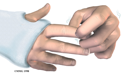
Surgery always means a certain amount of risk to your well being. Surgery is a deliberate, skillful injury to your body. It may take you several weeks to months to heal. Infections and blood loss are two possible complications that your surgeon will want to help you avoid.

You can do your part to make sure you heal well without problems. You can do this by choosing the most nutritious diet. Using supportive supplements for some weeks before having an operation is also helpful. Surgery is a big event. It makes sense to give yourself extra nutrition so you can replace any blood loss. Better nutrition can also help your incisions heal. Your diet and nutritional supplements will provide the raw materials your immune system needs to protect you against infection. These same nutritional elements are what you will use to repair your skin, nerves, blood vessels, muscle and bone. Getting good nutrition will help you make the best of your surgery.

This guide will help you understand

  • basic vitamin and mineral information
  • what specific vitamins and minerals do for you
  • what relation vitamins have to surgery

Information

Some vitamins are water-soluble. This means they dissolve in liquid or fluids. Since your body is mostly made up of fluid, you can quickly and easily absorb water-soluble vitamins. They also leave the body quickly through fluid loss such as urination. They must be taken two or three times every day. This way you’ll keep enough in your system to meet the demands of the day. Each vitamin in the water-soluble B-vitamin group must be taken together in order for each one to work properly.

Other vitamins are fat-soluble. They are absorbed by fat cells. These can be taken just once a day or even just a few times a week. Fat-soluble vitamins are used up fast when you are under high-stress. This is true when you are in pain, when you are fighting infection, or when you are healing an injury.

Our appetites change when we are stressed. We can’t always eat as well as we would like. Taking nutritional supplements when you are extra stressed makes good sense.

Vitamins and Minerals

What do vitamins and minerals do for you?

Vitamin B12

Vitamin B12 is the largest and most complex of all the vitamins. You need vitamin B12 so your body can create energy from your dietary fats and proteins. B12 is needed for you to make hemoglobin. Hemoglobin carries oxygen in the red blood cells. Vitamin B12 is needed for your nerves to function properly and for your moods to stay even. It even helps your memory and brain function.

Vitamin B12 deficiency affects about 15 percent of the people over the age of 60. There are several reasons people are low in B12. Most of these are related to changes in the stomach lining. These changes occur with age, from drinking alcohol, or from infection with bacteria that live in stomach ulcers. Certain drugs such as acid blockers used for gastric reflux or Glucophage used for Type 2 diabetes can also affect the stomach lining.

Folate

The terms folic acid and folate can both be used to refer to this B-complex vitamin.

Folate plays a vital role the work and the growth of all your body cells. Not having enough folate causes a number of different problems in the cells. Some of your immune system white blood cells will be affected. A shortage of folate for the rapidly dividing cells of the bone marrow will result in fewer but larger red blood cells. This causes a type of anemia called megaloblastic or macrocytic anemia. The large, immature red blood cells that result do not carry oxygen normally. If you do not take in enough folate you will experience fatigue, weakness, and shortness of breath. Megaloblastic anemia from folate deficiency is the same as megaloblastic anemia resulting from vitamin B12 deficiency. Never take a folate supplement without making sure you have enough Vitamin B12.

Vitamin B6

Vitamin B6 is needed for the proper function of about 100 essential chemical reactions in the human body.

Vitamin B6 is needed to make heme, a component of hemoglobin. Hemoglobin is found in red blood cells. It is vital to the their ability to transport oxygen throughout your body.

People who are low in vitamin B6 have impaired immune function. This is especially true for the elderly. Sleep, pain, mood, memory, and clear thinking are also affected by a shortage of Vitamin B6. The stress of hospitalization for surgery causes many people to lose sleep. Increased pain and mood changes are also common. You may find your appetite is changed when you are in the hospital. And you may not eat as well as you should. Taking a supplement that contains all of the B vitamin complex, including Vitamin B6, will help decrease the effects of your stress.

Vitamin B1 (thiamine or thiamin)

Vitamin B1 plays a critical role in making energy from food. It is needed for your heart, digestive system, and nervous system. Your muscles especially need vitamin B1 to work properly.

Thiamine deficiency is caused by not eating enough thiamine-rich foods. This occurs most often in low-income groups. Their diets are often high in carbohydrates. Alcoholism is linked with low intake of thiamine and other nutrients. Chronic alcohol use is the main cause of thiamine deficiency in higher income groups.

If you drink large amounts of tea and coffee (even decaf), you may end up with not enough thiamine. This is because of the action of certain enzymes in these drinks. Vitamin C and other antioxidants can protect your thiamine levels by preventing it from changing into an inactive form.

There are no known toxic effects from thiamine in food or from long-term oral supplementation (up to 200 mg/day).

Vitamin B2 (riboflavin)

Vitamin B2 is essential for changing the carbohydrates, fats, and proteins in your food into energy in your cells. It also helps manage drugs and environmental pollution in your liver.

If you do not have enough riboflavin you may not be able to absorb the iron you need to make hemoglobin in your red blood cells. Studies show that increasing your riboflavin intake will increase your red blood cell hemoglobin levels. Riboflavin improves your ability to prevent or recover from iron-deficiency anemia.

No toxic or adverse effects of high riboflavin intake in humans are known. High dose riboflavin therapy makes your urine a bright yellow color. This is a harmless side effect.

Vitamin B5 (pantothenate)

Pantothenic acid is essential to all forms of life. It is another of the vitamins you need to create energy from your food. Vitamin B5 is key to making the special fats that cover your nerves. This covering or lining is called a nerve sheath. It’s needed to pass messages along your nerves to all your tissues. Vitamin B5 helps regulate the production of cholesterol and your major hormones. B5 is needed to make hemoglobin to carry oxygen in your red blood cells. Your liver uses it to break down many drugs and environmental toxins.

Vitamin B5 will speed up wound healing. It can also increase the strength of scar tissue. Pantetheine is a form of vitamin B5 that can help lower your cholesterol and triglycerides if these are too high.

Pantothenic acid is not known to be toxic in humans. Diarrhea can occur with very high intakes (10 to 20 grams/day) of calcium D-pantothenate. Pantethine is generally well tolerated in doses up to 1,200 mg/day.

Vitamin B3 (niacin)

Vitamin B3 is also called nicotinamide or nicotinic acid. It is required for the proper function of more than 50 enzymes. Without it, your body would not be able to release energy or make fats from carbohydrates. Vitamin B3 is also used to make sex hormones and other important communicating molecules. People with high levels of vitamin B3 have less throat and mouth cancers. It can also help with cholesterol health. And it’s important for insulin-dependent diabetes. A severe lack of niacin will cause death from the disease called pellagra.

The usual advice is that you take a daily multivitamin/mineral supplement that will give you at least 20 mg of niacin daily. Higher doses of different forms of vitamin B3 can cause problems. This is true for people with liver disease, diabetes, or gout. It’s also true for anyone with active peptic ulcer disease, cardiac arrhythmias, inflammatory bowel disease, migraine headaches, and alcoholism.

Biotin

Biotin is one of the B-complex vitamins. It is needed for four important enzyme reactions in your body. These enzymes cause cell growth and immune system protection against bacterial and fungal infections. If you do not have enough biotin it can lead to depression, a sense of tiredness, and weakness. You may also notice a rash on your face and numbness and tingling of your hands or feet.

Pregnancy can cause a woman’s biotin levels to drop too low. This may cause birth defects. Not enough biotin can also cause problems with your blood sugar. This is especially true if you have diabetes. If you have brittle fingernails or notice increased hair loss, you may be low in biotin.

Biotin is not toxic even in higher doses. Biotin in capsule form is safe to take in doses up to 200 mg/day in people born with problems absorbing biotin. People with normal ability to digest biotin can take doses of up to 5 mg/day without problems.

Vitamin C (ascorbic acid)

Vitamin C is also known as ascorbic acid. It’s a water-soluble vitamin. Unlike most mammals, humans do not have the ability to make their own vitamin C. You have to get the vitamin C you need through what you eat and in the supplements you take.

Vitamin C is required to make collagen. Collagen is an important part of your blood vessels, tendons, ligaments, and bone. Vitamin C also plays an important role in making up your brain chemistry. Chemical balance is essential for how well you can think. Your moods may even be affected by changes in brain chemistry. Vitamin C is also needed to carry fat into cells for use as energy.

Vitamin C works well as an antioxidant. Even in small amounts vitamin C will protect essential molecules in your body. This includes proteins, lipids (fats), carbohydrates, and the genetic material of your cells. Vitamin C protects your cells from damage caused when you are exposed to toxins or pollutants. Powerful chemicals from smoking or medications can also damage cells.

Your ability to heal after surgery will depend on having proper blood flow to the surgical site. Vitamin C will help you heal from the trauma that is part of your surgery. Vitamin C allows blood vessels to be relaxed and open. You will deliver the most amount of blood to your injured tissues if you have enough vitamin C. Blood flow to injured areas is central to how well we heal. After surgery your circulation will be slowed by inactivity. If you have atherosclerosis or hardening of the arteries, your blood vessels are less able to relax. This adds to the challenge of recovering after an operation.

Adding vitamin C will improve the dilation(opening) of your blood vessels. This is important if you have certain health problems. This includes anyone with atherosclerosis, angina pectoris, congestive heart failure, high cholesterol, or high blood pressure. Improved blood vessel dilation can occur with a dose of 500 mg or more of vitamin C daily.

Vitamin A

Vitamin A is required for recovery from surgery. It is commonly known as the anti-infective vitamin. It is central to normal functioning of your immune system. Vitamin A is also needed to maintain the integrity and function of your skin and mucosal cells. Mucosal cells are the cells that line body cavities such as your mouth, intestines, and stomach. Vitamin A is important in making white blood cells. These are critical in the immune response that protects you against infection and promotes healing of your injuries.

All kinds of blood cells depend on vitamin A. Blood cells come from parent cells called stem cells. Stem cells need vitamin A to mature into normal red blood cells, white blood cells, and platelets. All of these types of blood cells are needed by your immune system to respond to surgery.

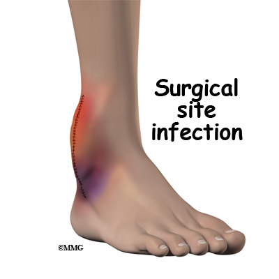
When you enter a hospital or clinic for surgery, many things will stress you. You will also be exposed to bacteria and viruses that your system is not used to. You are at much higher risk for infection at this time. Infection will quickly use up your vitamin A stores. In this way, infection starts a vicious cycle, because not enough vitamin A is related to increased severity and likelihood of death from infectious disease. It is important to go into surgery with a good supply of vitamin A in your tissues. You will need to continue taking enough vitamin A to keep those levels high.

Vitamin A is also needed to move the iron you have stored in your tissues to the developing red blood cells. This is important for making hemoglobin, the oxygen carrier in red blood cells.

Losing a large amount of blood in surgery can cause iron deficiency anemia. If you do not have enough vitamin A, you are more likely to develop iron deficiency anemia. If you supplement with a combination of vitamin A and iron you will be less likely to have anemia than if you take either iron or vitamin A alone. The usual recommended dose of vitamin A is 2,500 IU (International Unit, a measure used for vitamins) of vitamin A. A safe alternative is up to 5,000 IU of vitamin A when at least 50 percent comes from beta-carotene.

Vitamin E

The main function of vitamin E in humans is as an antioxidant. Oxidation damages tissues when free radical ions (unpaired electron in an oxygen atom) are formed in your body. Free radical ions occur during normal function. They also develop when you are exposed to harmful factors like cigarette smoke or environmental pollutants. Other foreign chemicals such as food additives can also cause free radicals. Free radical ions will destroy your cell walls. Vitamin E, in the form of mixed tocopherols, is best suited to stop the damage caused by free radicals.

Vitamin E has been shown to improve immune system functions that decline as people age. It helps increase blood flow. It does this by preventing blood clots and relaxing blood vessel walls.

Some surgeons might worry that vitamin E will cause too much bleeding during surgery. The Food and Nutrition Board of the Institute of Medicine established a tolerable upper intake level (UL) for vitamin E supplements. The Food and Nutrition Board reviewed the research that’s been done. They published a statement that 1,000 mg/day of vitamin E would be the highest safe dose. The scientists at the Institute of Medicine have found that doses of vitamin E as high as 1,000 mg daily are unlikely to result in bleeding in almost all adults. 1,000 mg of vitamin E equals about 1,500 IU. Most supplements contain between 200 IU and 800 IU.

Some doctors advise patients to stop taking supplements with vitamin E before surgery. This will decrease the risk of excess blood loss. Other doctors want their patients to take vitamin E supplements for optimal recovery after surgery. They are comfortable with doses that are well within safe ranges. The usual recommendation is for not more than 400 IU daily.

Beta Carotene

Beta Carotene is converted to vitamin A in the liver as your body needs it. It is a powerful protector against infection. It is an antioxidant that supports your immune system. It also protects your vision. Beta-carotene protects the body from the irritating effects of smoke and other environmental pollutants. It promotes tissue healing. It may be helpful in preventing mouth and stomach ulcers.

Recently, studies showed that synthetic (man made) beta-carotene caused an increase in lung cancer in smokers. When natural beta-carotene was used, no increase in cancer was seen. Natural beta-carotene has been shown to have antioxidant activity that isn’t found in the synthetic form. The suggested dose of beta-carotene is 5 to 6 milligrams (10,000 IU) up to 15 mg (25,000 IU) per day.

Vitamin D

Vitamin D is a fat-soluble vitamin needed for normal calcium metabolism. You make vitamin D in your skin when you are in the sun without clothing or sunscreen shielding you. You can also get vitamin D from some foods.

Cells that are dividing rapidly are said to be proliferating. This is important for growth and wound healing. Vitamin D makes sure cell growth occurs properly.

Vitamin D regulates your immune system function during times of stress. Adequate vitamin D levels are important for decreasing the risk of high blood pressure.

If you do not have enough vitamin D you will not absorb enough calcium. Then your body will steal calcium from your bones. This will increase your risk of osteoporosis and other health problems.

Vitamin D deficiency causes muscle weakness and pain. Taking 800 IU/day of vitamin D and 1,200 mg/day of calcium for three months will increase muscle strength. It can also decrease your risk of falling.

People with dark skin make less vitamin D on exposure to sunlight than those with light skin. The risk of vitamin D deficiency is high in dark-skinned people who live far from the equator. In the U.S., 42 percent of African American women between 15 and 49 years of age were vitamin D deficient compared to 4 percent of white women. The elderly are more likely to stay indoors or use sunscreen. This means they are less likely to make vitamin D in the skin.

Obesity increases the risk of vitamin D deficiency. Once vitamin D is made in the skin or ingested, it is deposited in body fat stores. Storage makes it less available especially to people with large amounts of body fat.

Osteoporosis has many different causes and not enough vitamin D is one of them. Without enough vitamin D, you will not absorb enough calcium. Decreased vitamin D may lead to bone fractures. In order for vitamin D supplementation to be effective in preserving bone health, it has to be taken along with 1,000 to 1,200 mg/day of calcium.

Medical researchers are discovering that vitamin D is more important, and less toxic than once thought. The importance of vitamin D is causing changes in what we consider a safe and healthy intake. Because sunlight exposure is so different for each one of us, it is impossible to decide on a Recommended Daily Allowance (RDA). Therefore, experts have decided to talk about adequate intake levels. They start by assuming that no vitamin D is being made in the skin. This is based on the idea that people simply do not get outside enough. They don’t get enough sunlight. When they do go outside, they tend to cover up with sunscreen. And they wear clothing that covers most of their body.

Multivitamin supplements for children generally provide 200 IU (5 mcg) of vitamin D. Multivitamin supplements for adults usually have 400 IU (10 mcg) of vitamin D. Single ingredient vitamin D supplements may provide 400-1,000 IU of vitamin D, but 400 IU is the most commonly available dose. A number of calcium supplements may also provide vitamin D.

Medical experts have set a vitamin D dose recommendation of 400 to 800 IU/day for children and adults (based on age). For example, infants up to one year should take 600 IUs of VitaminD daily. Children one year old and adults up to age 70 are advised to take 600 IUs. Adults over age 70 should bump the recommended daily allowance up to 800 IUs.

Research published since 1997 suggests that vitamin D toxicity is very unlikely in healthy people at intake levels lower than 10,000 IU/day. Some adults are advised to take higher dosages of vitamin D (up to 2000 IUs per day). You may need more if you have a history of malabsorption (e.g., celiac disease, inflammatory bowel disease, cystic fibrosis, gastric bypass surgery).

Monitoring for signs of vitamin D toxicity is recommended for anyone taking more than 4000 IUs per day. Signs of toxicity include gastrointestinal distress (e.g., nausea, vomiting, poor appetite), weakness, weight loss, increased urination, and heart palpitations. Vitamin D toxicity doesn’t result from sun exposure. Certain medical conditions can increase the risk of too much blood calcium in response to vitamin D. Check with your doctor to be sure you do not have a condition that increases your risk for too much blood calcium. This is important before you take more than 800 IU of vitamin D for a long time.

Iron

Iron is required for a number of vital functions, including growth, reproduction, healing, and immune function. You need the right amount of iron for hundreds of proteins and enzymes.

Heme is an iron-containing compound found in many biologically important molecules. Hemoglobin and myoglobin are heme-containing proteins. Hemoglobin has the vital role of carrying oxygen from your lungs to the rest of your body. Myoglobin is the molecule that supplies oxygen to your working muscles. These are needed for you to move and store your oxygen.

Cytochromes are heme-containing compounds that are needed for cellular energy production and therefore, to life. Cytochrome P450 is a family of enzymes that help make many important biological molecules. Cytochrome P450 helps your body use the drugs you need to take. It helps break down the pollutants you cannot avoid.

Copper

You must have enough copper in your system for normal iron metabolism and red blood cell formation. Anemia is a sign of copper deficiency. Copper is required for you to be able to move iron to your bone marrow for red blood cell formation.

Vitamin A deficiency will make iron deficiency anemia worse. Taking a combination of vitamin A and iron will protect you from anemia better than either iron or vitamin A alone.

A recent study in an elderly population found that high iron stores were much more common than iron deficiency. Do not take nutritional supplements containing iron unless you know for sure you are low in iron. This is important if you are older than 60. If you are in a low normal range, and you’re having surgery in which a lot of blood loss is likely, a brief period of supplementation prior to surgery is worth considering. The usual dose for supplementing iron is between 18 mg and 45 mg daily.

Calcium

Calcium is the most common mineral in the human body. About 99 percent of the calcium in your body is found in your bones and teeth. The other one percent is found in your blood and soft tissue. Calcium levels in your blood and body fluids must be kept within a very narrow range for normal physiologic functioning. The functions of calcium are so vital to survival that the body will steal calcium from your bones. It does this to keep blood calcium levels normal when your calcium intake is too low. Although this complex system allows for rapid and tight control of blood calcium levels, it does so by stealing from your skeleton.

Calcium plays many important roles. It is vital in controlling the constriction (closing) and relaxation (opening) of your blood vessels. It also aids proper nerve impulses, muscle contraction, and release of your hormones.

Calcium is necessary for optimal activity of many of your proteins and enzymes. The binding of calcium ions is required to cause your blood to clot when you are injured. Calcium is a key factor for good recovery from any surgery involving your bones.

Only about 30 percent of the calcium in your food is actually absorbed in your digestive tract. You lose a certain amount of calcium in your urine every day. This depends on how much caffeine you drink. Too much or not enough protein in your diet will affect your calcium absorption and the strength of your bones. Taking extra calcium makes sense when recovering from bone surgery as well as to prevent bone loss.

After adult height has been reached, the skeleton continues to build bone up to age 30. Adult men and women should consume a least 1,000 mg/day of calcium. This will make sure you make the best skeleton you can have, and limits how much bone you lose later in life.

To minimize bone loss, older men and postmenopausal women should consume a total of 1,200 mg/day of calcium. Taking a multivitamin/multimineral supplement containing at least 10 mcg (400) IU/day of vitamin D will help to make sure you absorb enough calcium.
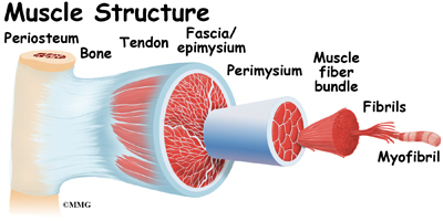
Magnesium

Magnesium plays important roles in the structure and the function of the human body. Over 60 percent of all the magnesium in your body is found in your bones. About 27 percent is found in muscle, while six to seven percent is found in other cells. Magnesium is required by many other nutrients, like vitamin D and calcium, to function properly.

Your system requires magnesium to turn fats and carbohydrates into energy in your cells. Magnesium is needed to create nerve impulses, muscle contractions, and the normal rhythm of your heart.

Proper wound healing after surgery requires the right amounts of calcium and magnesium in the fluid around the cells involved in the injury.

Several studies have found that elderly people tend to have low dietary intakes of magnesium. Intestinal magnesium absorption tends to decrease in older adults, too. And the amount of magnesium lost through urine tends to increase in older people. If you are older than 50 years and are extra stressed by injury, disease, or surgery, supplementing magnesium intake is important to be sure you have the nutrition you’ll need to recover fully.

If you are having surgery that involves your bones, magnesium is very important for the best outcome. The usual recommended dose of magnesium is 400 to 600 mg daily.

Boron

Boron is a trace mineral that is found in highest amounts in fruits, vegetables, nuts, and legumes (peas, beans, and lentils). It is required for mineral metabolism, brain function, and performance. It also helps maintain testosterone and estrogen hormone levels. Boron is important for the prevention of osteoporosis. It does this by affecting mineral metabolism and hormones needed for bone formation.

Research indicates boron improves the uptake of calcium and magnesium into bone. A lack of adequate boron is linked with arthritic changes in joints. Taking boron can decrease joint swelling and pain. It can also increase the levels of helpful sex hormones in post-menopausal women and older men.

The best dose of boron for prevention of osteoporosis and proper tissue function appears to be between 3-6 mg/day

Strontium Citrate

Strontium citrate is recommended for enhancing bone building when you have been diagnosed with fracture-prone brittle bones. Strontium will slow bone weakening and increase new bone formation. You are likely to see major increases in bone mineral density and enjoy reduced risk of fracture. Strontium is a unique, safe, and effective natural aid for osteoporosis resulting from multiple causes.

Strontium is absorbed best when taken away from food and calcium supplements. It is not advised for pregnant women or for those with kidney disease. The usual dose recommended is about 225 mg of elemental strontium three times daily.

Zinc

Zinc is an essential trace element for all forms of life. Zinc deficiency has recently been recognized by a number of experts as an important public health issue. A diet very high in grains like wheat can cause zinc deficiency.

Nearly 100 different enzymes depend on zinc to complete chemical reactions. Almost all of your tissues use enzymes that require zinc.

You must have enough zinc to keep your immune system healthy. If you don’t have enough zinc, you are more likely to become ill from a number of different infectious bacteria or viruses.

If you are taking iron pills, you may not absorb zinc as well as you should. This may occur if you are taking iron before surgery for your red blood cells. If this applies to you, you should take some extra zinc.

Copper assists in the formation of hemoglobin and red blood cells by increasing iron absorption. It is needed for protein metabolism, cell reproduction, and to make healthy nerve cells. Zinc and copper need to be in proper amounts to each other for each to work well. The recommended ratio of zinc to copper for best function is approximately 8:1, or zinc 30 mg and copper 4 mg.

Manganese

Manganese is a mineral element that is nutritionally essential especially for bone and joint strength.

It is part of the main antioxidant enzymes that work in your cells. Not enough manganese will cause abnormal skeletal development. Manganese is needed for the formation of healthy cartilage and bone.

It is also needed for healing after surgery. Wound healing is a complex process that requires increased production of collagen. Manganese is required for collagen formation in human skin cells.

If you are taking calcium, magnesium, or iron pills, you may not absorb manganese as well as you should. In certain situations, you may need to take some extra manganese. For instance, if you are supplementing with iron before surgery to increase your red blood cells, you may want to boost your manganese intake as well.

Women with osteoporosis have been found to have decreased blood levels of manganese. This improves when they take manganese supplements. A study in healthy postmenopausal women found that a supplement containing manganese (5 mg/day), copper (2.5 mg/day), and zinc (15 mg/day) in combination with a calcium supplement (1,000 mg/day) was more effective in preventing spinal bone loss than the calcium supplement alone.

Two recent studies have found that supplements with glucosamine, chondroitin sulfate, and manganese together will help to relieve pain due to mild or moderate osteoarthritis of the knee.

The usual recommended dose of manganese for adult women and men is between 5 and 10 mg daily.

Selenium

Selenium is a trace element that is essential in small amounts to the work of many other nutrients. This includes vitamin C, vitamin E, copper, zinc, and iron. It aids wound healing by regulating cell growth.

Your thyroid gland needs selenium for normal function. Thyroid hormone tells your body how fast to function. If your cells work too slowly, your healing from wounds and your daily tissue repair will not go well.

People who are low in selenium may be more likely to become ill when stressed by difficult events like surgery or exposure to bacteria and viruses. Taking selenium can improve the immune response even in people who have no symptoms of selenium deficiency.

Studies have shown over and over that people who live in areas with low soil selenium and low selenium in their food will have more death from cancer, especially cancer in men.

The usual recommendation for selenium supplementation is 400 mcg/day.

Chromium (polynicotinate)

Chromium (polynicotinate) is a nutritionally essential mineral. It acts to support your blood sugar and insulin functions. It also helps with your fat and protein metabolism. Normal insulin function is required to provide cellular energy and to prevent diabetes.

If you have kidney or liver disease, you may have problems with side effects from too much chromium. You should limit supplemental chromium intake to not more than 50 mcg daily.

Lysine

Lysine is an essential amino acid. It improves how well you absorb calcium and limits how much you lose through your kidneys. Lysine also contributes to strengthening bone.

There have been some reported cases in animal studies that high dose lysine may cause gallstones and increased cholesterol levels. Doses up to 1000 mg three times daily are commonly used for people who need extra lysine.

Muscle Cramps

A Patient’s Guide to Muscle Cramps

Introduction

You have over 600 muscles in your body. These muscles control everything you do, from breathing to putting food in your mouth to swallowing.

When it comes to muscle cramps, the most commonly affected muscles are the muscles of your upper arms, the muscles behind your thighs, and the muscles in the front of your thighs.

This guide will help you understand

  • what muscle cramps are
  • how the problem develops
  • what treatment options are available
  • how muscle cramps can be prevented

Anatomy

What parts of the body are involved?

Muscle Cramps

Muscles are composed of many fibers bundled together; the bigger, more frequently used muscles have more fibers than the smaller, lesser used ones. Among the muscles are voluntary and involuntary muscles. Voluntary, or striated muscles are those that we move by choice (for example, the muscles in your arms and legs). These muscles are attached to bones by tendons, a sinewy type of tissue. Involuntary muscles, or smooth muscles, are the ones that move on their own (for example, the muscles that control your diaphragm and help you breathe). The muscles in your heart are called involuntary cardiac muscles.

Muscle Cramps

When it comes to muscle cramps, the most commonly affected muscles are the gastrocnemius (calf muscles), triceps (the muscles in your upper arms), the hamstrings (the muscles behind your thighs), and the quadriceps (the muscles in front of your thighs).

Causes

What causes muscle cramps?

Muscle Cramps

To move your muscles, your brain sends signals to the voluntary muscles and coordinates the movements that you want. The voluntary muscles contract as they’re being used and they become tighter. The muscles then relax when the movement is complete. When the contraction/relaxation cycles are done repeatedly, as in exercising, the fibers become stronger and the muscles get larger and stronger. However, sometimes the muscles, or just a few fibers within the muscle, contract on their own, causing a muscle spasm or cramp. The difference between a spasm and a cramp is the force of the contraction. If it’s a quick contraction and release of muscle, without pain, it’s a spasm. If the contraction is prolonged and painful, it’s a cramp. Occasionally, cramps are so intense that you can’t use your muscle because it’s so tight and painful. Cramps can be short-lived, a minute or less, or as long as a couple of days. When researchers tested the cramping muscles of some athletes, they found rapid repetitive muscle firing, which could be described as the muscle fibers being hyperactive, in a sense.

Cramps can happen in one muscle, like the hamstring, or they can happen in a number of muscles together, like in your hands if you have writer’s cramp. They can happen once and then not again, or there can be a series of on-again-off-again cramping.

There are several reasons why muscle cramps may occur, including the most common one that is seen in both professional and weekend athletes. These are called exercise-associated muscle cramping or EAMC. These types of cramps fall under the category of paraphysiologic cramps. Those are cramps that affect normally healthy people but are brought on by an event, such as exercise.

Researchers have estimated that marathon runners and triathletes may have a 30 to 67 percent lifetime risk of developing these cramps. Although the exact cause of the cramping isn’t known, researchers do believe that they can be caused by inadequate stretching, muscle fatigue, or lack of oxygen to the muscle. Other causes can also include heat, dehydration, and/or lack of salt and minerals (electrolytes). New research suggests that there may be abnormal motor neuron (nerve) activity at the level of the spine.

Muscle Cramps

Researchers also have noticed that the athletes who suffer from a lot of cramping tend to be older, marathon runners, or have a high body mass index. They don’t stretch regularly, and have a family history of muscle cramps.

Cramps can also happen if you use the same muscles in the same way for too long a period. This could be as you crouch down to work in the garden, type on a keyboard, or write out long lists with paper and pen. The muscles contract and cause the pain.

Occasionally, these types of cramps seemingly come out of nowhere. For example, as we stretch, we often point our toes downward. This motion contracts the muscle in the calf of the leg and can cause a severe cramp or charley horse.

Women who are pregnant may also find that they get more muscle cramps during their pregnancy, but the reason why isn’t clear. Again, these are considered to be paraphysiologic cramps because they are brought on by the pregnancy. Along the same vein, seniors may also be prone to developing muscle cramps. Doctors believe this is due to loss of muscle mass as people age plus inactivity.

Muscle cramps can also occur as a side effect of medications. Diuretics, or water pills, cause you to eliminate fluid from your body. If too much fluid is eliminated too quickly, the resulting dehydration could cause muscle cramps. Other medications can also cause muscle cramps.

Skeletal problems can increase the chances of leg cramps. For example, people with problems like scoliosis (curvature of the spine) could have one leg longer than the other. This imbalance can cause cramping in the leg.

Symptomatic cramps are, as the name suggests, symptoms of an illness that may be causing the cramping. Examples of a few illnesses that can cause muscle cramping are: Parkinson’s disease, tetanus, diabetes, and heart disease. Atherosclerosis (hardening of the arteries) makes it hard for blood to circulate throughout the body as it should. Often, one of the first signs of atherosclerosis is a symptom called claudication or intermittent claudication. Someone who has atherosclerosis may start feeling cramping in one or both legs after walking for a while. At rest, the pain disappears, but it comes back when the person resumes walking.

Radiculopathy (irritation of the nerve root at the spine) is a known cause for muscle cramping, usually at night. Some other illnesses that can cause muscle cramping are cirrhosis of the liver, Black Widow spider bites, and malignant hyperthermia, among others.

Finally, idiopathic muscle cramps are cramps that have no known cause but they are symptoms of a disease, or can be inherited. Sudden nocturnal (occurring at night) leg cramps are an example of this type of cramping.

Symptoms

What do muscle cramps feel like?

Muscle cramps are painful, there’s no doubt about it. The symptoms of muscle cramps usually come on quickly and intensely. They can be so strong that you may have to stop what you’re doing, the discomfort of the cramping making it too difficult to continue. There are also cramps that occur after the fact. These delayed or nocturnal cramps can affect athletes.

The most obvious symptom of a muscle cramp is a sharp, acute pain in the affected muscle or muscles. If it’s a large muscle that is involved, like the one in the calf of your leg, you may be able to feel a knot or hard lump in the muscle, just under the skin.

Diagnosis

How do doctors identify muscle cramps?

Generally, people know what they are experiencing when they have a muscle cramp so they don’t seek medical help to find out what they are and what caused them. However, sometimes muscle cramps are more serious and they are frequent, lengthy, and unbearably painful. If they are caused by an illness, rather than overexertion, the reason for them will need to be found.

Your doctor will first do a physical examination and take your medical history. Let your doctor know is you have been ill recently with vomiting, diarrhea, or fever. Anything that may cause dehydration is important information and should be shared with the doctor.

You’ll be asked when the cramps began, how long have you been having them, how long they last, and what are you usually doing when they start. Other questions, such as are you pregnant, taking any medications (including over-the-counter and natural or herbal remedies), do you smoke, and how much alcohol do you drink, will be asked. Your doctor will also need to know how much and how often you exercise.

Your doctor will want to know exactly where the cramping is occurring. Let your doctor know if there are any other types of pain that happen at the same time. Your doctor will want to know if the cramps are always in the same place or if they occur elsewhere.

Further testing may recommended. Blood tests are usually first since muscle cramping may be caused by dehydration and depletion of salt and minerals (electrolytes). Since pregnant women can be more prone to muscle cramping, a pregnancy test may be ordered for women along with other blood tests.

Additional blood tests may be ordered to check if your thyroid and kidneys are working properly. Your thyroid is a small gland that is just below your voice box and is responsible for making and distributing hormones.

If the blood tests are all within normal range and negative for pregnancy, there are more tests that may be ordered. A vascular Doppler ultrasound uses ultrasound waves to make images on a screen. Using the Doppler (small machine), your doctor looks to see if there are any blockages in the blood vessels.

If your doctor thinks there may be a neurological (nervous system), disorder causing the cramping. One test that may be done is called an electromyography (EMG). To perform an EMG, your doctor will insert a needle into the muscle that has been cramping. The needle has an electrode that will relay to a recording device any electrical activity from your muscle. After the needle has been inserted, you’ll be asked to contract (flex, tighten) your muscle and then relax it.

A magnetic resonance imaging (MRI) scan may also be done. The MRI is a radiological test that uses magnetic waves and a computer to create pictures of the parts of the spine. To perform this test, you must lie in a tube for about an hour. No needles or dye are usually required. The machine takes pictures of the spine one slice at a time. It can do this in multiple directions. It allows the doctor to see the bones and soft tissues of the spine – including the nerves. Your doctor will be looking for anything that may show an injured disc, pinched nerve, or injured nerve that could represent a cause for the cramping.

Treatment

What can be done to relieve the pain from muscle cramps?

The type of treatments required for muscle cramps depends on what is causing them. If you’re having occasional muscle cramps from physical activity or overusing certain muscles, you can usually take care of the cramps yourself. Simply stopping the activity will stop the cramping. If the cramps continue, stretching the cramping muscle – although painful – should release the tension of the muscle. For example, if it is your calf muscle that is cramping, stand facing a wall or solid object that you can hold on to for balance. Keeping your heel of the sore leg as close to the floor as possible, tilt your body (slowly) to the wall or object, stretching the calf muscle. If you’re lying in bed when the cramping starts, you can try pointing your toes straight up towards the ceiling, or grab hold of your toes and pull your foot up towards you.

Muscle Cramps

Some people find that using ice packs can help relax the tense muscles, others have better luck with heat such as from heating pads, warming packs, even warm towels. Be careful when applying ice or heat to a sore part of your body. Ice should never be held directly on the skin. Ice should always be buffered with at least one layer of cloth. Heating pads can get very warm and can cause burns, so be sure to monitor the heat level and keep a layer of fabric between the heat and the skin. Massage may help as well, although it can be painful as the knot is being worked out. If the cause of the cramping is dehydration, then fluids with electrolytes (sports drinks, for example) are essential to balance the fluid loss.

For athletes who experience a lot of cramping good nutrition is important. Adequate fluid and electrolytes may help limit the cramping. This could mean meeting with a dietitian to discuss diet and eating habits.

Some muscle cramps can be caused as a side effect of certain medications. Talk to your doctor. Adjusting the dosages or changing the medication may help the cramping problem. Don’t change any of your prescription drugs or stop taking them without your doctor’s knowledge and approval.

If the cramps are caused by an illness, they should subside by treating the illness. Medications are generally not recommended or used for muscle cramps because of their side effects. Most muscle cramps are short-lived. By the time the medication has started working the cramping has already stopped. There are some cases where doctors may use Botox® to stop cramping in certain muscles. This is decided on an individual basis and depends on the cause and the impact of the cramping.

Because muscle cramps come on so quickly and suddenly, usually resolving just as fast as they came, the best treatment is prevention. Anyone who is about to do something strenuous or athletic should warm up and stretch their muscles first. If you’re moving furniture, digging up a garden, painting, or doing general maintenance that you’re not used to, the muscles you will be using can get fatigued. By stretching them, this should be prevented.

It’s also important to stretch correctly. Don’t stretch quickly. Stretch slowly and hold each stretch for 30 seconds. Any shorter than 30 seconds and there’s no benefit. There is also no benefit for holding it longer than 30 seconds. Some experts suggest that athletes continue to stretch daily. This may keep the muscles flexible. Also remember to stretch after the activity to allow the muscles to cool down.

Other important tips include staying hydrated; drink enough fluids to keep your body’s electrolytes from depleting – but don’t overdo the fluids either. Finally, don’t overdo the exercising, especially in hot weather.

Summary

Since exercise-associated muscle cramping occurs most often in healthy individuals, the important issue is to control cramping frequency and intensity. This can be done through preventative measures, such as proper preparation and stretching, and ensuring adequate fluid intake before undertaking physical exercise or activity.

For those who experience muscle cramps due to illness, their frequency and intensity may be affected by the treatment for the illness. If side effects from medication are causing the cramps, this may be avoided by changing or adjusting medications, dosage, or medication combinations when possible. Ask your doctor if a change in your prescription medication may help.

Nutraceuticals (Dietary Supplements or Vitamins)

A Patient’s Guide to Nutraceuticals (Dietary Supplements)

Introduction

Nutraceutical is a new word, invented by Dr. Stephen DeFelice in 1989. It is two words put together: nutritional and pharmaceutical. Nutraceuticals are dietary supplements that are also called functional foods.

According to the U.S. Food and Drug Administration (FDA), a nutraceutical is any substance that is a food or a part of a food that has medical or health benefits. Nutraceuticals help prevent and treat disease. These products can be single nutrients like Vitamin C. Or they can be dietary supplements such as a multiple vitamin/mineral combined together. A nutraceutical can also be a genetically engineered designer food. You can get nutraceuticals from herbal products. Nutraceuticals come in processed foods like cereals with iron added. Even sports drinks with electrolytes have nutraceuticals in them. The definition of nutraceutical also includes special diets. One example is a diet used to treat chronic inflammation.

This guide will help you understand

  • what a nutraceutical is
  • why nutraceuticals help
  • how effective are nutraceuticals

What is a nutraceutical (dietary supplement)?

Nutraceuticals are most often thought of as a chemical product taken from foods. They have been shown to have health benefits. They also provide protection against chronic disease. There are some nutraceuticals other than vitamins and minerals that are good for spine and joint problems. These include glucosamine sulfate, chondroitin sulfate, fish oils, and the herb Boswellia.

The U.S. Congress defined the term dietary supplement in 1994. It�s part of the Dietary Supplement Health and Education Act (DSHEA). It says that a dietary supplement is a product taken by mouth that contains any chemical ingredient meant to add to what a person gets in their usual diet. The dietary ingredients in these products may include: vitamins, minerals, and herbs or other plant materials. They also include amino acids and substances such as enzymes or organ tissues. Dietary supplements can be extracts or concentrates. They may be found in many forms such as tablets, capsules, soft gels, gel caps, liquids, or powders. They can also be in other forms, such as a snack bar. If they are offered as a snack food, then information on the label must say the product is not food and not the only item of a meal or diet.

How do nutraceuticals and dietary supplements work?

Supplemental nutraceuticals work by giving you extra nutrition. Whenever your diet is not able to meet all the nutrients needed by your body, supplemental nutrients may be helpful. Illness, injury, or extra hard work can increase the amount of nutrients your body needs.

Nutrients are the chemical elements that make up a food. Nutrients are the basic elements of what you eat that give your body what is needed for running the show. That process is what we call metabolism. Certain nutrients such as carbohydrates, fats, and proteins give us energy. Other nutrients like water, electrolytes, minerals, and vitamins are needed for healthy metabolism.

Metabolism is the work your body does to change the food you eat into the tissues and organs of your body. Metabolic processes help the body make the hormones and other chemical messengers. These products of metabolism signal your organs to work properly. Metabolism also refers to the way your cells change the chemical energy in nutrients into mechanical energy or heat. Metabolism is the work of your cells, fluids, tissues, and organs of your body.

You can think of nutrients and metabolism as a kind of bucket brigade. You have a health problem. For this example, we’ll say it’s like having a house on fire. The goal is to put out the fire (heal the health problem). You have a line of people, passing buckets of water to toss on the fire. These are like the nutrients that do the work of metabolism. If there are not enough people in the line to stretch between the water source and the fire, then the process will not be complete. In our example, this means the fire will burn up the house. What if you have some, but not quite enough of the proper elements of nutrition? You may not heal a wound, or repair an injury as well or as soon as you could. If you had all the nutrients you needed for the job, then wound healing and tissue repair would go faster.

Good health and proper function requires a good diet. It must contain all the elements you need for metabolism needed to do the job. A person who is climbing a mountain will need to metabolize more than a person sitting at a desk all day. A person who is healing from surgery needs extra nutrition to meet the demands of tissue repair.

Why are nutraceuticals prescribed?

Nutraceuticals are prescribed because a healthy diet is hard to find. Many people like to think that they eat a healthy diet. They think they have everything their bodies need for good health. Studies have shown that many people can describe a healthy diet. But when they write down what they actually eat, it’s not a nutritionally complete diet.

Other studies have shown that the standard American diet has more than enough calories. But the food does not have the right amounts of vitamins and minerals. It doesn’t have enough fiber, carbohydrate, or protein. All of these things are needed to really be healthy.

Supplemental nutrition is needed because most people do not eat an ideal diet. Over time, your body will begin to show the effects of a less-than-perfect diet. Feeling tired or having colds too often are some of the signals of nutritional deficiencies in your body tissues. Regular joint stiffness and body aches can also be signs of decreased nutritional health. When you are faced with extra challenges like injury or surgery, nutraceuticals are a quick and reliable way to flood your system with chemical elements. For a short time, you will get all you need to restore, repair, and return to excellent health.

When should I consider taking nutraceuticals?

You should use nutraceuticals when your diet does not give you all the nutrition needed for your situation. Different life events need varied nutritional support. For example, most people know that a pregnant woman needs to eat more. She needs to eat a special diet because she is in a special situation. Pregnancy is a state that requires better nutrition for a better outcome. We know that if a woman does not have enough folic acid, she is much more likely to have a baby with certain birth defects.

Too much of something can be as bad as not enough. A woman who eats too much sugar when she is pregnant can develop a kind of diabetes that will also make her baby sick at birth. This is called gestational diabetes. It increases the child’s risk of being overweight. The child is also at greater risk of diabetes as an adult.

In the same way, many other situations require better nutrition for a better end result. If you are injured, you need more nutrition to give your body the raw materials it needs to rebuild your damaged tissues. If you are sick with an infection, you need extra dietary chemistry to help your immune system. Good nutrition helps the immune system fight the virus or bacteria that is making you sick. And if you are having an operation, you will have an injury that needs to be repaired.

Inflammation is part of chronic conditions like arthritis and degenerative disc disease. The inflammatory process can result in destructive, painful diseases. Inflammation is caused in part by poor nutrition. Correcting your nutritional intake can relieve it. Osteoporosis is a condition in which bone is weakened because of nutritional deficiency. Taking supplemental minerals and vitamins can help improve the strength of brittle bones.

Once a disease is started, it is very hard for most people to eat enough of the right foods and to digest them properly to help. It takes a special effort to get the extra nutrition needed to get over an illness. Dietary supplements are used like medicine in this case. They are used to get the needed doses of nutrition in the right amounts required for healing damaged tissue.

Surgery requires good nutrition for the best possible recovery. In a hospital there are many extra challenges to the immune system. There are unusual bacteria and viruses your body must protect itself from. There is the stress of being away from home. Having pain and surgical wounds to heal are also stressors.

Spine surgery requires a lot of blood building. Your nutrition is the source of all the chemistry you need to make new red blood cells. You will have an excited immune system with a lot of work to do to protect you and help you recover. You will need an extra supply of nutritional elements to help you replace the blood you lost during surgery.

How do we know that nutraceuticals work?

At this point, there have been years of research on this topic. Medical researchers have published many thousands of articles. They tell us how individual nutrients and herbal medicines work in the bodies of animals and humans.

This research is carried out in the same way that drugs are studied. There are strengths and weaknesses in using this method to figure out how any substance will work in the human body. We can study how a single chemical will affect a human cell. That information helps us make a reasonable decision about whether it will be useful for that cell’s function or not.

We can also study how having too much or not enough of a certain substance affects large groups of people. This includes vitamins or minerals or a certain dietary item like fat. We know that groups of people who do not have enough of the mineral selenium in their diet get sick more often with everything from infections to cancer. These studies do not tell us if a particular person is sick because they do not have enough selenium. The study only suggests that if that person is part of the group of people studied, there is a good chance they are low in selenium. This puts them at risk of being sick more often. Common sense tells us that selenium is an important nutrient for protection against disease for all people.

Studies have shown that many people in the U.S. do not get enough Vitamin D. More than 90 per cent of people with chronic muscular or skeletal pain, limb pain, and low-back pain are lacking in Vitamin D. Research makes it clear that taking supplements with vitamin D will reduce inflammation. It will also safely treat musculoskeletal pain for many people.

In the same way, research has shown that people with the brittle weak bones of osteoporosis can be helped by supplements. These are used to rebuild and restore the strength of their bones. Minerals and vitamins containing calcium, magnesium, Vitamin D, and L-lysine are used.

Glucosamine sulfate and chondroitin sulfate are two more nutritional elements. They can be used to build connective tissue like the cartilage that cushions your joints. Studies have shown positive results for people with osteoarthritis of the hands, hips, knees, jaw, and lower back. They get relief from painful symptoms when they take regular doses of chondroitin and glucosamine sulfate.

What are the right nutraceutical supplements for me to take?

No drug or herb is a single, simple chemical item. And no single item, like Vitamin E for instance, can be swallowed by two different people and be digested and absorbed in exactly the same way. There is actually no way we can know exactly what a single chemical, drug, vitamin, mineral, or herb is going to do in any one person’s body. It all depends on how a person digests things. How well their liver and kidneys work makes a difference. And they must have all the other chemical elements that the pill they are taking needs in order to work once inside the body.

Research has given medical specialists a general idea about what amounts of different nutrients will be helpful to most people. It makes sense to take recommended amounts of nutraceuticals. This is especially true in situations where you know you will need to fight infection or heal a wound. In fact nutraceuticals can help your body face any extra stress on your system.

Eating right and adding nutraceuticals will make sure you have all the raw materials you need. This combination will help you repair everyday wear and tear. It gives your body a much better chance to deal with damage from disease, accidental injuries, or surgery.

A healthy diet and the right nutraceuticals will:

  • help you control chemical irritants inflaming your tissues
  • reduce nerve pain stimulation
  • decrease tightening and stiffening of muscles, connective tissue, and joints
  • make sure you have all the raw materials you need to heal a wound or prevent loss of bone or tissue function

Septic Arthritis

A Patient’s Guide to Septic Arthritis

Introduction

Septic arthritis is an infection inside a joint that is caused by bacteria. (Septic describes an infection in the body caused by bacteria.) It comes on suddenly and causes very noticeable pain and swelling in the affected joint. It can affect anyone, including infants and children. But it is most likely to affect people over 60 and people with certain health or joint problems.

This guide will help you understand

  • how septic arthritis develops
  • how doctors diagnose the condition
  • what can be done for septic arthritis

Anatomy

Where does septic arthritis develop?

In septic arthritis, the synovial fluid–the lubricating fluid inside a joint–becomes contaminated, or infected, with bacteria. In response to the infection, the body produces white blood cells that try to fight and kill the bacteria. This creates pus that mixes with the synovial fluid inside the joint. The white blood cells that fight infection produce chemicals that may not only kill the bacteria but may also damage the joint surfaces. Once this process begins, the breakdown of the joint can continue even after the infection goes away and the bacteria are all dead.

About 80 percent of cases of septic arthritis involve only one joint. Roughly half of the time a knee is affected. The other most common sites are the hip, shoulder, wrist, ankle, elbow, and small joints in the hands and feet. When more than one joint is affected, the knees are usually involved.

Causes

Why do I have this problem?

The cause of septic arthritis is known. It is caused by a bacterial infection in a joint. Bacteria can get into a joint in many ways. The bacteria may be introduced into the joint as a result of injury, such as a laceration that goes into the joint. Surgery on the joint can introduce bacteria into the joint. There is always a small chance of causing an infection if a needle is placed in the joint to either remove synovial fluid for testing or to inject medications into the joint. The joint infection can also come from infections in the skin around the joint or infections in the bones around the joint. The bacteria can also be carried by the blood from an area of infection somewhere else in the body. Once in the joint, synovial fluid provides a good place for the infection to lodge and grow.

Although they do not cause septic arthritis, several risk factors make it more likely to develop. These risk factors include a suppressed immune system, drug abuse, and other diseases. Other diseases that go along with septic arthritis include rheumatoid arthritis, diabetes, cancer, sickle cell disease, anemia, lupus, liver disease, skin infections, and hemophilia.

Artificial joints and recent joint surgery or arthrocentesis (inserting needles into the joint) are also risk factors for septic arthritis. However, the risk from these sources is small. The rate of septic arthritis from artificial joints is less than one percent. The rate following corticosteroid injections is less than 0.01 percent. Rates of septic arthritis following arthroscopy (surgery conducted using a small TV camera inserted into a joint) range from 0.04 percent to four percent. Septic arthritis caused by arthroscopy usually begins within two weeks of the procedure.

The biggest risk factor in septic arthritis is advanced age. Roughly half the cases of septic arthritis occur in people over age 60. In these patients, 75 percent of the infections occur in joints that have already been affected by arthritis, especially the hips, knees, and shoulders.

Symptoms

What does septic arthritis feel like?

Septic arthritis causes moderate to severe joint pain that comes on from a few hours to a few days. (Doctors refer to this as acute onset.) The pain is much worse when you move or touch the joint. The joint is usually warm and red, and it can’t move easily. You may have other evidence of infection, including a fever, chills, flu-like symptoms, and high white blood cell counts. However, 20 percent of patients with septic arthritis run no fever. Even fewer people over 60 run a fever or have high white blood cell counts when they have septic arthritis.

Diagnosis

How do doctors identify the condition?

The sooner a diagnosis of septic arthritis is made, the better. The more time the infection goes unchecked, the more damage it can do to the joint.

If you have acute onset in one joint and a risk factor of any kind, your doctor will probably test for septic arthritis. A needle will be inserted into the joint and synovial fluid removed and sent to the lab. Your doctor will perform several tests on the synovial fluid. The most important test is probably the measuring the white blood cell count in the fluid. An infection in the joint causes the white blood cell count to be extremely high. Other tests are also useful especially making sure there are no crystals in the fluid that could indicate the presence of gout. A gout attack can easily be mistaken for an infection, and vice versa.

The fluid will be examined under a microscope to look for bacteria. Your doctor will also have the lab perform a culture of the synovial fluid to try and grow the bacteria. In this test a small sample of the fluid is placed in a special container and allowed to incubate in a warm cabinet. The test usually takes from 24 to 36 hours to see if any bacteria grow. This test can also help determine the specific type of bacteria that is causing the infection. If bacteria grow in culture the lab can also test different antibiotics on the bacteria to see which antibiotic will work the best.

Your doctor may ask you to have an X-ray. The X-ray will be used to look for any problems in the joint. It will also provide a baseline of information so your doctor can see the changes in your joint over the course of the infection.

Treatment

What can be done for septic arthritis?

Your doctor will prescribe an antibiotic right away, taking into account your medical history, other diseases or infections you have, and the lab test results. The antibiotic may be changed as your doctor gets information from the testing of the synovial fluid. You will probably be started on intravenous (IV) antibiotics. You will need to continue on oral antibiotics for up to six weeks.

Antibiotics alone may not get rid of septic arthritis. The fluid in the joint may need to be drained at least once a day to remove the pus. This can be done using a large needle. In larger joints such as the knee, arthroscopy may also be used to wash the joint out and remove dead or damaged tissue. Surgery may be necessary to drain and clean joints such as the hip and shoulder. Surgery may also be necessary in joints with bone infections and in joints where the infection can’t be easily controlled.

As your infection begins to clear up, your doctor may suggest that you begin strengthening and range of motion exercises. They will help your joint regain its strength and flexibility. A physical or occupational therapist may oversee your exercise program.

How well your septic arthritis responds to treatment will depend on a number of factors. Most important is how soon you got treatment. Only about 25 percent of patients who wait a week to see a doctor can hope for a complete recovery. So you may feel as though your doctor is rushing you through tests and treatment, but it is for a very good reason.

Tendonitis

A Patient’s Guide to Tendonitis

Introduction

Chronic, or long-term, tendon problems are common. Tendon problems are especially common in people who play certain types of sports. Tendon problems account for almost 30 percent of all running injuries and 40 percent of all tennis injuries.

We use the term tendonitis, which means inflammation of the tendon, to refer to these chronic tendon problems. Doctors now know that the tendon does not always become inflamed when it is injured. Other changes in the tendon can cause tendon pain. However, tendonitis is still the most commonly used term.

This guide will help you understand

  • how tendonitis develops
  • how doctors diagnose the condition
  • what can be done for tendonitis

Anatomy

Where does tendonitis develop?

Tendonitis

Tendons connect muscle to bone. A tendon is made of material called collagen. Collagen is a key building block of the body. Collagen is considered a connective tissue because it forms tough strands that are like the strands of a nylon rope. Like the strands in a rope, the strands of collagen line up. The more strands, and the better they line up, the stronger they are. The tendon is wrapped in a thin, slippery covering called the tendon sheath. The tendon sheath allows the tendon to slide easily against the tissues around it.

Tendonitis

Many parts of the tendon can be injured. Tendon problems can involve the area where the tendon attaches to the bone, the tissue that surrounds the tendon (the tendon sheath), or the main tissues of the tendon. Doctors use different terms to refer to injuries of different parts of the tendon.

Tendon injuries can show up anywhere in your body. Doctors see tendonitis most often in certain sites.

Achilles Tendonitis

Tendonitis

The tendon sheath, the tissues of the tendon, and the attachment to the bone can all become injured in the Achilles tendon, found in the lower leg. Damaged Achilles tendons carry a higher risk of rupturing because of the weight they bear while standing and walking.

Related Document: A Patient’s Guide to Achilles Tendon Problems

Posterior Tibial Tendonitis

Tendonitis

Tendonitis along the inside edge of the ankle and into the instep of the foot is called posterior tibial tendonitis. It is usually caused by age-related degeneration. If this tendon breaks, it can cause the arch of the foot to become flat and painful.

Related Document: A Patient’s Guide to Posterior Tibial Tendon Problems

Patellar and Quadriceps Tendonitis

Tendonitis

Problems in the tendons of the knee occur mostly in people whose exercise involves running or jumping. Patellar tendonitis is also called jumper’s knee.

De Quervain’s Disease and Trigger Finger

Tendon problems are common in the hand and wrist. De Quervain’s disease causes pain in the wrist just above the thumb. Trigger finger generally causes pain in the palm just below the knuckles, but it eventually causes problems with movement.

Related Document: A Patient’s Guide to De Quervain’s Tenosynovitis

Related Document: A Patient’s Guide to Trigger Finger and Trigger Thumb

Lateral Epicondylitis

Lateral epicondylitis, also called tennis elbow, affects the area where the tendons of the elbow attach to bone on the outside of the elbow. It causes pain when using the wrist and hand.

Tendonitis

Related Document: A Patient’s Guide to Lateral Epicondylitis

Tendonitis

Medial Epicondylitis

Medial epicondylitis, also called Golfer’s elbow, affects the area where the tendons of the elbow attach to bone on the inside of the elbow. It causes pain when using the wrist and hand.

Related Document: A Patient’s Guide to Medial Epicondylitis

Rotator Cuff Tendonitis

Tendonitis

Rotator cuff problems of the shoulder range from mild damage to complete tears. They can cause pain even when resting.

Related Document: A Patient’s Guide to Rotator Cuff Tears

Often, the muscles or other tissues of the joints become tight, misaligned, or weak around the area of tendon injury. Some of the pain and swelling of tendonitis may actually be in the surrounding tissues.

Causes

Why do I have this problem?

Doctors don’t know exactly what causes most tendon problems. They think that repetitive stress on the tendon is the most common cause. The tendon can be injured by the repetitive pounding of running and jumping, or by the stress caused by lifting heavy loads over and over again. Tendonitis usually builds up over weeks or months.

If the tendon is too damaged, or if it doesn’t get time to heal, the problem becomes chronic (long-lasting). In general, the heavier the load or the more often the stress is repeated, the more likely you are to develop tendonitis.

Too much stress on the tendons can be made worse by other factors. Lack of flexibility or weakness in your muscles can make tendonitis more likely. Shoes that don’t fit right, poor equipment, or incorrect technique can also increase your risk of tendon injury. Don’t underestimate the benefit of upgrading your equipment. The improved design of athletic shoes over the past few decades seems to have decreased tendon injuries.

Aging seems to cause tendon damage in some cases. As we age, the tissues of the tendon can break down, or degenerate. Age-related tendon problems do not seem to cause inflammation. The tendon material itself is more affected in these conditions and some surgeon’s refer to this type of tendon problem as tendonosis.

Tendonitis

Some researchers think that a decreased blood supply to the tendons can cause the tendon damage in tendonosis. The decreased blood supply does not allow the tendon to get enough oxygen from the blood. This leads to a condition where the tendon degenerates. The collagen material that makes up the tendon actually becomes weaker and loses its nylon rope appearance. This type of degeneration has been noted in the rotator cuff around the shoulder, in the Achilles tendon in the heel and in the tendons of the elbow.

Many factors can work together to cause tendonitis. For instance, a woman in her forties who takes up running may have tendonitis caused jointly by the degeneration of aging and the mechanical stress of running.

Symptoms

What does tendonitis feel like?

Tendonitis causes pain. This is the primary symptom of tendonitis and tendonosis. The affected tendons are sometimes swollen. In some cases this swelling occurs from actual thickening of the tendon itself. In other cases the swelling comes from thickening or swelling of the tendon sheath. Tendon problems often cause pain after resting, such as when you first get up in the morning. This pain usually goes away within minutes, or even seconds.

Tendonitis

The pain or swelling in your tendon may make your joint hard to move. Some types of tendon problems cause crepitus, a crackling feeling when the joint moves. In rare instances the weakened tendon may actually rupture, or break, with a sudden force. This may require surgery to repair.

Diagnosis

How do doctors identify tendonitis?

Your doctor will take a detailed medical history, including many questions about your activities, your job, and your symptoms. Your doctor will also physically examine the sore area. The probing and movement may cause pain, but it is important for your doctor to know exactly where it hurts.

X-rays do not usually show tendon damage. Your doctor may still ask you to get an X-ray or another imaging test to rule out other problems. Sometimes tendon injuries and other joint or muscle problems occur together. In some cases your doctor may recommend a magnetic resonance imaging (MRI) scan to look at the tendons. The MRI scan is a test that uses magnetic waves instead of X-rays. This test shows the tendons and other soft tissues of the body. It can show the damage in the material that makes up the tendon.

In rare cases it is difficult to find the exact source of your pain. In these cases, your doctor may ask you to go through more sophisticated imaging tests. Your doctor may also inject a local anesthetic into the tendon suspected of causing the pain. If the pain goes away, you have found the right tendon.

Treatment

What can be done for the problem?

Tendon problems can be difficult to treat effectively. They can last for many months to several years, even with treatment. You should expect your treatment to take from six to nine months. Even if treatment is effective, your pain may come back. The exact treatment your doctor recommends depends on which tendon is affected.

Your doctor will probably recommend nonsteroidal anti-inflammatory drugs (NSAIDs), such as aspirin and ibuprofen, to help control the inflammation and pain. NSAIDs are usually used for a short time with tendon problems. Your doctor may also suggest ice or heat treatments.

If nothing helps relieve the pain, corticosteroid injections around the tendon are sometimes used. Doctors disagree on whether to use corticosteroid injections in tendons. Even if your doctor does use injections, they are not safe in all cases. Injections into tendons can cause more injury–in some cases, the tendons actually tear. It is generally recommended to give no more than three corticosteroid injections, at least three months apart. Patients need to avoid heavy activity for a few weeks after the injection.

You will need to rest to give your body time to heal. If playing tennis injured your tendon, stop playing tennis until it is completely healed. You should only take part in activities that don’t strain the injured tendon.

Your doctor may refer you to a physical or occupational therapist. A therapist will teach you stretches and exercises to help your tendon heal and regain its strength. A therapist can also assess your work site and athletic equipment and recommend changes to reduce the strain on your tendon. Depending on your type of injury, you may be asked to try such special equipment as arch supports, heel lifts, and splints.

Most people with chronic tendon problems can find ways to relieve the pain and take part in their normal activities, even if the problem doesn’t completely go away. In a few cases, patients can’t find ways to manage the pain even after six months. For these patients, surgery may be necessary. Surgery can be very successful in relieving the pain of chronic tendonitis.

Viral Arthritis

A Patient’s Guide to Viral Arthritis

Introduction

We know that some viruses cause joint pain and inflammation (swelling, redness, and heat). Researchers have wondered for a long time whether some kinds of arthritis with unexplained causes may be the result of a virus. So far no one knows, partly because the virus itself may be long gone before a patient ever develops the pain and inflammation of arthritis.

This guide will help you understand

  • how viral arthritis develops
  • how doctors diagnose viral arthritis
  • what can be done for the condition

Anatomy

Where does viral arthritis develop?

Most viral infections in the body cause a limited illness. Then the body’s immune system destroys the virus, and the symptoms of the illness go away.

In viral arthritis, the immune system’s response to the virus causes inflammation in the joints. Even after the virus is eliminated from the body, the changes in the joint can continue to cause pain and swelling. The joint may even become permanently damaged.

Causes and Symptoms

Which viruses cause viral arthritis?

Several viruses are known to cause problems with joint inflammation and pain. Some of the most common are listed below.

Parvovirus B-19

Parvovirus B-19 is a common virus. About 60 percent of adults have been infected with it at some point in their lives. Parvovirus B-19 causes the illness called fifth disease, which causes a rash on the face and body. People usually get fifth disease as children. About 15 percent of children with fifth disease have some kind of joint pain, which usually goes away quickly. Almost 80 percent of adults who get fifth disease report sore joints within three weeks of the infection. Any joint can be involved, but usually it affects the foot, hand, knee, wrist, and ankle joints on both sides of the body. Most of the time the joint pain clears up in about two weeks, but it has been known to come and go for as long as ten years.

Hepatitis B

Hepatitis B infection, which causes severe inflammation of the liver, can cause a severe and sudden form of arthritis that affects many joints on both sides of the body. The hands and knees are the most common sites, but the wrists, ankles, elbows, shoulders, and other large joints are also affected. The arthritis often starts before the jaundice (yellowness of the skin) of hepatitis, and it may last for several weeks after the jaundice is gone. For patients with chronic (meaning long-lasting) hepatitis, joint pain may come and go.

Rubella

Rubella, a mild but highly infectious viral disease, causes joint pain in many adults, especially women. Joint symptoms tend to appear within a week of the rash common with this disease. The joints are usually not inflamed, but they are stiff and painful. The hands, knees, wrists, ankles, and elbows are most commonly affected. The joint pain of rubella usually goes away within two weeks, but in some cases it can last for several years.

The rubella vaccine also causes joint pain in about 15 percent of people. Joint stiffness occurs about two weeks after the shot and lasts for about a week. The vaccine has been known to cause more severe joint stiffness in some people, however, which can last for more than a year.

HIV

Human immunodeficiency virus (HIV) is the virus that causes AIDS. It is connected to several different forms of arthritis. When people are newly infected with HIV, they often have flu-like symptoms and joint pain. About 10 percent of HIV patients have severe joint pain that comes and goes, mostly in the shoulders, elbows, and knees. Patients with HIV are much more likely to develop reactive arthritis, Reiter’s syndrome, and psoriatic arthritis. In these cases, doctors aren’t sure whether HIV actually causes these forms of arthritis, or whether the arthritis occurs separately. Up to 30 percent of HIV patients also suffer from fibromyalgia.

In some cases, viruses that cause arthritis type symptoms can be carried by insects. Alphaviruses, one such family of viruses, are carried by mosquitoes in Africa, Australia, Europe, and Latin America. All can cause arthritis symptoms.

Diagnosis

How do doctors identify viral arthritis?

There is no specific test for any type of viral arthritis. Your doctor will make the diagnosis based on other symptoms. Usually blood work and X-rays don’t help. In many cases the diagnosis of viral arthritis is made after a search for other causes turns up negative and your symptoms or history suggest a virus as the cause.

Treatment

What can be done for the condition?

In most cases, viral arthritis runs its course fairly quickly. Your doctor may recommend a medication to help with the discomfort. Most of the time this will be an over-the-counter pain reducer like acetaminophen (Tylenol) or a nonsteroidal anti-inflammatory drug (NSAID), such as aspirin or ibuprofen. Your doctor may also recommend use of hot or cold packs on the inflamed joints and rest.

Osteoporosis

A Patient’s Guide to Osteoporosis

Introduction

Osteoporosis is a very common disorder affecting the skeleton. In a patient with osteoporosis, the bones begin losing their minerals and support beams, leaving the skeleton brittle and prone to fractures.

In the U.S., 10 million individuals are estimated to already have the disease and almost 34 million more have low bone mass, placing them at increased risk for osteoporosis. Of the 10 million Americans affected by osteoporosis, eight million are women and two million are men. Most of them over age 65.

Bone fractures caused by osteoporosis have become very costly. Half of all bone fractures are related to osteoporosis. More than 300,000 hip fractures occur in the United States every year. A person with a hip fracture has a 20 percent chance of dying within six months as a result of the fracture. Many people who have a fracture related to osteoporosis spend considerable time in the hospital and in rehabilitation. Often, they need to spend some time in a nursing home.

This guide will help you understand

  • what happens to your bones when you have osteoporosis
  • how doctors diagnose the condition
  • what you can do to slow or stop bone loss

Anatomy

What happens to bones with osteoporosis?

Osteoporosis

Most people think of their bones as completely solid and unchanging. This is not true. Your bones are constantly changing as they respond to the way you use your body. As muscles get stronger, the bones underneath them get stronger, too. As muscles lose strength, the bones underneath them weaken. Changes in hormone levels or the immune system can also change the way the bones degenerate and rebuild themselves.

As a child, your bones are constantly growing and getting denser. At about age 25, you hit your peak bone mass. As an adult, you can help maintain this peak bone mass by staying active and eating a diet with enough calories, calcium, and vitamin D. But maintaining this bone mass gets more difficult as we get older. Age makes building bone mass more difficult. In women, the loss of estrogen at menopause can cause the bones to lose density very rapidly.

The bone cells responsible for building new bone are called osteoblasts. Stimulating the creation of osteoblasts helps your body build bone and improve bone density. The bone cells involved in degeneration of the bones are called osteoclasts. Interfering with the action of the osteoclasts can slow down bone loss.

In high-turnover osteoporosis, the osteoclasts reabsorb bone cells very quickly. The osteoblasts can’t produce bone cells fast enough to keep up with the osteoclasts. The result is a loss of bone mass, particularly trabecular bone–the spongy bone inside vertebral bones and at the end of long bones. Postmenopausal women tend to have high-turnover osteoporosis (also known as primary type one osteoporosis). This relates to their sudden decrease in production of estrogen after menopause. Bones weakened by this type of osteoporosis are most prone to spine and wrist fractures.

In low-turnover osteoporosis, osteoclasts are working at their normal rate, but the osteoblasts aren’t forming enough new bone. Aging adults tend to have low-turnover osteoporosis (also known as primary type two osteoporosis). Hip fractures are most common in people with this type of osteoporosis.

Secondary osteoporosis describes bone loss that is caused by, or secondary to, another medical problem. These other problems interfere with cell function of osteoblasts and from overactivity of osteoclasts. Examples include medical conditions that cause inactivity, imbalances in hormones, and certain bone diseases and cancers. Some medications, especially long term use of corticosteroids, are known to cause secondary osteoporosis due to their impact on bone turnover.

Osteoporosis creates weak bones. When these weak bones are stressed or injured, they often fracture. Fractures most often occur in the hip or the bones of the spine (the vertebrae). They can also occur in the upper arm, wrist, knee, and ankle.

Causes

What causes osteoporosis?

Aging is one of the main risk factors for osteoporosis and osteoporotic fractures. If you are lucky enough to live a long life, you are much more likely to develop weakened bones from osteoporosis. In women, the loss of estrogen in menopause causes bone loss of up to two percent per year. White women over age 50 have a lifetime risk of fracture of about 50 percent. This figure increases with increasing age.

A number of factors contribute to osteoporosis:

  • advanced age
  • female gender
  • low body weight or a thin and slender build
  • recent weight loss
  • history of fractures
  • family history of fractures
  • tobacco use
  • alcohol abuse
  • lack of exercise
  • extended use of certain medications (e.g., corticosteroids, anticonvulsants, and thyroid medicine)
  • Asian or Caucasian race

These risk factors are just as important as a measurement of low bone mass in determining how likely you are to have a fracture. People with low bone mass but no additional risk factors often don’t develop fractures. People with small amounts of bone loss but many risk factors are more likely to eventually develop fractures.

Symptoms

What does osteoporosis feel like?

Fractures caused by osteoporosis are often painful. But osteoporosis itself has no symptoms. That is why it is especially important to get tested if you are a woman past menopause and have any of the above risk factors. Women over 65 should be tested whether or not they have other risk factors. People with other bone problems or who take drugs that weaken the bones should also be tested. An initial screening for osteoporosis is painless and easy.

Diagnosis

How do doctors diagnose osteoporosis?

Free osteoporosis screenings are available in many drug stores and malls. Most of these screenings use a machine that scans the bone in the heel of your foot. It is a fast and simple way to get an idea of your bone density. However, this test is not entirely accurate. Because the heel bears a lot of weight, the test may show normal bone in the heel, even though the hipbones or spine may have low bone density. If the foot scan shows a low bone mass, you should talk to your doctor.

Your doctor will take a detailed medical history to help weigh your risk factors for osteoporosis. Information about your lifestyle and diet will also help your doctor develop a plan to help you build or maintain bone density.

Your doctor may also recommend more precise testing. Dual-energy X-ray absorptiometry (DEXA) is the most common method of measuring bone mass. A DEXA test uses special X-rays of the bones of your hip and spine to show your bone mass in these areas. The bone mass is then compared to that of a healthy thirty-year-old, called a T score. If you are within one standard deviation (SD) for bone density, you have normal bone. (SD is a statistic to measure variations in how a group is distributed.) If you are between one and 2.5 SDs below ideal levels, you are considered to be osteopenic. This means you have a mild form of osteoporosis. If the bone mass is more than 2.5 SDs below ideal levels, you have osteoporosis.

Be aware that DEXA scans are not perfect. Different equipment or different technicians can get somewhat different readings. If you need to have more precise data, your doctor may recommend additional types of bone scans or ultrasound tests.

A single DEXA scan also can’t show your doctor whether your bone mass is stable, increasing, or decreasing. Your doctor may have you take certain medications that create markers in the blood or urine to show what is happening in your bones. These tests will tell your doctor if you have high-turnover or low-turnover osteoporosis.

If bone density tests show that you have weakened bones, your doctor will need to rule out other causes. In some cases, problems with bone marrow or hormone levels can cause bone loss. Blood tests can show these conditions.

In other cases the bone weakening is actually from a condition called osteomalacia. Osteomalacia involves a softening of the bones caused by a lack of vitamin D. Vitamin D in your body comes from food and sunlight. Due to a lack of sunlight, almost 10 percent of people with hip fractures in the northern parts of the world have osteomalacia rather than osteoporosis. Urine and blood tests can help rule out osteomalacia.

In some cases, your primary care physician may refer you to a specialist. If you still have significant bone loss while on medication to prevent bone resorption, you may need to see a specialist. Referral is also advised for patients who have recurring fractures during therapy or repeated, unexplained fractures. Your doctor will help you find the right specialist for your situation.

Treatment Options

What can be done for osteoporosis?

The goal of your treatment plan will be to prevent fractures. This is especially important if you’ve already suffered a fracture from osteoporosis. To prevent fractures, you need to increase your bone mass. If you have high-turnover osteoporosis, you also need to prevent rapid bone reabsorption.

You need to take several steps to increase your bone mass

  • Make sure you get enough calcium and vitamin D. (Vitamin D helps your body absorb calcium.) Researchers think that increased calcium intake alone could reduce the number of fractures by 10 percent. More and more of us don’t get enough calcium and vitamin D, especially as we age. It is difficult to get recommended levels from the food we eat, so supplements are probably necessary. Talk to your doctor about what kind to buy. Calcium comes in many forms–for example, calcium carbonate, calcium citrate, calcium phosphate, and calcium from bone meal. Some forms of calcium need to be taken with food, and others need to be taken with certain types of food. Taking extra calcium and vitamin D improves the effectiveness of all other treatments for osteoporosis.
  • Eat enough calories to maintain a healthy weight. Being too thin increases your risk of osteoporotic fractures. Weight loss can be a cause of bone loss.
  • Exercise. Your bones are constantly adjusting to the demands you put on them. Even low levels of exercise can help you maintain better bone mass. Low-impact exercises like fast walking, stair climbing, and safe forms of dance help stimulate osteoblasts, slowing down reabsorption. Muscle-strengthening exercises, using light weights, can help keep the bones underneath the muscles strong. Balance training can help you prevent the falls that can cause fractures. Your doctor may recommend seeing a physical therapist to help you develop an exercise program with all three kinds of exercises. (See below.)
  • Premenopausal women should avoid overtraining and certain eating disorders, which can cause missed periods (amenorrhea).
  • If you smoke, quit immediately.
  • If you drink alcohol, do so moderately.

Medication

If you follow these recommendations and still have significant bone loss, your doctor may prescribe medications to slow down your body’s reabsorption of bone.

Many drugs are now available for the prevention and/or treatment of osteoporosis. Finding the right drug for each patient takes into consideration benefits and risks of the drug. These are matched against specific patient characteristics and risk factors. Ultimately, the best drug is the one most likely to be taken consistently and/or correctly by the patient. Osteoporosis management is most effective when drugs are taken in such a way that they have their full benefit.

If you are past menopause, hormone replacement therapy can be very effective. Bisphosphonates and calcitonin can also slow your body’s reabsorption of bone.

Studies have shown that 80 percent of women actually build bone mass up to two percent per year while on estrogen replacement therapy. Estrogen has been shown to decrease the occurrence of fractures in the vertebrae by 50 percent and fractures in the hip by 25 percent. Studies have also shown that hormone replacement therapy can also lower rates of coronary artery disease, relieve some symptoms of menopause, and maybe even prevent or postpone Alzheimer’s disease.

Hormone replacement therapy worries many women. Studies have shown that it may increase the risk of breast cancer. For women with a family history of breast cancer or who have had a stroke or thrombophlebitis (blood clots), hormone replacement therapy is probably not appropriate. Other women should at least consider taking estrogen. Its effects on osteoporosis are dramatic. Researchers estimate that, if estrogen were widely used, it could reduce all osteoporotic fractures by 50 to 75 percent.

Hormone replacement therapy must be continued to be effective, however. When a woman stops taking estrogen, she’ll start to lose bone at a very fast rate again. Within seven years, her bone density will be as low as that of a woman who never took estrogen.

Doctors often prescribe calcitonin to patients with fractures. Calcitonin is a non-sex, non-steroid hormone. Calcitonin binds to osteoclasts (the bone cells that reabsorb bone) and decreases their numbers and activity levels. Calcitonin used to be given only by injection, but now it is available in a nasal spray and a rectal suppository. Nasal calcitonin is used most often for women with osteoporosis who are five years or more past menopause and unable to take other approved agents. For unknown reasons, calcitonin seems to relieve pain. Calcitonin from salmon is much more effective than calcitonin from humans.

You and your doctor need to work together to monitor the effects of calcitonin. It is a new drug, and its long-term effects and benefits are still not fully known. More than 20 percent of patients develop a resistance to calcitonin over time, and it stops working for them.

Bisphosphonates also slow reabsorption by affecting the osteoclasts. The FDA has approved a variety of bisphosphonates for the treatment of osteoporosis. You may have heard the names of some of these such as Alendronate (Fosamax), Risedronate (Actonel), or Ibandronate (Boniva).

Some bisphosphonates are taken orally (pill form) on a daily basis. Others are available in weekly or monthly doses. A new injectable bisphosphonate (Zoledronate) can be given annually (once a year). Boniva comes in pill form and can also be injected once every three months. The injectable forms of this drug are used in the management of postmenopausal osteoporosis.

Studies have shown that bisphosphonates increase bone mass and prevent fractures. No one is sure how well bisphosphonates work when used for a long time. But stopping the drug doesn’t seem to cause the rapid bone loss that happens when someone stops taking estrogen. Because there can be some side effects with these medications, you need to work closely with your doctor if you take them.

The FDA is currently studying several drugs that may be used to treat osteoporosis. Some of these drugs, such as sodium fluoride, can be helpful in low-turnover osteoporosis. These drugs affect your osteoblasts in ways that cause them to create more bone. Sodium fluoride may be available in the near future. Other FDA-approved drugs are now available for the treatment of osteoporosis. For example, Raloxifene (Evista) is an anti-estrogen. Anti-estrogens are also called selective estrogen-receptor modifiers (SERMs). SERMs improve bone density and prevent fractures similar to estrogen, yet without increasing the chances of hormone-related cancer. Their main benefit over hormone replacement therapy is that they do not increase the risk of breast cancer.

Raloxifene is used most often for postmenopausal women younger than 65. They must not be at risk for blood clots or have cardiovascular disease. Men may be prescribed the only anabolic agent (Teriparatide/Forteo) approved for the management of osteoporosis. Anabolic usually refers to hormones that build up muscle or bone mass. Forteo is a form of parathyroid hormone used for patients at high risk of fracture. It is usually followed by an agent with antiresorptive effects such as a bisphosphonate.

Lifestyle changes, hormone replacement therapy, exercise prescription, and recent advances in drug therapy can help you take control of your osteoporosis. You and your doctor should be able to find ways to help you prevent the debilitating fractures of osteoporosis.

Physical Therapy

Many patients benefit from working with a physical therapist. People learn safe ways of moving, lifting, and exercising. Treatments also help people gain muscle strength and improved posture.

The physical therapist relies on your test results and the information received by you and your doctor. The therapist also looks at your body height, posture, body movements, strength, flexibility, balance, and your risk for having a fall.

Accurately measuring and recording your body height is a key part of the evaluation. It can give your therapist an idea of how osteoporosis is affecting your bones and posture, and comparing the recordings over a period of time can help track your success with treatments.

Posture exercises are used to help you “be tall”, regaining body height commonly lost with osteoporosis. This training can help patients who have stooped posture, called kyphosis, in the upper part of the spine. In healthy spine posture, the head is balanced on top of the spine rather than jutted forward.

In posture exercises, the goal is to get your body lined up from head to toe, with weight going through your hips. In people with advanced osteoporosis, the upper body is commonly bent forward at the hips. This prevents the hip bones from getting the right amount of stress and weight on them. As a result, the bones weaken and become more prone to fracture.

Your therapist will explain ways you can put good posture into practice. This is called body mechanics–the way you align your body when you do your activities. Remember that healthy posture is balanced with the body aligned from the head to toes. The same posture should be used when you bend forward to pick things up. Instead of rounding out your shoulders and upper back, keep the back in its healthy alignment as you bend forward at the hip joint. This keeps your back in a safe position and prevents the vertebrae from pinching forward. When bones are weakened from osteoporosis, rounding the spine forward when bending and lifting increases the risk of a spine fracture. As the back rounds forward, it pinches the front section of the vertebrae and can cause a fracture.

Your therapist will work with you in designing a safe program of exercise. Weight-bearing exercise strengthens existing bone and the muscles around joints. These types of exercises include walking outdoors or on a treadmill, doing safe forms of dance, and performing resistance training.

Some of the keys to safe exercise for osteoporosis include using good body alignment, avoiding bending or heavy twisting of the trunk, building up the amount of weight and number of repetitions gradually, and being consistent with your exercise program. Avoid exercises that curl your trunk forward such as stationary bike riding, sit-ups, toe-touches, and knee-to-chest exercises. Don’t exercise using abdominal crunch machines or rowing machines. Emphasize exercises that promote upright posture of the spine, such as walking. And do upper body exercises with your back supported in optimal alignment.

Your physical therapist will also check to make sure you have good balance. Poor balance can lead to a hazardous fall. When people with osteoporosis fall, they often end up fracturing a bone–a potentially life-threatening situation. Exercises to improve balance can be as simple as standing on one foot. As your balance gets better, more challenging types of exercises may be given.

People with balance problems can also benefit from practicing tai chi, an exercise form originating in China. In addition to gaining better balance, people who use the exercise movements show improved posture, flexibility, and strength.

Your therapist will continue to compare your test results of body height, posture, balance, and strength to see how well you are improving. The therapist’s goal is to help you become proficient and safe with your exercises, to improve you stature, strength, and flexibility, and to give you tips on how to avoid future problems.

When patients are well underway, regular visits to the therapist’s office will end. The therapist will continue to be a resource, but patients will be in charge of doing their exercises as part of an ongoing home program.

Arthroscopy

A Patient’s Guide to Arthroscopy

Introduction

Until recently, surgery on the inside of any joint meant making a large incision and opening the joint to do even the most minor procedure. Twenty years ago, fiber optics began changing all that and is continuing to change how orthopedic surgeons operate on joints in the body.

  • What is arthroscopy?
  • How is it used?
  • Why is it better?
  • What joints are being scoped?
  • What goes on during an arthroscopy?
  • What are the risks of arthroscopy?
  • What should I do after my arthroscopy?

What is it?

The term arthroscopy basically means to look into the joint. (Arthro means joint, and scopy means look.) So the common phrase scope the joint means to insert an arthroscope into the joint and have a look. In the early days before the development of miniature video cameras, about all the surgeon could do was take a look.

Over the past several years, the development of very small video cameras and specialized instruments have allowed surgeons to do more than simply take a look into the joint. The arthroscope is now used more and more for actual surgical procedures.

How is it used?

Using the arthroscope to assist with joint surgery usually involves making smaller incisions into the joint than those made in a regular open-incision surgery. Once the arthroscope is inserted into the joint, it is used first to try to see the problem. In this way, the problem can be confirmed before making any large incisions and causing any damage unnecessarily. Using the arthroscope as his eyes the surgeon can then use small specialized instruments inserted into the joint through other small incisions to perform the operation. As surgeons have become familiar with this type of surgery, more surgical procedures that were once done with large incisions are now being done arthroscopically.

Why is it better?

All surgical procedures that are done result in damage to tissues that are otherwise normal, because an incision must be made to see the problem. This is particularly bothersome for joints because to enter a joint, the joint capsule and ligaments must be incised (cut into). For minor surgical procedures inside the joint, it is not unusual for the recovery time to be much longer. This is because the normal tissues that were cut must also heal. Also, large incisions into the joint to perform surgical procedures increase the chances for infection. Long procedures where the joint is open to the air can lead to injury to the articular cartilage (the smooth surface of all joints) because it dries the cartilage out.

Arthroscopy causes less damage to normal structures by requiring much smaller incisions through the joint capsule and ligaments around the joint. Arthroscopy also allows the joint to remain closed and reduces the risk of infection and drying out of the articular cartilage. Because of this, the healing time for most arthroscopic procedures is greatly reduced. Rehabilitation is faster, and unnecessary damage to normal structures is avoided.

Arthroscopy has also greatly expanded orthopedic surgeon’s understanding of problems around the joints. In many cases, conditions which were completely unknown before the invention of the arthroscope have been discovered and are now being treated very effectively with arthroscopic surgery.

What joints are being scoped?

Just about every joint in the body has been scoped, but the vast majority of surgical procedures done with the arthroscope are done on the knee and the shoulder. The knee was the primary site of arthroscopic procedures in the early days of arthroscopy and continues to be a common target of the scope.

Probably the most common use of the arthroscope initially was to remove loose bodies from the knee joint or to remove a torn cartilage or torn meniscus. A loose body is a fragment of bone or cartilage that moves around inside the joint and can get caught between the two bones of the joint and cause pain. The menisci (or cartilages) of the knee are two small pieces of tissue that sit between the two bones of the knee joint and act similar to a gasket. A torn meniscus is a common problem that involves these structures and can get caught between the knee and cause pain.

Very complex surgical procedures are now done inside the knee with the aid of the arthroscope. For example, the anterior and posterior cruciate ligaments inside the knee are now almost always reconstructed without opening the knee joint. The procedure is done by using the arthroscope to help see where to drill holes in the bone and to place the ligament graft. Even fractures that involve the joint surface of the lower leg bone (the tibia) are being fixed by watching the fracture through the arthroscope while screws are inserted through small incisions in the skin–a procedure that once required a large incision into the joint.

The arthroscope is continuing to change the way orthopaedic surgeons deal with shoulder surgery as well. The arthroscope is now being used to repair torn tendons around the shoulder, to reconstruct the shoulder in patients who dislocate their shoulder and have instability, and to identify problems in the shoulder that were unknown prior to shoulder arthroscopy.

Other common joints that may be treated with arthroscopy include the ankle, wrist, elbow, and hip.

What goes on during an arthroscopy?

Preoperative

Your doctor will meet with you to explain what the surgery is intending to accomplish and what you can expect. You will be asked to sign a surgical consent form that describes the operation and the potential risks. It is a record that you understand the procedure. If you have any questions, now is the time to ask.

You will also need to review your other medical problems with the doctor in case you have a medical problem that will require evaluation before surgery. If you have serious lung or heart problems, you may need to see your family physician to get medical clearance before the procedure.

Admission

Most arthroscopic surgical procedures are done on an outpatient basis, which means you will probably be able to go home the same day. (Some arthroscopically assisted procedures still require larger incisions to be made around the joint, and these larger procedures may require a night or two stay in the hospital.) Generally, you are asked to not eat or drink anything after midnight on the evening before surgery. If you commonly take medications in the morning, ask your doctor whether you should take them or not.

Anesthesia

There are many different types of anesthesia that can be done, depending on your wishes, your doctor’s recommendations, and, of course, which joint is involved. In general, anesthesia may be local, regional, or general. Each of these types of anesthesia has advantages and disadvantages, but all are quite safe.

Local Anesthesia

Local anesthesia involves injecting lidocaine (or a similar medication) into the joint and in the areas where the incisions are made. This is the same type of anesthesia that is typically used to sew up a laceration, for example. The benefits of this type of anesthesia are that it is relatively safe, has less effect on the rest of your body, and returns to normal quickly after the procedure. The disadvantages are that you may still feel some discomfort during the procedure. This type of anesthesia is not generally used for shoulders or hips.

Regional Anesthesia

Regional anesthesia is probably the most common type used for arthroscopy. It is often referred to as a block (for example, a spinal block). The block is done by injecting lidocaine around the nerves that go to an extremity (in a spinal block, these are the nerves that go to the legs). The advantages to this type of anesthesia are that it does not affect the function of the lungs (a concern if you have lung problems). It does not cause as much nausea as general anesthesia, and it is relatively safer than general anesthesia. You are also awake and can watch the procedure on the TV monitor. The disadvantages are that it takes longer to take affect than going to sleep, and the actual block involves an injection–which may be uncomfortable for a short period of time.

General Anesthesia

General anesthesia is commonly referred to as going to sleep. It is commonly used for arthroscopic procedures–especially procedures which may be long and complicated. The advantages of general anesthesia are that you are not aware of anything that occurs during surgery. The disadvantages are that you may have a hangover with nausea and vomiting due to the drugs used, and you may have a higher risk of lung problems after surgery.

Surgery

Once the anesthesia is working, the operating room nurses and your doctor will prepare the equipment for arthroscopy. There are lots of electronics and equipment that need to be assembled. The surgical limb will be positioned to make it easier for the surgeon to do the surgery. If the procedure is going to be done on the knee, ankle, elbow, or wrist, a tourniquet may be used to stop the blood flow temporarily during the procedure. This makes it easier to see inside the joint.

When the surgery starts, several small incisions are made into the joint. These are usually about one-quarter of an inch long. The number varies from one to about six depending on what will be done. The joint is then filled with clear fluid (usually similar to the salt solution used in intravenous fluids). This expands the joint and allows the camera to work better. The fluid flows through the joint continuously to wash away blood and other material that is present during the procedure.

Once the surgery is under way, various instruments are used to perform the procedure. The camera is used to view the inside of the joint while the other instruments are used to either remove or repair the problem. If you are awake, you can watch the TV monitor and see exactly what your doctor is seeing. Still pictures or video can be taken through the arthroscope to record the condition of the joint. Your doctor performs the procedure while watching the TV monitor and guiding the instruments by what he or she sees on the TV. That’s why arthroscopy is like a very sophisticated video game.

Recovery

After surgery you will be taken from the operating room into the recovery room. There you will be closely monitored by the nurses until they are sure you are not having any problems from either the surgery or the anesthesia. You will probably be able to have something to drink if you are not nauseated. Normally, you will remain in the recovery room thirty to sixty minutes.

Discharge

Once you are ready to be released from the recovery room, you will probably return to where you started–the outpatient surgery department. Here you can wait with your family or friends until you are ready to be released to go home. You can probably have something to eat at this point and prepare to go home. You will be given some instructions at this point about what you should expect and what you should, and should not, be doing at home.

What are the risks of arthroscopy?

Like all surgical procedures, arthroscopy has potential complications. (Some of the more complicated procedures have specific risks.)

In general, arthroscopy requires anesthesia, and there are risks associated with anesthesia. Different types of drugs are given during the procedure that may cause a reaction–even death in very rare instances.

The arthroscopy procedure itself can result in damage to the joint structures or damage to nerves and blood vessels around the joint. Infection of the joint or soft tissues around the joint can occur after arthroscopy and may require additional operations to drain or treat the infection. Infection rates are generally less after arthroscopic procedures due to smaller incisions, shorter operations, and the fact the joint is constantly being flushed out during the procedure.

View animation of pulmonary embolism

Thrombophlebitis (blood clots), sometimes called deep venous thrombosis (DVT), can occur after arthroscopy (usually after arthroscopy of the hip or lower extremity joints). DVT occurs when the blood in the large veins of the leg forms blood clots within the veins. This may cause the leg to swell and become warm to the touch and painful. If the blood clots in the veins break apart, they can travel to the lung, where they get lodged in the small blood vessels of the lung and cut off the blood supply to a portion of the lung. This is called a pulmonary embolism. (Pulmonary means lung, and embolism refers to a fragment of something traveling through the vascular system.) Most surgeons take preventing DVT very seriously. There are many ways to reduce the risk of DVT, but probably the most effective is getting you moving as soon as possible.

What should I do after my arthroscopy?

You will be given specific instructions about what to do after your arthroscopy depending on what type of procedure you had performed. In general, you should take it easy for several days following surgery. You should watch for any signs of infection such as fever or chills, redness around the incisions, or increasing swelling or fluid build-up around the joint. If you have nausea and vomiting that persists for more that twenty-four hours after returning home you may want to discuss a medication change with your doctor. For specific instructions for your procedure you should consult your doctor.

Cast Care

Taking Care of Your New Cast

Cast Care Instructions

Unless you have a waterproof cast, you should keep your cast dry. However, even waterproof casts should be dried out thoroughly after getting them wet. If you don’t dry your cast out after getting it wet, the skin underneath stays damp and can become moldy and smelly. Also, don’t swim with your cast on unless you have the waterproof kind.

To keep your cast dry in the shower, you can enclose it in a plastic garbage bag. Tape the open end of the bag so that water can’t get in. You can also buy a special cast protector.

Casts don’t completely harden for about two days. Be especially careful with your cast during this time. Don’t rest the full weight of the cast on a hard surface during these first two days. Doing so can dent the cast and can cause pressure sores on the skin under the cast. If the cast involves your foot, don’t walk on it for forty-eight hours, even if you have what is known as a walking cast. Walking on a soft cast may cause it to crack or dent.

Keep the casted or splinted limb elevated (propped up) above the level of your heart when you’re able to do so. This will reduce the swelling and help to keep the cast from becoming too tight.

Avoid too much activity and situations that may re-injure you or damage your cast. Remember your cast is there to help your arm or leg heal. It can’t do its job without your cooperation.

Warning Signs

If you have any of the following warning signs after your cast is placed or changed, you should consult your doctor.

Cast Care

Severe Pain

Your injury will probably cause some pain, but if the pain becomes steadily worse after the cast has been placed or changed, that may be a sign that the cast is too tight.

Bluish Nailbeds

Cast Care

The area under the fingernails and toenails is called the nailbed. Normally the nailbeds are pink. When the nailbed is pinched and released, it turns white for a few seconds and then pink again. This is because small blood vessels under the nail are squeezed shut. When you release the pressure, the blood vessels quickly open back up, turning the nail bed pink once again. This is called blanching. If the nailbed has a blue color and doesn’t turn pink again after being pinched and released, this may be a warning sign that the cast is too tight.

Numbness or Tingling

Cast Care

You may feel some numbness after a broken bone has been straightened or fixed if any type of anesthesia (such as a regional block or local anesthesia) was used. This should wear off in a few hours. But if you feel constant numbness or tingling in the fingers or toes of the casted arm or leg, this may indicate that the cast is too tight or that a nerve has been injured.

Immobility of Fingers or Toes

Cast Care

Because of your injury, you may have pain that makes it difficult for you to move your fingers or toes. But if you have no ability to move the fingers or toes of the casted arm or leg, it may be a signal that the cast is too tight and the muscles or nerves aren’t working properly.

Severe Coolness of Fingers or Toes

Many people notice a difference in the temperature of the casted arm or leg. Usually, this difference is minor. But if you have any other problems mentioned here that suggest that the circulation is not good, and the hand or foot is cool, it may be a sign that the cast is too tight.

Severe Swelling

Cast Care

If you have severe swelling above or below your cast, try keeping the casted limb propped up above the level of your heart. Most injuries cause at least some swelling. But if the swelling continues to get worse after the cast is put on, it may be either a sign that the cast is too tight or that something is going on under the cast that needs to be checked.

Dented or Cracked Cast

Cast Care

If your cast is dented or cracked, or starts falling apart before you are due for a checkup, this could lead to problems with healing. Small cracks are probably alright, but if the cast isn’t doing what it is supposed to, it could affect the healing of the injury. You should also check with your doctor if you think the cast isn’t holding the arm or leg still enough.

Cumulative Trauma Disorder

A Patient’s Guide to Cumulative Trauma Disorder

Introduction

Cumulative trauma disorder (CTD) is a broad category that includes many common diseases that affect the soft tissues of the body. CTD in itself is not a disease. Doctors use the concept to understand and explain what may have caused, or contributed to, certain conditions. Examples of the conditions that may be caused or aggravated by cumulative trauma include carpal tunnel syndrome, tennis elbow, and low back pain.

Other terms are often used to describe the concept of CTD. These include repetitive stress injury (RSI), overuse strain (OS), and occupational overuse syndrome (OOS). This document will refer to these categories generally as CTD.

This guide will help you understand

  • what factors may contribute to CTD
  • how doctors diagnose conditions related to CTD
  • what treatment options are available
  • how to prevent CTD

Causes

What causes CTD?

Opinions abound as to what may cause CTD, but there is very little agreement. Some of the theories about how CTD starts are described below. The theories include

  • overuse
  • muscle tension
  • nerve tension
  • psychosocial factors
  • mind-body interaction
  • contributing factors

Overuse

Using muscles and joints after they have become fatigued, or overly tired, increases the likelihood of injury. Overloaded muscles and soft tissues without proper rest have no chance to recover fully. This problem often hampers athletes who have to throw, jump, or run repeatedly. It can also affect people who work in jobs where they keep doing the same action again and again, such as typing, gripping, and lifting.

All body tissues are in a constant state of change. Minor damage occurs continuously, which the body must repair in the normal course of a day. But the damage can occur faster than the repair mechanisms can keep up with it. When this happens, the tissues become weaker. They may begin to hurt. The weaker the tissues become, the more likely they will suffer even more damage. A cycle begins that looks like a spiral–constantly downward.

Muscle Tension

Some doctors think muscle tension causes CTD. To function, or work properly, the body and each of its parts needs a steady supply of blood, rich in oxygen and nutrients. Nutrients are the body’s fuel–glucose, for example. Cutting off or slowing the blood supply harms the tissues of the body.

Tense muscles are believed by some to actually squeeze off their own flow of energy and fuel. Muscles can get energy without oxygen, but the process produces a chemical called lactic acid. This chemical can be a potent pain-causing chemical. Lactic acid is a chemical that can produce a burning feeling when muscles are overexercised. Some physicians believe that lactic acid produced by tense muscles may cause some of the symptoms of CTD.

As pain develops, muscles tighten even further because they attempt to guard the surrounding area. Guarding is a term that is used to describe a reflex that all muscles in the body share. When pain occurs anywhere in the body, muscles around the painful area go into spasm (they tighten uncontrollably) to try to limit the movement in the area. As a result, blood flow is slowed down even more. The muscles begin to ache more. The nerves that have their blood supply reduced and squeezed by muscles begin to tingle or go numb.

Nerve Tension

This theory suggests that nerves become extra sensitive when they’ve become shortened and irritable. It is thought that poor postures used over long periods causes muscles to bulk up and interfere with blood flow. The nerves that course through the body then become shortened and may begin to stick to the nearby tissues. Moving the arm or leg puts tension on the nerve and can cause pain to radiate along the limb. The problem is thought to get worse from stress because the muscles and nerves tense up and become even tighter. Also, when the same activities are done over and over again, the tight nerve is pulled and strained to the point that it can’t heal and eventually becomes a chronic source of symptoms.

Psychosocial Factors

Problems with CTD tend to be more common among people who suffer from boredom, who have poor working relations, who aren’t satisfied with their jobs, and who have unhappy social circumstances. Reasons why this is so are unclear. The number of CTD cases reported may also be influenced by state worker’s compensation rules. States where claims are processed quickly and with greater benefits tend to have higher volumes of CTD cases. Both of these findings suggest that many cases of CTD may be highly influenced by the patient’s perception of the overall situation. Some patients may subconsciously, or consciously, rationalize their symptoms due to many factors that are not medical but have to do with their overall job and social situation.

Mind-Body Interaction

A newer theory suggests that there isn’t really an injury going on in the soft tissues where symptoms are felt. Instead, the problem is said to be coming from influences within the mind. It is theorized that the brain starts producing pain signals as a cover-up for deep-rooted feelings of past emotional pain or problems. Though the idea sounds hard to believe, practitioners using this approach claim they have had success rates as high as 95 percent. Their patients are reported to have gotten swift relief from treatments aimed at the underlying and unconscious emotional triggers.

Contributing Factors

The way people do their tasks can put them at risk for CTD. Some risk factors include

  • force
  • awkward or static postures
  • poor tool and equipment design
  • fatigue
  • repetition
  • temperature
  • vibration

One of these risk factors alone may not cause a problem. But doing a task where several factors are present may pose a greater risk. And the longer a person is exposed to one or more risks, the greater the possibility of developing CTD. Many different symptoms can arise from the accumulation of small injuries or stresses to the body. CTD is not so much a disease as it is a response to excessive demands these factors can place on our bodies without giving them adequate time to recover between.

Symptoms

What does CTD feel like?

The symptoms of CTD usually start gradually. Patients usually don’t recall a single event that started their symptoms. They may report feelings of muscle tightness and fatigue at first. People commonly report feeling numbness, tingling, and vague pain. Others say they feel a sensation of swelling in the sore limb. Some patients with arm symptoms sense a loss of strength and may drop items because of problems with coordination. Symptoms often worsen with activity and ease with rest.

Diagnosis

How do doctors diagnose CTD?

Your doctor begins the evaluation by taking a history of the problem. You’ll probably be asked questions about your job, such as the type of work you do and how you do your job tasks. Other questions give your doctor information about your work conditions, such as the postures you use, the weights you have to lift or push, and whether you have to do repetitive tasks. You may be asked about how you like your job and whether you get along with your supervisors and coworkers.

Your doctor will do a thorough physical examination. Your description of the symptoms and the physical examination are the most important parts in the diagnosis of CTD. Your doctor will first try to determine what conditions are affecting you. For example you may have symptoms of carpal tunnel syndrome or tennis elbow that need to be treated. Second, your doctor will try and determine if cumulative trauma is playing a role in your condition. If so, part of the treatment will be to try and eliminate the source of the cumulative trauma.

There are no specific tests that can diagnose CTD. There are many tests that may be ordered as your doctor looks for specific conditions.

Treatment Options

What treatments are available?

Nonsurgical Treatments

Many nonsurgical treatment approaches are used by physicians, physical and occupational therapists, and other health providers to reduce the symptoms of CTD-related conditions. A splint may be suggested initially to protect and rest the sore area. Anti-inflammatory drugs are often used together with therapy treatments such as ultrasound, cold packs, or electrical stimulation. A series of exercises may be suggested to help tissues move safely while healing.

Surgical Treatment

Surgery is rarely indicated for CTD. Specific conditions that can occur as a result of CTD may require surgery. Unless the doctor is quite sure there is a structural problem, such as a pinched nerve or severely inflamed tendon, then surgery is not usually suggested.

Prevention

How can I help prevent problems of CTD?

The best medicine for treating CTD is to prevent the problem from occurring in the first place. Key items to consider when attempting to prevent problems with CTD are listed below.

Use healthy work postures and body alignment. Posture can have a significant role in CTD. Faulty alignment of the spine or limbs can be a source of symptoms. Using healthy posture and body alignment in all activities decreases the possibility that CTD will strike. Incorrect posture may lead to muscle imbalances or nerve and soft tissue pressure, leading to pain or other symptoms. Most people spend many hours at their work place, and using unhealthy posture during these long hours increases the likelihood that CTD will develop.

Ergonomics

Assessing where and how a person does work is called ergonomics. Even subtle changes in the way a work station is designed or how a job is done can lead to pain or injury.

Rest and Relax

Rest and relaxation (R and R) have recently become front-line defenses in the prevention of CTD. Methods can be as simple as deep breathing, walking, napping, or exercising.

This strategy is useful during work and off hours. Whether at home or work, our bodies need time to recover, which simply means giving them a chance to heal. Rest and relaxation allow the body to recover and provide a way of repairing these injured tissues along the way, keeping them healthy.

The following ideas may be used to foster rest and relaxation at work:

  • Be relaxed. Try to work with your muscles relaxed by pacing your work schedule, staying well ahead of deadlines, and taking frequent breaks.
  • Stop to exercise. Gentle exercise performed routinely through the day helps keep soft tissues flexible and can ease tension.
  • Change positions. Plan ways to change positions during work tasks. This could include using a chair rather than standing or simply readjusting your approach to your job activity.
  • Rotate jobs or share work duties. This can be fun by offering a new work setting, and it allows the body to recover from the demands of the previous job task.
  • Avoid caffeine and tobacco. These can heighten stress, reduce blood flow, and elevate your perception of pain.

Rehabilitation

What can I expect with treatment?

Getting treated right away for symptoms of CTD can shorten the time it takes to heal. Symptoms can sometimes go away within two to four weeks when steps are taken quickly to address the factors that may be causing your symptoms. However, people who keep doing activities when they have symptoms and don’t seek help right away may be headed for a long and frustrating recovery time, perhaps as long as a year or more.

Your doctor may have you work with a physical or occupational therapist to speed your recovery. Your therapist will want to gather more information and will further evaluate your condition. The answers you give and the results of the examination will guide the therapist in tailoring a treatment program that is right for you.

Therapists often teach patients ways to relax. Relaxation ideas often include helping you learn to breathe deeply by using your diaphragm muscle. Taking the time to relax and breathe deeply eases tense muscles and speeds nutrients and oxygen to sore tissues.

Your therapist may apply treatments such as heat, ice, ultrasound, or gentle hands-on stretches to reduce pain or other symptoms. Muscle stretching may be used to restore muscle balance and to improve your posture and alignment. Therapists sometimes apply stretches that are designed to help nerves glide where they course from the spine to the arms or legs.

Strengthening exercises are used to restore muscle balance and to improve your ability to use healthy postures throughout the day.

Therapists pay close attention to your posture and movement patterns. You may receive verbal instruction and hands-on guidance to improve your alignment and movement habits. Helping you see and feel normal alignment improves your awareness about healthy postures and movements, allowing you to release tension and perform your activities with greater ease.

Your therapist will spend time helping you understand more about CTD and why you are feeling symptoms. You may be given tips on how to combat symptoms at work using rest and relaxation. You may also be given specific stretches and exercises to do at work. Your therapist may visit your work place to analyze your job site and to watch how you do your job tasks. Afterward, your therapist can recommend changes to help you do your job with less strain and less chance of injury. These changes are usually inexpensive and can make a big difference in helping you be more productive with less risk of pain or injury.

Your therapist’s goal is to help you understand your condition, to look for and change factors that may be causing your symptoms, and to help you learn how to avoid future problems. When patients are well underway, regular visits to the therapist’s office will end. Your therapist will continue to be a resource, but you will be in charge of practicing the strategies and exercises you’ve learned as part of an ongoing program.

Postsurgical Infection

A Patient’s Guide to Postsurgical Infection

One of the greatest advances in surgery during the twentieth century has been in understanding how to reduce the risk of infection during and after surgery. Any surgical procedure, no matter how small, carries the risk of infection. Simple procedures, such as placing a needle into a joint to remove fluid or inject medications, have a very low risk. More complex operations that require larger incisions and leave the skin open for longer have a higher risk. Operations done to insert something into the body, such as metal plates or artificial joints, must be done carefully to ensure that the foreign material that is left in the body does not carry infection with it. Preventing infection is one of the most important parts of any operation. All members of the health care team take preventing infection very seriously.

Infection prevention begins before surgery. Your doctor will make sure that no infections exist that may spread to cause problems with your surgery. If you have a skin infection, a bladder infection, or some other type of infection, your surgery may be postponed until the infection is treated and under control.

The morning before surgery, you may be asked to shower with antibacterial soap to reduce the number of bacteria on the skin. Before surgery begins, the skin around the incision site will be cleansed and prepped with a disinfectant, such as iodine. Before surgery, you may be given a dose of antibiotics. This is not true for all types of surgeries. Operations that have a low risk of infection usually do not require antibiotics as a precaution. Many orthopedic operations do require antibiotics, especially those operations where something artificial is left in the body.

During surgery, great care is taken to ensure that the entire operation is done under sterile conditions. Surgery is performed in a sterile room that is designed to prevent the spread of infection. All the items needed for surgery are sterilized.

Your wound dressing will also be applied under sterile conditions before you leave the operating room. The wound dressing is an important barrier against infection until the incision seals itself. This usually occurs in the first few days. If an incision is still seeping any type of drainage, this can be an avenue for germs to enter the wound. The dressing will be kept in place until all drainage stops and the incision seals itself.

After surgery, you may continue on antibiotics for 24 to 72 hours. This is to help reduce the chance of infection.

While you are in the hospital or surgery center, your health care team will make sure your bandage stays dry and clean. A bandage that has soaked through can create an entryway for germs to enter the wound. Alert your nurse if this occurs. Before leaving the hospital or surgery center, you will be taught how to take care of your incision. The same instructions apply if you see extra drainage after your return home. Change the dressing, and call to let your nurse know.

Notice that the health care team always washes their hands before changing the bandage. You should do the same. If you develop an infection anywhere else in your body after surgery, let your doctor know. Some infections can spread and cause problems with the surgery.

Follow these guidelines:

  • Wash your hands before changing your bandage.
  • Change your bandage as instructed.
  • Don’t get the incision wet unless your doctor tells you to.
  • Don’t put any medication on the incision unless your doctor tells you to.
  • Don’t scratch or pick at the incision.

If you develop any of the following warning signs of infection, contact your doctor:

  • fever
  • increased redness around the incision
  • increased swelling around the incision
  • drainage from the wound for more than five days after surgery
  • drainage from the wound that is cloudy, yellow, or foul smelling
  • pain that increases and becomes constant.

Fibromyalgia

A Patient’s Guide to Fibromyalgia

Introduction

Fibromyalgia, a common painful disorder among women in their middle years (40 to 60 years old) is no longer considered a “disease” but rather a syndrome. The term “syndrome” is used to represent a group of symptoms that tend to occur together either at the same time or in close proximity to one another. Sometimes fibromyalgia is referred to as fibromyalgia syndrome (FMS).

The most common symptom is widespread pain throughout the body, with especially tender spots near certain joints. The pain stops people with fibromyalgia from functioning normally, partly because they feel exhausted most of the time. Fibromyalgia is a chronic (meaning long-lasting) condition that usually requires many years of treatment. It can occur along with other forms of arthritis or all by itself. It can occur after an injury or out of the blue.

This guide will help you understand

  • how doctors diagnose fibromyalgia
  • what can be done for the condition

Anatomy

Where does fibromyalgia develop?

Pain in fibromyalgia is present in soft tissues throughout the body. Pain and stiffness concentrate in spots such as the neck, chest, shoulders, elbows, knees, buttocks, and lower back. The tender spots don’t seem to be inflamed. Most tests show nothing out of the ordinary in the anatomy of people with fibromyalgia.

Causes

Why does fibromyalgia develop?

The causes of fibromyalgia are unknown, but one thing is for sure: you’re not making it up. Many sufferers have been told that it’s all in your head by family members or other doctors. It is true that people with fibromyalgia are often depressed, and that stress worsens symptoms. But depression and stress don’t seem to be the driving forces behind the disease.

Scientists haven’t been able to unlock all of the secrets behind fibromyalgia syndrome (FMS). Right now, the main theory is that FMS occurs when something goes hay wire in the nervous system. That something may be what’s called central sensitization syndrome. It means your nervous system is ramped up to react too soon, too often, and for too long.

With a dysregulation of the central nervous system like this, there appears to be some kind of mistake within the nervous system in how it recognizes and transmits pain messages. Somehow, the nervous system seems to think even the simplest touch is a noxious (painful) stimuli.

Nervous system dysregulation of this type is likely caused by biochemical abnormalities, altered brain blood flow, and problems with the pain processing mechanisms. Sufferers have lower pain thresholds and lower levels of serotonin, a brain chemical involved in pain, sleep, and mood.

Sometimes FMS occurs as a result of an injury or some other medical condition. For example, patients with rheumatoid arthritis or Lyme disease (inflammatory diseases), metabolic dysfunction (e.g., thyroid problems), or cancer often develop a type of FMS referred to as reactive fibromyalgia. It’s important to identify whether or not the FMS is primary (the main problem) or secondary (caused by other problems).

Folks who have fibromyalgia syndrome (FMS) often have certain triggers that seem to bring on (or increase) symptoms. The triggers vary from person to person but may include degenerative (spinal) disc disease, headaches (all kinds), irritable bowel syndrome, reflux (heart burn), trigger points of the muscles, and poor posture. Anxiety, depression, and post-traumatic stress disorder also seem to be linked with FMS. Having a bipolar illness increases the risk of developing fibromyalgia syndrome (FMS) dramatically.

About 80 percent of all fibromyalgia patients report serious problems sleeping. Because fibromyalgia is so strongly connected to sleep disturbance, in some cases it is possible that the sleep disturbance may be a major contributing factor. In fact, studies have produced fibromyalgia-like symptoms in healthy adults by disrupting their sleep patterns.

New evidence suggests that fibromyalgia is really caused by a dysregulation of the central nervous system. There appears to be some kind of mistake within the nervous system in how it recognizes and transmits pain messages. Somehow, the nervous system seems to think even the simplest touch is a noxious (painful) stimuli. It’s like a ten-alarm fire signal is sent to the brain when a breeze blows by the barn. Nervous system dysregulation of this type is likely caused by biochemical abnormalities, altered brain blood flow, and problems with the pain processing mechanisms. Sufferers have lower pain thresholds and lower levels of serotonin, a brain chemical involved in pain, sleep, and mood.

Symptoms

What does fibromyalgia feel like?

The symptoms of fibromyalgia are long lasting and intense. However, they can vary from day to day. Some of the most common symptoms include

  • pain and stiffness throughout the body, with especially tender points along the back of the neck, top of the shoulders, center of the chest, elbows, knees, low back, and buttocks
  • a feeling of exhaustion that sleep often does not help
  • sleep problems
  • tension headaches
  • numbness or tingling in the arms and hands
  • a feeling of swelling in the hands, although this is not confirmed in physical exams
  • constipation and diarrhea along with abdominal pain (known as irritable bowel syndrome)
  • intense PMS pains in women
  • depression

Diagnosis

How do doctors identify fibromyalgia?

Blood tests and X-rays don’t show fibromyalgia in your body. However, your doctor may do these tests to rule out other conditions. Doctors have only two tools to diagnose fibromyalgia. One is your history of symptoms. The other involves putting pressure on eighteen tender point sites. If you feel pain in eleven of these eighteen sites, you are considered to have fibromyalgia. (However, it is still possible that you can have the disease with pain in fewer sites.)

Fibromyalgia

In some patients, doctors may recommend X-rays to look at the bones near painful spots. The X-rays will not show fibromyalgia but are used to make sure there are no other causes of your pain. Other special tests such as electromyograms, which measure the contraction of muscles, may be used to try to determine if the muscles show abnormalities. Most of the time these tests are negative. A sleep history, and possibly a sleep study, could be important to the diagnosis.

Other conditions can occur along with fibromyalgia that can confound the diagnosis. In some cases, other problems such as Lyme disease, Epstein-Barr virus, viral hepatitis, HIV infection, and thyroid problems mimic fibromyalgia making the diagnosis more difficult. Chronic fatigue syndrome (CFS) may need to be ruled out. CFS is another disease that is difficult to diagnose and has puzzled doctors for many years. CFS and fibromyalgia share many symptoms, especially the severe exhaustion. The major difference is that CFS causes flu-like symptoms, such as low-grade fevers, sore throats, and swollen lymph nodes.

Treatment

What can be done for the condition?

There have been some significant breakthroughs in our understanding (and therefore treatment) of fibromyalgia syndrome (FMS). Today’s modern approach is multimodal, meaning many different treatment options are pursued at the same time. Combining medications with exercise, behavioral counseling, and alternative medicine have made it possible to live a more normal life for those who suffer with this condition.

Usually the first step in the treatment of fibromyalgia is to help patients understand this complex and frustrating disease. Many patients are relieved to learn that the disease is not all in their head. After that, the task is to manage the pain and exhaustion.

Until the exact pathologic pathways are understood, treatment will be more of a management approach. The first-line treatment for fibromyalgia includes medications and a variety of other nonpharmacologic (nondrug) treatment. There isn’t one magic pill patients can take to wipe away the pain, improve sleep, or restore energy. Instead, a wide range of medications are available that can act on the nervous system in a variety of ways. These include tricyclic antidepressants, selective serotonin reuptake inhibitors (SSRIs), selective serotonin-norepinephrine reuptake inhibitors (SSNRIs), and anticonvulsants (also known as antiepileptics).

And for the first time, there are some medications now FDA approved from these categories specifically for fibromyalgia (e.g., pregabalin, duloxetine, milnacipran). In the past, many of the medications were used off-label. This means the medications were intended for something else (like seizures or depression) but were found to be effective for fibromyalgia.

Two of these drugs (duloxetine and milnacipran) are antidepressants. Studies have shown that these medications don’t work because they improve the person’s mood (reduce depression). The chemical pathway of the drug seems to impact pain signals directly.

Neither one of these drugs seems to improve sleep. They do improve energy levels, physical functioning, and cognitive function — probably because they reduce pain, a symptom that can bring a person down in all these areas.

Pain relievers, whether over the counter or prescription, are generally not effective by themselves. Many pain medications are addictive and should be used with caution. Mild pain medications may help in combination with other treatments. Opioid (narcotic) pain relievers, corticosteroids,and nonsteroidal antiinflammatories (NSAIDs) are no longer recommended.

Like many chronic diseases, the symptoms of the disease can be controlled or managed. The successful treatment of fibromyalgia is very much a joint effort between doctor and patient. You must be willing to make lifestyle changes as well as give attention to your psychological health to help control the symptoms. Other treatments or lifestyle changes your doctor may recommend include

  • exercise (aerobic and strength training)
  • biofeedback
  • electrical stimulation
  • electromagnetic wave tharapy
  • nutritional counseling
  • meditation
  • acupuncture, trigger point injection
  • hypnosis
  • pain medication
  • massage
  • heat for temporary pain relief
  • behavioral cognitive therapy

All of the research so far confirms the need to treat this problem with a multidisciplinary approach. A multidisciplinary team of professionals includes doctors, nurses, physical therapists, psychologists, pharmacists, nutritionists, and other practitioners in the healing arts.

You’ll notice that exercise is listed first in the list above. That’s because there is a lot of evidence from good scientific studies that any form of exercise but especially isometrics (contract, hold, relax individual muscles) can be helpful. Pilates-based stretching, yoga, and low-impact aerobic exercise have the greatest benefit.

Anyone with fibromyalgia syndrome (FMS) must be very careful when trying weight-lifting, rowing, or jogging. In fact, these are not really recommended during painful flare-ups. Many people with FMS don’t have any real trouble during exercise. It’s the painful joint and muscle “after shock” that is the worst. Some can barely get out of bed the next day after what seems like a mildly strenuous work out.

That’s why it’s important to have a physical therapist evaluate you and prescribe the optimal mode (type), frequency, intensity, and duration of exercise on an individual basis. There isn’t a one-size-fits-all type of program because of the wide range of physical abilities and disabilities among adults with this condition.

Patients must learn as much as they can both about this condition as well as about themselves and what works best for them. That’s easier said than done. Many times the pain and fatigue keep patients from getting the exercise they need. They become deconditioned and weak, which adds to their pain and loss of function.

This is another place where medications can be very helpful. Medications can help get the pain under control so that the person can move and exercise again. Sometimes several medications are combined to get the most relief of symptoms with the fewest adverse effects. Some medications address the pain or sleep issues, while others deal with the anxiety, depression, or other psychologic disorders.

Early recognition, diagnosis, and treatment can provide a faster resolution of symptoms and much improved prognosis. Reducing and managing symptoms, improving quality of life, and decreasing distress are reasonable goals. But the patient must understand that at the present time, there is no one-best treatment that works for everyone with fibromyalgia. In the ideal plan, the patient is really the manager who consults with these other experts to formulate the most effective plan. Treatment (or more accurately, the management plan) will likely last for many years.

Patients do get better. In the ideal plan, the patient is really the manager who consults with these other experts to formulate the most effective plan. In fact, there is new evidence that half of all adults diagnosed with fibromyalgia early in the development of their disease (and who are adequately treated) no longer have this problem two years later. Many others have reduced their pain to tolerable levels.

Arthritis

A Patient’s Guide to Arthritis

What Is Arthritis?

Arthritis means inflammation of the joints. Inflammation generally includes symptoms of redness, heat, swelling, and pain. Many different diseases can result in inflammation of the joints. Arthritis is therefore a general term that describes more than one hundred different diseases of the joints of your body.

In some types of arthritis, the cause of the disease is known, but in others it is still unknown. Some types of arthritis come on suddenly, and others develop slowly. Any joint can be affected, including your knees, hips, neck, shoulders, and fingers.

The diseases that cause arthritis can also attack muscle and connective tissue around joints. Some diseases may even damage other organs of the body, such as the kidneys, intestines, and heart. Because the diseases inflame the joints, most arthritic conditions and related diseases involve chronic (long-term) pain. Over time, they may cause increasing damage to the joints or soft tissues of your body.

Arthritis

Your joints are beautifully designed to minimize stress and damage while you move. Nearly all joints of the body are synovial joints. Most synovial joints occur where two bones come together and must rub against one another to allow motion. Smooth, slick articular cartilage covers the end of the bones so they don’t rub together. Synovial fluid lubricates the joint. Around the joint, connective tissue forms a watertight sack that is called the joint capsule. Small, fluid-filled sacks, called bursae, cushion parts of the joint. Ligaments connect the bones together and tendons connect the muscles to bones. A problem with any one of these parts can lead to joint pain and inflammation–arthritis.

Arthritis

Many people of all ages suffer from arthritis. It is estimated that forty million Americans–that’s one in seven of us–have arthritis. Almost two-thirds of arthritis patients are women, but for some types of arthritis most sufferers are men.

Arthritis and related diseases are often painful to live with and difficult to treat. But your doctor can help you find treatment and pain management strategies that work for you. The method of treatment will vary depending on the specific disease.

Gout

A Patient’s Guide to Gout

Introduction

Gout is a disease that involves the build-up of uric acid in the body. About 95 percent of gout patients are men. Most men are over 50 when gout first appears. Women generally don’t develop gout until after menopause. But some people develop gout at a young age.

This guide will help you understand

  • how gout develops
  • which parts of the body are affected by gout
  • what can be done for the condition

Anatomy

What is gout?

Gout was the first disease in which researchers recognized that crystals in the synovial fluid could be the cause of joint pain. Synovial fluid is the fluid that the body produces to lubricate the joints. In gout, excess uric acid causes needle-shaped crystals to form in the synovial fluid. Uric acid is a normal chemical in the blood that comes from the breakdown of other chemicals in the body tissues.

Everyone has some uric acid in his blood. As your immune system tries to get rid of the crystals, inflammation develops. For the person with too much uric acid, this inflammation can cause painful arthritis.

Gout

The first attack of gouty arthritis usually happens in just one joint. Half of the time, gout affects the metatarsophalangeal (MTP) joint. This is the joint at the base of the big toe. Eventually, 90 percent of people with gout will have pain in the MTP joint. Other joints that are commonly affected include the mid-foot, ankle, heel, and knee joints. Less commonly gout affects the fingers, wrists, and elbows.

Over time, patients with gout can develop tophi, or lumps that grow around crystal deposits in joints or near pressure points. Tophi most often occur in the fingers, wrists, ears, knees, elbows, forearms, and heels. Tophi can also grow in the kidneys, heart, and eyes.

Causes

Why does gout develop?

Hyperuricemia

The underlying condition that causes gout is called hyperuricemia. It means that you have high levels of uric acid in your blood. This can happen for two reasons: (1) your body creates too much uric acid, or (2) your kidneys don’t excrete the uric acid effectively. Whether or not you will develop gout is related to how bad your hyperuricemia is over time.

For people who create too much uric acid, the cause is usually genetic. Some rare genetic and metabolic disorders can cause overproduction of uric acid, which can eventually lead to gout. The breakdown of purines in the body also releases uric acid. Purines are ingested through certain types of food such as sweetmeats (e.g., liver, kidney, brain) and seafood. The increased intake of fructose-sweetened soft drinks has also been linked with an increased risk of gout. Usually the excess uric acid is then passed out of the body through the urine.

More than 90 percent of people with gout have kidneys that don’t effectively get rid of uric acid. Sometimes this is caused by certain kinds of drugs, such as diuretics, cyclosporine, and low-dose aspirin. Other medical conditions, such as obesity, hypertension, and diabetes, can also make some people more likely to develop gout.

Many gout patients have a combination of overproduction and under-excretion of uric acid. Their bodies create too much uric acid and have problems getting rid of it. This combination of problems happens with drinking alcohol, especially beer. The more alcohol the patient drinks, the worse the problem is. Alcohol both raises uric acid levels in the body and impairs the kidneys’ ability to excrete the buildup.

Acute Causes

Attacks of gouty arthritis seem to be caused by sudden increases in the amount of urate (a solid form of uric acid) in your synovial fluid. This rapid change can be caused by injury to the joint, alcohol use, or use of certain drugs.

An injury that can trigger gout can be very slight. Even gentle exercise can cause inflammation in the joint, although you may not notice it. Once the joint is at rest, the body absorbs some of the water in the synovial fluid. This leaves the synovial fluid more concentrated with urate, which may allow crystals to grow.

Other Factors

Heredity plays a role in gout. In some families, hyperuricemia tends to develop into gout, while in other families it doesn’t. But genes alone don’t account for gout. The rising number of people with gout since World War II suggests more than just a genetic or hereditary basis for this condition.

Dietary changes may be the major difference over the last 60 years. Food scarcity during the 1940s was followed by an increased intake of carbohydrates and especially carbohydrates containing high-fructose corn syrup. The increase in obesity (another risk factor for gout) during the same time supports this idea.

There are several other risk factors for gout. These conditions do not cause gout, but they are closely related to severe hyperuricemia. The risk factors include metabolic syndrome, kidney problems, high hemoglobin levels, high triglyceride levels, and hypertension (high blood pressure). About 14 percent of hypertension patients have gout. A combination of factors such as eating lots of organ meat, a sedentary lifestyle without exercise, and drinking lots of alcohol increases the risk for symptomatic gout.

It is important to note that hyperuricemia alone doesn’t cause gout. Most people with high levels of uric acid in their blood never develop any symptoms of gout. At least five percent of Americans have at least one period of hyperuricemia as adults without showing any symptoms of gout. And most people can tolerate fairly high levels of uric acid in their bloodstream without damage to their kidneys.

Symptoms

What does gout feel like?

Gout causes attacks of very painful joint inflammation. This pain is often described as burning pain. Early gout attacks usually affect only one joint. This joint is most commonly the MTP joint at the base of your big toe. The joint becomes swollen, warm, and red within eight to 12 hours. Most of the time the attacks happen at night. Patients say the pain is so bad the joint can’t even stand the slightest touch. Even the weight of a sheet causes excruciating pain. Walking and standing are almost impossible if the legs or feet are affected. Many patients have flu-like symptoms, including fever and chills. The pain may go away on its own in a few hours, or it may take a few weeks.

Gouty arthritis attacks come and go. There may be months between attacks. Over time the attacks happen more often, last longer, and involve more joints. Eventually the pain doesn’t ever completely go away. The joints stay swollen and tender even between flare-ups, and the flare-ups start to happen every few weeks. Eventually, some patients develop tophi on joints or pressure points and kidney stones.

Diagnosis

How do doctors identify the condition?

Diagnosis is important because crystals within the joint can lead to joint damage. This can happen without you knowing it. Patients with arthritic episodes that come and go may not seek medical help. Some patients are medically evaluated but complete testing is not done. They are misdiagnosed with rheumatoid arthritis. Either of these situations will delay treatment and increase the risk of erosive damage to the joint.

The diagnosis begins with a history of your symptoms and a physical exam. Your doctor will need to look at synovial fluid from the affected joint to identify the needle-like crystals. This is the most important part of the diagnosis. To get a sample of the synovial fluid, the physician performs an arthrocentesis. A long, thin needle is inserted into the affected joint and a small amount of synovial fluid is aspirated or removed. The fluid is sent to a laboratory where it is viewed under a special polarized light microscope to determine if uric acid crystals are present.

Gout

If there are uric acid crystals, then you have gout. But only 80 per cent of the tests are positive when the person really has gout, so this test is not completely accurate. In some cases (such as the midfoot), it isn’t easy to aspirate fluid. Without the use of fluoroscopy (a special X-ray imaging) or ultrasound to guide the needle, aspiration isn’t done. In these situations, the diagnosis is made without joint aspiration when the patient responds favorably to therapy.

Ultrasonography may be helpful in the diagnosis because the crystals form into the shape of rosary beads inside the hyaline cartilage and this can be seen in the ultrasound pictures. Hyaline cartilage coats the ends of the bones to protect them. Ultrasonography can also show the double contour sign. This sign looks like a top covering or extra coating of the joint surface when crystals are deposited in the hyaline cartilage. Ultrasound studies do not replace fluid removal and examination under a microscope because ultrasound does not confirm infection.

The diagnosis must rule out the presence of infection, which can be a hidden problem. Your doctor may also get a blood test to look at the levels of uric acid. However, uric acid levels rise and fall depending on many complex factors in your body. It is possible to have a normal uric acid level while you are having severe gout pain.

If you have tophi, your doctor may want to biopsy one of the lumps.

Your doctor will need to rule out other forms of arthritis. Gout can occur with other forms of arthritis, such as septic arthritis and rheumatoid arthritis. There are also other diseases that cause different kinds of crystals to form in the synovial fluid.

X-rays don’t show doctors much in the early stages of gout. X-rays can help monitor your disease, and they may be needed to rule out other problems.

Treatment

What can be done for the condition?

Gout cannot be cured, but it can be very successfully treated. The main goal of treating gout is to reduce the amount of urate in your blood. Joint crystals will not dissolve or go away unless the serum urate concentration is below six mg/dL.

During the acute or early phase of a gouty attack, doctors prescribe medicines called colchicine, certain nonsteroidal anti-inflammatory drugs (NSAIDs), and corticosteroids to decrease swelling and relieve pain. All of these drugs work quickly and are very effective. The sooner they are given after an attack starts, the faster the pain goes away. These drugs may be given by mouth, through an intravenous line into your bloodstream, or injected directly into the joint. There are some potential adverse side effects of these medications. It may take a bit of time to find the most effective drug with the least intolerable side effects for some patients. But it is important to start treatment during the first few days of an attack to get the best results.

Your doctor may also decompress the affected joint. Aspiration of synovial fluid immediately decreases the pressure in the joint. And the needle leaves a pathway or track that acts as a vent for continued drainage after the needle is removed.

Lifestyle changes can help you manage intermittent gout without using drugs every day. Your doctor may ask you to do the following:

  • Change your diet. Diets that are lower in meat, shellfish, and some other foods can help decrease the amount of uric acid in your body. Avoid fructose sweetened foods and beverages.
  • Quit taking drugs such as diuretics.
  • Lose weight.
  • Quit drinking alcohol.
  • Avoid activities that stress your joints.
  • Drink plenty of fluids to help your kidneys work more efficiently.

If your gout is severe, prolonged, or chronic, you may need to take daily serum uric acid-lowering (SUA) medication to reduce your uric acid levels. Your doctor will put you on the lowest dose possible of medications such as uricosuric drugs or xanthine oxidase inhibitors. Doctors usually prescribe allopurinol (Zyloprim, a xanthine oxidase inhibitor) for patients who overproduce urates or have tophi, kidney disease, or kidney stones. Allopurinol is useful in preventing recurrence of gouty attacks. It blocks the production of uric acid and decreases the formation of purine. For patients who have difficulty getting rid of uric acid through the kidneys, medications to help the kidneys remove more uric acid from the blood may be prescribed as well. Probenecid is one of the commonly prescribed drugs that increase the removal of uric acid in the urine.

Another, serum uric acid-lowering (SUA) medications that has been shown to
reduce the risk of occurrence is Uloric (Febuxostat). Febuxostat has been approved by the FDA for patients with mild to moderate kidney disease. It lowers uric acid slowly enough to avoid flaring up the gout. It isn’t processed by the kidney, so it’s possible patients with kidney disease may be able to take it. But it is metabolized by the liver. Anyone with a liver problem or who abuses alcohol may not be able to take this drug.

As with all medications, you should report any side effects to your doctor right away. Watch for skin rashes, itching, fever, nausea, vomiting, diarrhea, or other new symptoms not present before taking the serum uric acid medications.

Sometimes patients experience a flare-up after taking urate-lowering agents. This reaction can come as a surprise, since you expect your pain and swelling to get better. Flares of this kind mean that old deposits of crystals stored in the tissues are being released. The increase in symptoms is not a sign that new crystals are forming. Don’t stop taking your medication without first checking with your doctor. Getting rid of the old crystals can help protect the joint from further damage.

Doctors seldom treat hyperuricemia without symptoms of gout. However, if hyperuricemia is at least moderately bad over several years, it is more likely to lead to gout. In this case, a doctor may begin treatments to prevent gout. This is called prophylaxis.

A program to control uric acid levels and manage symptoms often includes daily colchicine and allopurinol or probenecid (usually both are not taken at the same time) along with dietary restrictions. Regular follow-up with your physician and blood tests to detect serum uric acid concentration that are above the six mg/dL target level are important in maintaining good control and preventing joint erosion.

So far, there are no drug treatments to prevent the formation and deposit of these crystals in the joints. Researchers continue to look for pharmacological and other biologic therapies that might prevent, if not cure, gout for those who suffer from symptomatic outbreaks.

Joint Injections for Arthritis

A Patient’s Guide to Joint Injections for Arthritis

Doctors recommend joint injections of corticosteroids (also commonly known as cortisone) for many arthritis patients. Cortisone is a powerful anti-inflammatory medication that can reduce joint inflammation. Because the medication is injected directly into the joint, the effects of the medication are concentrated on the painful joint. The injected cortisone can bring the inflammation in the joint under better control and decrease the swelling and pain.

These injections involve putting a needle directly into the joint. Through the needle, your doctor can remove excess synovial fluid (the lubricating fluid found in joints) and inject corticosteroid medication to help reduce the inflammation, pain, and swelling.

This process may sound risky. It is actually safe and fast. It involves little or no pain. And therapeutic injections have important benefits. They deliver the medicine to the exact spot that needs it. They also allow you to use lower and fewer doses of oral steroids, which are highly toxic.

Most doctors give only three to four injections per year in large, weight-bearing joints. This includes joints in your knee and hip. However, patients with arthritis pain that cannot be controlled in other ways can get injections more often.

Complications

The most common side effect from injections is a temporary increase in pain and swelling. Rest, cold packs, and anti-inflammatory drugs help this pain go away within four to twenty-four hours. Studies have shown that about 6 percent of arthritis patients who receive injections in their joints experience this passing pain. It is probably caused by the body’s reaction to the corticosteroid crystals in the medicine. If you have problems with pain and swelling after injections, your doctor may want to change the type of corticosteroid in your next injection.

Another fairly common complication is mild, temporary flushing (sudden redness of the skin) and agitation. Injections can also make diabetic symptoms worse.

There is a chance that the injection can introduce an infection into the joint. However, the odds of this are very slight. Studies show infections following injections happen from 1 in 1000 to 1 in 1600 times. Still, infections in the joint can be very serious. It is important to have an experienced professional perform the injection.

Some doctors and patients have wondered if the cartilage and other tissues of the joints are damaged by injections into the joint. Studies have not shown this to happen.

Systemic Lupus Erythematosus

A Patient’s Guide to Systemic Lupus Erythematosus

Introduction

Systemic lupus erythematosus (also called SLE, or lupus) is an autoimmune disease of the body’s connective tissues. Autoimmune means that the immune system attacks the tissues of the body. In SLE, the immune system primarily attacks parts of the cell nucleus.

SLE affects tissues throughout your body. Five times as many women as men get SLE. Most people develop the disease between the ages of 15 and 40, although it can show up at any age.

This guide will help you understand

  • how SLE develops
  • how doctors diagnose the disease
  • what can be done for the condition

Anatomy

Where does SLE develop?

SLE causes tissue inflammation and blood vessel problems pretty much anywhere in the body. SLE particularly affects the kidneys. The tissues of the kidneys, including the blood vessels and the surrounding membrane, become inflamed (swollen), and deposits of chemicals produced by the body form in the kidneys. These changes make it impossible for the kidneys to function normally.

The inflammation of SLE can be seen in the lining, covering, and muscles of the heart. The heart can be affected even if you are not feeling any heart symptoms. The most common problem is bumps and swelling of the endocardium, which is the lining membrane of the heart chambers and valves.

SLE also causes inflammation and breakdown in the skin. Rashes can appear anywhere, but the most common spot is across the cheeks and nose.

Causes

Why do I have this problem?

Doctors and researchers know quite a bit about the changes in the bodies of SLE patients. But the cause of SLE is a mystery.

Heredity plays a role in SLE. If you have a close relative who suffers from SLE, you are much more likely to develop the disease yourself. However, genes alone do not seem to cause SLE. It seems to be triggered in unknown ways. Not everyone with a tendency toward SLE will develop the disease. Researchers think some of the triggers that set off SLE may be infections, stress, diet, and toxins, including some kinds of prescription drugs. These triggers may also help explain why SLE has a cycle of flare-ups and remissions.

Symptoms

What does SLE feel like?

The symptoms of SLE come on in waves, called flares or flare-ups. In between flares, patients may have almost no symptoms. Almost every SLE patient suffers from general discomfort, extreme fatigue, fever, and weight loss at some point. In addition to these general symptoms, SLE produces different symptoms in different body systems.

Skin

Rashes caused by SLE are red, itchy, and painful. The rash can show up on any part of the body. The most typical SLE rash is called the butterfly rash, which appears on the cheeks and across the nose. SLE also causes hair loss. The hair usually grows back once the disease is under control.

People with SLE tend to be very sensitive to sunlight. Being in the sun for even a short time can cause a painful rash. Some people even get a rash from fluorescent lights at work.

Muscles and Bones

Systemic Lupus Erythematosus

Almost everyone with SLE has joint pain or inflammation. Any joint can be affected, but the most common spots are the hands, wrists, and knees. Usually the same joints on both sides of the body are affected. The pain can come and go, or it can be long lasting. The soft tissues around the joints are often swollen, but there is usually no excess fluid in the joint. Many SLE patients describe muscle pain and weakness, and the muscle tissue can swell.

In its late stages, SLE can cause areas of bone tissue to die, called osteonecrosis.. Osteonecrosis can cause serious disability. It can be caused at least in part by using high doses of corticosteroids over a long time. Corticosteroids help control the symptoms of SLE.

Kidneys

People with SLE usually don’t notice any problems with their kidneys until the damage is severe. Sometimes kidney problems aren’t noticed until the kidneys are actually failing.

Nervous System

SLE can cause headaches, seizures, abnormal blood vessels in the head, and many other problems with the nervous system. SLE can also cause organic brain syndrome. This disorder involves serious problems with memory and concentration, emotional problems, and severe agitation and hallucinations. Any of these symptoms may show up alone, without any other symptoms of SLE.

Membranes

In the body, membranes surround your internal organs. The membranes around your lungs, heart, and the organs in the abdomen become inflamed in SLE. This is called serositis and can be seen on X-rays. Many SLE patients develop symptoms of pleurisy (swelling of the membrane around your lungs). The pericardium, the membrane around your heart, is often affected as well.

Digestive System

Problems with the stomach and intestines are common. Symptoms include abdominal pain, loss of appetite, nausea, and sometimes vomiting. In most cases this is caused by serositis in the membrane around the organs in your abdomen.

Lungs

SLE can cause many lung problems.

  • Inflammation of the lungs, called Lupus pneumonitis, can come on suddenly or slowly. It has many of the same symptoms of pneumonia.
  • A hemorrhage (burst blood vessel) can occur in the lungs.
  • A blood clot can form in the artery going to the lungs.
  • The blood vessels in the lungs can begin to contract.
  • Shrinking lung syndrome involves scarring of the lungs due to long standing inflammation decreases the lungs’ capacity to take in air. It seems that the lungs can no longer hold normal amounts of air.

Blood

SLE causes very low levels of red and white cells in your blood. SLE often does not directly cause low levels of red blood cells, called anemia. Anemia is instead caused by blood loss, kidney problems, or the drugs taken to control the disease.

You may have few of these symptoms, almost all of them, or any combination in between. The disease affects different people in very different ways. There is even a group of patients considered to have latent lupus. They have some chronic SLE symptoms, but the disease never seems to progress into true SLE.

A few patients have drug-induced lupus. In these cases, SLE symptoms come on suddenly while taking certain kinds of drugs. The symptoms are usually milder than in true SLE, and the symptoms go away when the patients stop taking the drug.

Pregnancy

Because so many SLE patients are young women, pregnancy is a major concern. Women with SLE can get pregnant. The disease can be managed during pregnancy if it has already been brought under control. However, the chances of miscarriage, premature birth, and death of the baby in the uterus are high.

Diagnosis

How do doctors identify the condition?

Your description of symptoms and a detailed medical history will help your doctor make a diagnosis. Your doctor will also do a complete physical exam. You will be asked to give a blood sample.

The exams and tests your doctor will do to diagnose SLE depend on your symptoms. You may be asked to have X-rays taken of joints or organs. You may need to have an ultrasound of your heart or kidneys. Many other tests and exams can help diagnose SLE. Because SLE can develop and change over time, your doctor may ask you to do some of these tests again, to help monitor your disease.

Treatment Options

What can be done for SLE?

Treatment options for SLE have improved dramatically since the 1970s. SLE cannot be “cured” and will most likely be a lifelong disease that will require management and attention. You and your doctor should be able to find ways to manage flare-ups.

Lifestyle Alterations

One of the most important parts of dealing with SLE is learning about your own disease. The more you know, the better you can help yourself. Many patients find support groups helpful, both to learn about the disease and to meet others with SLE.

The skin of many SLE patients is extremely sensitive to sunlight. To avoid painful rashes, avoid going outside during the middle of the day, use sunscreen, and wear hats and long clothing.

Infections are common with SLE. If you come down with a fever, talk to your doctor right away. This is especially important if you are on certain drugs, including high-dose corticosteroids and cytotoxic drugs, those that are destructive to cells.

Birth control is important in women with SLE. It is especially important in women with kidney disease. Many of the drugs used to treat SLE can harm a growing fetus, so any pregnancy must be planned with the help of your doctor.

Medication

Several types of drugs can be used to treat the complications of SLE. What your doctor prescribes for you will depend on your symptoms.

  • Nonsteroidal anti-inflammatory drugs (NSAIDs) are used to treat muscle, bone, and joint pain, and mild cases of serositis (inflammation of internal membranes). They may also be used for fevers.
  • Corticosteroids are used in many forms. Creams are rubbed into rashes, and oral or intravenous forms are used to treat flare-ups and to keep the disease under control. Corticosteroids can be injected directly into painful arthritic joints. These drugs are very toxic, however. It is important to use them only when absolutely necessary.
  • Antimalarial drugs (hydroxychloroquine, chloroquine, and quinacrine) work to manage the skin problems of SLE. They can also help treat other symptoms. These drugs can hurt your eyes. As a precaution, you may need to get regular eye exams if you take antimalarial drugs.
  • Methotrexate in low weekly doses can help manage arthritis, rashes, serositis, and other symptoms.
  • Cyclophosphamide, given intravenously, is often used when SLE is affecting the kidneys, heart, and lungs. This is an extremely toxic drug, with many side effects. Patients often experience severe nausea and vomiting and almost total hair loss. The hair does grow back, even when patients must continue taking the drug.
  • Azathioprine can be used instead of cyclophosphamide to treat kidney disease. Doctors consider it less effective, but it is also far less toxic. It can also be used instead of steroids.

SLE progressively damages the kidneys over time. In late stages of the disease, kidney failure requires dialysis or kidney transplants.

SLE is a very serious disease. Its effects on the kidneys, heart, and lungs can cause many long-term problems. Although doctors now have better ways to help you live with SLE, there are not many options to help prevent or reverse the damage to your organs.

Lyme Disease

A Patient’s Guide to Lyme Disease

Introduction

Lyme Disease Lyme disease is an inflammatory disease caused by tick bites. It is the most common tick-borne disease in North America, Europe, and Asia. Connecticut has the highest annual rate of new cases of Lyme disease each year. The name Lyme disease was used because of the number of children in Lyme, Connecticut who first developed this problem back in the late 1970s.

More than 90 percent of the Lyme disease cases in the United States continue to occur in Connecticut and nine other states including New York, New Jersey, Rhode Island, Massachusetts, Pennsylvania, Wisconsin, Delaware, Maryland, and Minnesota.

It is possible to visit these areas and have the infection show up later, when you are back home. And although children between the ages of five and nine seem to be affected most often, adults between the ages of 55 and 59 seem to have a peak incidence as well. The summer months are the peak time when infection may occur, but cases have been reported in all 50 states year-round.

This guide will help you understand

  • how Lyme disease develops
  • how doctors diagnose the condition
  • what treatments are available

Anatomy

What parts of the body are affected?

Lyme disease can cause inflammation in many systems of the body if left untreated. It mostly affects the skin, joints, the heart, and the nervous system. When the joints are affected, the knee is involved most often. Lyme-associated arthritis has also been reported in the shoulder, elbow, hip, and ankle.

Causes

Why do I have this problem?

Lyme disease is caused by Borrelia burgdorferi, a spiral-shaped organism transmitted through the bite of an infected tick. The human body mounts a reaction to the infecting organism that triggers production of inflammatory agents throughout the body. These inflammatory agents are the chemicals produced by the body’s immune system that normally fight off infection.

Symptoms

What does Lyme disease feel like?

Doctors divide the symptoms of Lyme disease into three phases: early localized, early disseminated, and late disease. The three phases of symptoms can overlap. Many patients never show early symptoms at all.

Early localized disease refers to a red rash in the area of the tick bite. This rash shows up two to 30 days after the tick bite. The bite itself is usually found near the waist or belt line, or in other warm, moist areas of the body. In children, the rash is seen first on the head and neck, arms and legs, or back and less often on the abdomen, groin, or chest. The bite may burn, itch, or hurt. The rash usually grows over a period of days. The rash may be in a bulls-eye pattern, red with a white spot in the middle, or completely red.

Most patients with early localized Lyme disease also complain of flu-like symptoms, such as headaches, fatigue, and muscle and joint pain.

Early disseminated disease shows up days to months after the tick bite. Patients may not remember any rashes or bites. The most common symptoms of early disseminated disease are cardiac and nervous system problems. About eight percent of patients who have the rash and are not treated with antibiotics develop heart problems referred to as Lyme carditis. Heart effects include heart blockage and weakening of the heart muscle. The patient may experience dizziness, fainting, fatigue, shortness of breath, and/or heart palpitations. Most of the time, the heart symptoms clear up on their own or after treatment.

About ten percent of patients who don’t receive antibiotics develop damage to the nervous system. Neurologic symptoms also tend to go away on their own, but very slowly. These neurologic symptoms can include meningitis and headaches. Individual nerves can be affected causing numbness, weakness, and pain in the areas the damaged nerve travels.

Late disease symptoms develop months or even years after the tick bite. Some patients who have late disease symptoms have never had any other symptoms of Lyme disease. Late disease causes arthritis pain that comes and goes in many joints. About ten percent of people with late Lyme disease develop chronic (long-lasting) arthritis of one knee. Other joints can be affected (e.g., hip, shoulder, elbow, ankle) but the number of patients with other joint involvement is considerably less.

Many patients who have had Lyme disease describe headaches, fatigue, and joint pain that can last for months after treatment. These problems generally go away without any extra treatment, but very slowly.

Diagnosis

How do doctors identify the condition?

Doctors diagnose Lyme disease based on your health history and a physical exam. Your doctor may order blood tests, but they are only used to confirm the diagnosis. The techniques used to test your blood are called ELISA and Western blot. Both tests can sometimes give false positive or unclear results. If you have had the infection for less than six weeks, your body may not even be making enough antibodies to be detected in the tests.

Lyme disease affecting the knee must be differentiated from septic (infectious) arthritis, which has both a different cause and a different treatment. The two distinguishing features of septic knee arthritis that set it apart from Lyme knee arthritis are refusal to put weight on the knee and fever (more then 101.5 degrees Fahrenheit). Patients with Lyme disease may have a low-grade fever and pain on weight-bearing but do not exhibit the high fever and refusal to put weight on the affected leg observed more often with septic knee arthritis.

When trying to rule out septic arthritis, the synovial fluid (the lubricating fluid of a joint) or spinal fluid may need to be analyzed. Studies show that patients with septic (infectious) knee arthritis are 3.6 times more likely to have a high synovial fluid cell count compared with patients with Lyme disease. But some patients with Lyme disease have elevated synovial fluid cell count, too so this test is just one of many tools used to diagnose the problem. The fluid can also be cultured to identify the presence of bacteria such as staphylococcus aureus (staph infection), streptococcus pneumonia (strep infection), or other less common types of bacterial infections. Bacteria associated with septic arthritis help rule out a diagnosis of Lyme disease.

Treatment Options

What can be done for Lyme disease?

Two to four weeks of antibiotics almost always cures Lyme disease. Early treatment usually prevents later heart, nerve, and joint symptoms. If you have symptoms of early disseminated or late disease, your doctor will probably start you on intravenous (IV) antibiotics.

In a small number of cases, it may be necessary to surgically drain the joint of fluid. This is especially true if there is a suspicion of septic arthritis that cannot be confirmed or differentiated from Lyme disease. In cases of true Lyme joint disease that does not respond to medical treatment, the surgeon may have to remove some of the damaged joint synovium. The procedure is called a synovectomy.

Headaches, fatigue, and muscle and joint pain may continue for months after finishing the antibiotics. Neurologic damage may take even longer to go away, as the nervous system regenerates only one or two millimeters each day. This does not mean that you need more antibiotics. Your body just needs more time to heal. If the antibiotics do not help your symptoms at all, you and your doctor should consider that Lyme disease may not be the cause of your symptoms and look for other possible causes.

The risk of getting Lyme disease is very small, even if a tick has bitten you. Doctors do not treat you just in case. You must show symptoms.

The best treatment for Lyme disease is prevention. Check for ticks at the end of a day outdoors. Wear light clothes so you can see ticks. Wear long clothing, and tuck your pants into your socks. And finally, use a bug spray that contains DEET (a chemical used in some types of insect repellents) to keep the ticks away.

Osteoarthritis

A Patient’s Guide to Osteoarthritis

Osteoarthritis (OA) is the most common form of arthritis. In fact, more than 75 percent of people older than fifty-five show the joint deformations of OA on X-rays. But most of these people have no symptoms. For people who do have the joint pain and stiffness of OA, it can become a crippling disease. Some people suffer from OA in just one joint, while others have it in several joints. It affects more women than men, and most OA patients are over 45.

This guide will help you understand

  • how OA develops
  • how doctors diagnose the condition
  • what can be done for OA

Anatomy

Where does OA develop?

Osteoarthritis

OA is most common in the small joints of the hands, the spine, the knees, the hips, and certain toe joints. OA primarily affects the articular cartilage, the slippery, cushioned surface that covers the ends of the bones in most joints and lets the bones slide without rubbing. Articular cartilage also functions as a shock absorber.

In OA, the articular cartilage becomes damaged or worn away. As this happens, the joint no longer fits together well or moves smoothly. In the early stages of OA, the cartilage actually becomes thicker as your body tries to repair the damage. The repaired areas are more brittle than the original cartilage, and these brittle areas begin to wear away and become thin. They may even wear away entirely. This eventually leads to a condition called eburnation, in which the bones become thick and polished as they rub together. X-rays can show these changes in the cartilage and bones.

But OA is not just a disease of the cartilage. The damage to the cartilage seems to start a sort of chain reaction that involves all the parts of the joint. Bone spurs, or outgrowths, often begin to form around the edges of the joint. The joint capsule (the watertight sack around the joint) can become thickened and lose its stretch. The synovial membrane that lines the inside of the joint capsule may become inflamed (swollen, red, hot, and painful), and crystals may form in the synovial fluid. The tendons and ligaments around the joint can also become inflamed.

Even the muscles around the joint can lose their strength. This usually occurs as a result of under-use of the muscles due to pain in the joint. When something hurts we subconsciously change the way we use the joint to avoid the pain. This causes the muscles to become weakened. Because cartilage itself does not have nerves to feel pain, the pain of OA probably comes from these other changes in and around the joint.

Causes

Why do I have this problem?

The exact cause of OA is not known. There are probably different causes in different people. Doctors think of OA in two different categories, primary OA and secondary OA. Primary OA refers to breakdown of a joint from a disease process. Secondary OA means that something else was wrong–an infection in the joint or a fracture for instance–that caused damage to the joint. Even when the original problem clears up, the chain reaction effect of OA can cause the disease to progress.

Major injuries and repetitive stress both seem to cause OA. A person who breaks an ankle is likely to develop OA in that same ankle. Just like any machine, a joint that is damaged and unbalanced wears out faster. People who consistently put heavy stress on the same joint, such as jackhammer operators or baseball pitchers, are more likely to develop OA in that joint.

OA of the knee and hip occurs much more often in people who are seriously overweight. A study that followed overweight young adults for thirty-six years found that being overweight at a young age was closely related to developing OA later in life. The same study also showed that losing even small amounts of weight decreased the odds of developing OA.

Heredity–your genes–may also play a role for some people, especially women. OA in the fingers, which affects ten times more women than men, shows up much more often among women in the same family. Researchers do know that some genes cause problems with cartilage formation.

In some cases, rare metabolic disorders or other problems with the bones or joints can lead to OA. But the primary factor in most patients with OA seems to be age. If you’re lucky enough to live a long life, you are much more likely to develop OA.

Symptoms

What does OA feel like?

Patients with OA have one or more joints that are painful and stiff. The pain is a deep, dull ache that usually comes on gradually. Pain gets worse when the joint is used and gets better with rest. The joint is stiff after waking up or after not being used for some time, but the stiffness usually goes away fairly quickly. Over time the pain and the stiffness become almost constant.

No matter which joints are affected, OA patients report many of the same symptoms

  • Most patients say that the pain is worse in cool, damp weather.
  • Many OA patients feel or hear crackling or popping in the affected joints (called crepitus). This is most common in the knees.
  • Joints enlarge or change shape. The enlarged areas are often tender to the touch.
  • In most cases the affected joints can’t move through a normal range of motion.
  • In other cases the joints have become so unstable that they can actually move too much or in the wrong direction.

Some symptoms depend on the affected joint. Patients with knee OA may have problems with the joint locking up, especially when they are stepping up or down. Patients with OA of the hip often limp. OA of the hands can affect the strength and movement of fingers and make simple tasks such as getting dressed very difficult. OA of the spine can cause neck and low back pain as well as weakness and numbness.

Diagnosis

How do doctors identify the condition?

It may seem that diagnosing OA would simply involve a few X-rays. However, it is very important that your doctor rule out other forms of arthritis. Your doctor will also need to figure out if your OA was caused by another problem (secondary OA). And even if OA is the main problem, the breakdown of cartilage may have caused problems in other parts of the joint that need to be addressed.

Your doctor will ask you many detailed questions about your health history. Explaining the nature of your pain will be important. Following the history the doctor will thoroughly examine the affected joints. X-rays will most likely be taken. Blood samples and samples of the synovial fluid in the joint may be taken to try to identify other problems.

Treatment Options

What can be done for the problem?

There is no cure for OA. It is a chronic but very treatable disease. The goals of treatment are to relieve your pain and to improve or maintain the movement of the joint.

Nonsurgical Treatment

Much of the treatment for OA involves no prescriptions at all. Your doctor will encourage you to take some steps to help manage your symptoms:

  • Get aerobic exercise.
  • Do strengthening and range of motion exercises. These are most often taught and monitored by physical or occupational therapists.
  • Lose weight.
  • Use heat and cold packs.
  • Tape the knee, if it is affected.
  • Wear wedged insoles in your shoes, if the hip or knee is affected.
  • Receive massage.
  • Use equipment to help take pressure off your joints, such as a cane or special gadget to open jars.
  • Participate in education programs or support groups.

Drugs are available to help alleviate your pain. Your doctor will probably start with an over-the-counter pain reliever, such as acetaminophen (Tylenol). If this doesn’t work, nonsteroidal anti-inflammatory drugs (NSAIDs), such as ibuprofen and aspirin, may be prescribed. The main problem with NSAIDs is that they can be very hard on your stomach and kidneys over time, and you will be probably be taking these drugs over many years. In rare cases of extreme pain doctors may prescribe stronger pain medications, but these can be addictive and must be used with caution.

Related Document: A Patient’s Guide to Medications for Arthritis

All these medications can interact with other drugs. You must let your doctor know what other medicines you are taking, and you must work closely with your doctor to set up dose amounts and schedules.

In recent years, two unique compounds have been used by people with OA. These compounds are gaining greater acceptance among many doctors. Glucosamine and chondroitin sulfate are dietary supplements taken in pill form that have shown benfits of reducing pain and increasing joint mobility. These treatments are controversial. Yet some doctors feel there are enough benefits to encourage their patients to supplement with these compounds.

Related Document: A Patient’s Guide to Glucosamine and Chondroitin Sulfate for Osteoarthritis of the Knee

Steroid injections directly into the joint can help ease the pain in some cases. Capsaicin cream rubbed into the joint sometimes provides pain relief. Occasionally doctors may recommend tidal joint lavage, which involves rinsing the joint with saline. Alternative approaches, such as pulsed electromagnetic fields and electrical nerve stimulation, have been tested.

Surgery

Osteoarthritis

When pain cannot be relieved and joint function cannot be maintained, your doctor may recommend surgery. While this option may sound scary, surgery can be very effective in treating OA.

Many types of surgical procedures have been designed to treat OA of different joints. Perhaps the most well known treatment is artificial joint replacement. Artificial joint replacement is the final answer to OA after the joint is totally destroyed, but other surgical procedures have been designed to treat osteoarthritis in early stages to reduce symptoms and slow the progression of the disease.

It will take some work to manage your OA, but it is possible. OA doesn’t always worsen over time. In many patients the disease stabilizes. In some patients, especially those with OA of the knee, the disease can actually reverse itself. And even when the OA does continue to progress, it often moves very slowly.