Arthritis Rehabilitation

A Patient’s Guide to Rehabilitation for Arthritis

Introduction

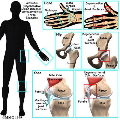
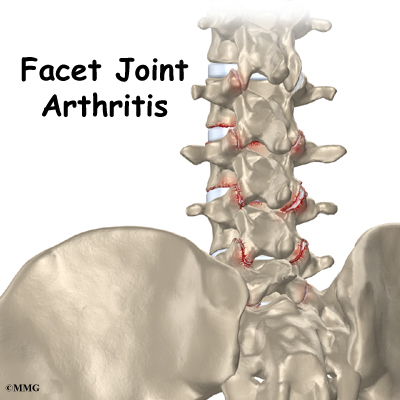
Arthritis is the most common cause of chronic disability. There is no cure for most forms of arthritis. But with some effort, you don’t need to lose all the movement in your joints. A rehabilitation program can help you maintain and even improve your joints’ strength and mobility. With some help from specialists and special equipment, arthritis won’t always stop you from doing the things you enjoy or the things you need to do.

Rehabilitation is a hands-on form of care and relies on your participation and effort. It involves exercising, learning how to care for sore and swollen joints, and figuring out ways to minimize the stress on your joints. In the early stages of arthritis, the goal of rehabilitation is to maintain or improve your joint strength and range of motion. If your joint is severely damaged, rehabilitation will focus on managing your pain and finding special equipment to help you with necessary tasks. Rehabilitation also helps people recover from joint surgery. Your rehab program will involve managing your symptoms, exercise, and lifestyle changes.

Rehabilitation requires patience. It takes time to strengthen your joints and learn how to do familiar tasks in new ways. But the result can be a greatly improved quality of life.

Most doctors refer their patients to physical or occupational therapists for rehabilitation. Many other types of medical professionals are involved in caring for people with arthritis; rehabilitation nurses, vocational rehab counselors, recreational therapists, and sometimes even medical social workers, speech therapists, and psychologists. No matter what kinds of specialists you see, rehabilitation is a team effort–you, your doctor, and your therapists.

Your First Visit

What happens on the first visit to the therapist’s office?

The first step in your rehabilitation is for your therapist to learn more about you and your joint problems.

History of the Problem

Your therapist will ask questions about your disease history, your day-to-day activities, and what you have problems doing. You may be asked to rate your pain on a scale from one to ten. Your answers will help guide your therapist’s examination. Below are some other questions your therapist may ask you.

  • What makes your pain or symptoms better, and what makes them worse?
  • How do your symptoms affect your daily activities?
  • What treatments have been helpful for you?

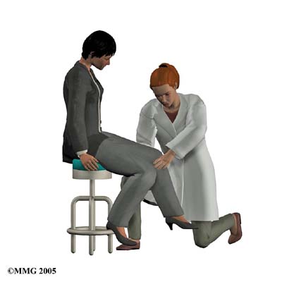
Physical Examination

After reviewing your answers, your therapist will do an examination that may include some or all of the following checks.

Posture and Joint Alignment

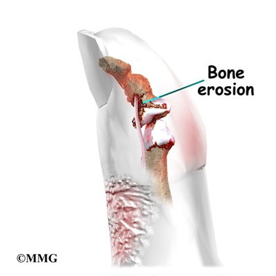
By checking your overall posture and joint alignment, your therapist can see if you have swelling or other signs of inflammation. Your therapist will also look to see if you have any nodules or other changes around a joint that may be present with various forms of arthritis.

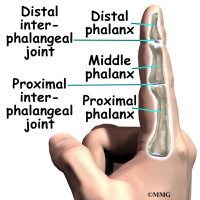
Range of Motion (ROM)

Your therapist will check the ROM in your sore joints. This is a measurement of how far you can move the joint in different directions. Your ROM is written down to compare how much improvement you are making with the treatments.

Strength

Your strength is tested by having you hold against resistance as your therapist tests the muscles around the sore area. Weakness and pain with these tests may be expected due to the presence of arthritis.

Manual examination

Carefully moving the joint in different positions can give your therapist an idea of the stiffness in your joints.

Palpation

Your therapist will feel the soft tissues around the sore areas. This is called palpation. Through palpation, the therapist checks for changes in skin temperature and any swelling. The therapist also pinpoints sore areas and looks for tender points or spasm in the muscles around the sore area. Palpation is important in helping your therapist decide which treatments to recommend.

Planning Your Care

What goes into a rehabilitation plan?

All the information you give the therapist, along with the results of the manual exam, will be used to create a rehab program especially for you. Your therapist will put together a treatment plan that describes the goals you and your therapist have for the treatment. The plan lists the exercises and treatments that will be used, and it includes an estimate of how many visits you will need over what period of time. Your therapist will also let you know what results to expect from the program.

Therapy Treatments

What kind of treatments and activities might the therapist recommend?

Controlling Your Symptoms

Rehabilitation therapy, combined with drugs and other treatments prescribed by your doctor, can help you manage the pain and swelling in your joints. Your therapist’s recommendations will depend on your specific symptoms and needs and may include one or more of the following treatment choices.

Rest

Knowing when to rest painful joints can help ease arthritis pain. Rest is especially important during flare-ups. As a common sense rule, if a certain activity or movement causes severe pain, avoid doing it. If you can’t avoid it, do it less, or take breaks that let your joints rest.

Your therapist may make you a special resting splint to support your sore joint when you’re not using it. A resting splint keeps the joint properly aligned, which limits pain and prevents joint deformity.

Heat

Heat makes blood vessels expand, which is called vasodilation. Vasodilation helps flush away chemicals that make your joints and muscles hurt. It also helps your muscles relax. Moist hot packs, heating pads, and warm showers or baths are the most effective forms of heat therapy. Heat treatments usually involve applying heat to the sore area for fifteen to twenty minutes. Paraffin baths or warm whirlpools can be especially helpful for joints of the hands or feet. You may find you have less pain and better mobility after applying heat.

Be cautious when using heat. While heat can be very helpful at times, heat can make serious inflammation worse. And even when heat is the best treatment for your discomfort, hotter is not better. Your skin can overheat and even burn. Sleeping with an electric hot pad is a bad idea. The prolonged heat can actually burn your skin.

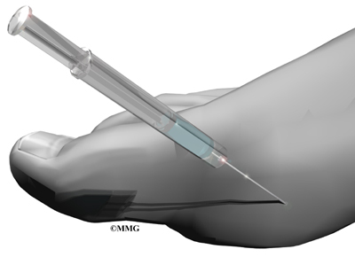
Electrical Stimulation

Gentle electrical currents through the skin can help ease pain and decrease swelling. Electrical stimulation eases pain by replacing pain impulses with the impulses of the electrical current. Once the pain lets up, the muscles begin to relax, making movement and activity easier.

Topical Creams

Certain creams rubbed on the skin can give temporary relief to sore joints. The rubbing is relaxing, and the creams create feelings of warmth or coolness that are soothing. Capsaicin, a cream derived from the common pepper plant, has been shown to effectively relieve arthritis pain. With all creams, you need to wash your hands after using them. What feels good on your sore joint does not feel good in your eyes.

Therapeutic Exercise and Functional Training

Whether at work, home, or play, your capabilities depend on your physical health and function. Specialized treatments and exercises can help maximize your physical abilities, including movement, strength, and general fitness. Therapists also use functional training when you need help doing specific activities with greater ease and safety.

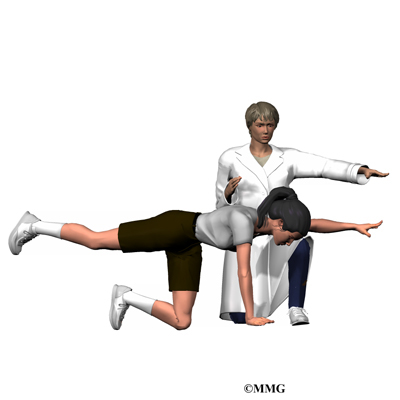
Exercise is safe for arthritis patients. In fact, it’s necessary if you want to improve or maintain joint function. Avoiding exercise just makes your arthritis worse. The less a joint is used, the weaker and stiffer it becomes. This leads to even more pain. Even if you don’t have much range of motion in a joint, your therapist can help you find ways of stretching and moving that can help strengthen your joint. There are some specific types of exercises that therapists recommend especially for people with arthritis.

Stretching

Gentle stretching lengthens muscles and helps the joint maintain its shape and mobility. Therapists teach specific stretches for different types of joints.

Strengthening

Your therapist will teach you exercises that have been adapted especially for arthritic joints. Isometric exercises involve tightening muscles without moving joints. This allows you to keep the muscles strong without stressing your joints. Isometrics can often be done even during flare-ups.

Muscles themselves are not part of joints. But strong muscles around a joint help joints move with less pain. Toned muscles act as shock absorbers in protecting the joint.

Stabilizing

There are also specific stabilization exercises to help keep your joints aligned. When your joints are positioned correctly, there is less rubbing or overstretching, and therefore less pain. Correct alignment also helps prevent joint deformities.

Pool Therapy

When you exercise in a swimming pool, the water bears some of your weight. This puts less stress on the joints of your feet, ankles, knees, and hips. The water’s buoyancy lets you move easier, and the water’s warmth can relax your muscles. You will probably start pool therapy in a group led by an instructor. If it is helpful, you may continue the exercises on your own. The warmth of the water can help relax muscles, improve circulation, and ease soreness.

Aerobic Exercise

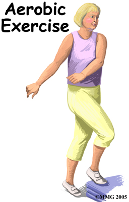
Your therapist and doctor will probably also recommend that you do some kind of aerobic exercise. Doctors generally recommend thirty minutes of moderate activity, at least five days a week. People with arthritis can safely try exercises such as walking, swimming, stationary biking, and low-impact aerobics. Your doctor or therapists can suggest an exercise program based on your condition and your overall health. Keeping your body fit is important for your general health and can help keep your arthritis under control.

Aerobic exercise also helps you manage your weight. Weight control is especially important for people with arthritis in the hips, knees, feet, and spine. Keeping your weight down can do a lot to help you control your symptoms.

No matter what type of exercise you do, you should not feel extra pain in the joints while you exercise. Your joints may be sore after exercising, but the soreness should be mild and go away within a short period of time.

Lifestyle Management and Functional Training

It is important that you be very open with your therapist about the ways your disease affects your daily activities. Your therapists can suggest ways to help you reduce the effort it takes to do difficult tasks.

Special Devices

There are many different kinds of equipment available to help you minimize the stress on your joints while you do daily tasks around the house or at work. What kind of equipment you need depends on which joints are affected. Canes and walkers help ease the stress on your weight-bearing joints. Raising the height of chairs and toilet seats can make it easier for you to sit down and stand up. Reachers or grabbers can help you pick up items from the floor without having to bend or stoop. There are devices to help with buttoning, putting on socks, or using zippers. A rolling cart is easier to haul around with arthritic fingers than a hand-held briefcase.

Your therapist may also suggest special splints or braces. A working splint keeps the joints aligned as you go about your daily activities. Splints are made for specific joints and specific activities.

Your therapist may also recommend simple changes in equipment. For example, a good pair of shoes can help reduce shock. If you walk or stand for long periods of time, you should try to do it on soft surfaces. As another example, women may choose a shoulder bag or a small backpack to take the place of a clutch purse or brief case if they have problems with the joints in their hands.

Ergonomics

When they hear the word ergonomics, most people think of the way their desk and computer are set up at work. The meaning is larger than that. Ergonomics considers the way you use your body when you take part in certain activities.

Rehabilitation therapists examine your workstation to help determine if you need to make changes. Your therapist will pay special attention to your posture, the repetitions involved in your work, rest times, the amount of weight you are working with, and which activities seem to cause you the most problems. Your therapist will look at the heights of your chair and desk, alignment of computer monitors, lighting, and any special equipment you use.

After evaluating your work site, your therapist will make recommendations. If changes are suggested, they are usually small and inexpensive, such as changing the height of your chair or standing in a different position. But even these minor changes can make big differences in your discomfort on the job.

The ideas behind ergonomics can also be applied to the tasks you do at home. If you have problems with specific jobs or hobbies, talk to your therapist. Together you may come up with a plan or some simple devices that can help.

Pacing Yourself

Plan to take breaks. Pace your activities so that you don’t get too tired or have to force your joint to function through pain.

Taking Care of Your Mind

Not all your work will be physical. Dealing with the pain and loss of function of arthritis can be emotionally draining. Make sure you take care of yourself mentally, and try to bolster your coping skills. Breathing exercises, naps, visual imagery, and meditation can all help you relax. Learning more about your condition can help you feel more in control of your disease. Many people find support groups helpful.

Home Program

Your therapist’s goal is to help you figure out ways to keep your pain under control and improve your strength and range of motion. When you are well under way, your regular visits to the therapist’s office will end. Your therapist will continue to be a resource for you, but you will be in charge of your own ongoing rehabilitation program.

Arthritis Medications

A Patient’s Guide to Medications for Arthritis

Doctors have only a few kinds of drugs to help treat the many different kinds of arthritis. The three classes of arthritis medications doctors prescribe are nonsteroidal anti-inflammatory drugs (NSAIDs), corticosteroids, and disease-modifying antirheumatic drugs (DMARDs).

Nonsteroidal Anti-Inflammatory Drugs (NSAIDs)

Uses of NSAIDs

The most commonly used drugs are NSAIDs. The over-the-counter drugs aspirin, ibuprofen, and naproxen are NSAIDs. Your doctor must write a prescription for other NSAIDs. Almost all of the NSAIDs are taken in pill form. Your doctor will probably prescribe only one form of NSAID at a time.

Inflammation is your body’s response to an injury. In normal circumstances, inflammation is the process that helps the injury heal. In arthritis, the inflammatory response gets out of control and actually causes damage to the tissues. NSAIDs help reduce inflammation. They also decrease pain and fever. However, they are short-acting drugs. After NSAIDs have passed through your body, inflammation, pain, and fever can return quickly.

NSAIDs can be very effective against inflammation, but they do not prevent tissue damage. Even when NSAIDs are controlling the inflammation, the joint or organ damage of arthritis can continue to get worse. NSAIDs only lessen your pain and discomfort. They do not affect your underlying disease.

Complications of NSAIDs

NSAIDs are safe drugs. However, they have many side effects. The side effects happen more often when they are used over long periods of time, which is common in arthritis patients. Some of the side effects can become very serious. It is important to use the lowest doses possible to control your symptoms.

GI Effects

NSAIDs irritate the gastrointestinal (GI) tract (the digestive system–your esophagus, stomach, and intestines). They increase the production of gastric acid, and they harm the gastric lining. NSAIDs aggravate ulcers and GI bleeding. Up to 5 percent of people who use NSAIDs for a year develop ulcers, bleeding, or tears in the GI tract. The risks are higher for older patients, patients with a history of GI problems, and patients with heart disease.

Blood Effects

NSAIDs make it harder for the platelets in your blood to clump together at the site of an injury. This can cause bleeding problems. Aspirin especially has this effect. Before you have surgery, you should stop taking aspirin for two weeks to prevent bleeding problems.

Liver Effects

NSAIDs can be toxic to your liver. You will not feel this, but elevated levels of certain liver enzymes can easily be seen in blood tests. Liver function almost always returns to normal when you stop taking NSAIDs.

Kidney Effects

NSAIDs can make it hard for your kidneys to get rid of some kinds of wastes. If you have a history of kidney problems, or if your disease may affect your kidneys, your doctor will use NSAIDs with caution.

Other Effects


Some people get skin reactions and rashes from NSAIDs. Some get a combination of runny nose, polyps in the nose, and asthma. Different kinds of NSAIDs can have different side effects. Salicylates can cause problems with hearing. Other kinds of NSAIDs can cause headaches and confusion, especially in elderly patients. Many of the possible side effects depend on your health and the disease for which you are being treated.

Individuals can react very differently to the same NSAIDs. You and your doctor must work together to find the type and dose of NSAID that controls your symptoms without causing unwanted side effects.

Corticosteroids

Uses of Corticosteroids

Corticosteroids are chemical copies of hormones that occur naturally in your body. The most commonly used corticosteroids are prednisone, prednisolone, and methylprednisolone. Corticosteroids can be given orally or put directly into the bloodstream through an intravenous needle. They can also be injected directly into an inflamed spot. Corticosteroid cream can be rubbed on the skin.

Corticosteroids are powerful drugs. They drastically decrease inflammation. But they are also highly toxic. Doctors have different opinions about how corticosteroids should be used.

Corticosteroids can’t cure your disease. But they do seem to affect the development of some diseases, including rheumatoid arthritis (RA).

Complications of Corticosteroids

Corticosteroids can have many unwanted effects on your body. Whether or not you develop these complications depends on many factors: what type of corticosteroid you take, your dose, the length of time you are on it, and how sensitive your body is to these hormones. The most common side effects are.

Osteoporosis

All corticosteroids slow bone growth and create conditions that lead to osteoporosis, a disease process that results in reduction of bone mass. Compression fractures of the vertebrae can happen with long-term corticosteroid use. Men and women past menopause are most likely to develop osteoporosis. Your doctor may recommend that you take calcium and vitamin D pills while you take corticosteroids.

Infections

High levels of corticosteroids hinder your body’s ability to fight bacterial infections. High-dose corticosteroids can even mask the symptoms of some types of infections, such as abscesses and bowel tears. Most viral infections are not a problem, except for herpes.

Adrenal Insufficiency

This means that your pituitary and adrenal glands can’t produce enough of certain kinds of hormones. This can happen after taking corticosteroids in moderate doses for only a few days. Adrenal insufficiency is most likely to happen as you are reducing the dosage. It can be a problem if you need surgery or if you get an infection or serious injury.

Withdrawal

When you stop taking corticosteroids, the doses will be slowly reduced over a period of days or weeks. Even if you have only been taking steroids for a few weeks, you will still need to taper off. Corticosteroid withdrawal can be very difficult for your body. In many patients, the disease symptoms become worse. Some people experience a sickness that includes fevers, nausea, vomiting, low blood pressure, and low blood sugar. Others have withdrawal symptoms that include muscle and joint pain, weight loss, fever, and headaches. If you have problems coming off corticosteroids, your doctor will have you taper off the drug more slowly.

Different people, and different diseases, react very differently to corticosteroids. You and your doctor will need to find a dose that controls your symptoms but minimizes unwanted effects.

Disease-Modifying Antirheumatic Drugs (DMARDs)

Uses of DMARDs

DMARDs are primarily used to treat rheumatoid arthritis (RA) and other systemic diseases. In the past twenty years, DMARDs (which are also called slow-acting antirheumatic drugs) have become much more widely used.

The idea behind using DMARDs is to prevent joint damage. This means you start using them early on, and you switch drugs or doses when your current drugs stop working. Using DMARDs requires you to be alert for possible side effects. You also need to be patient. DMARDs take some time to work. But they can be very effective at slowing the course of your disease.

DMARDs do not cure disease. Very few patients see their disease go into a complete remission. Most patients find that their symptoms come back after months, or sometimes years, of improvement on DMARDs.

Doctors often prescribe DMARDs, corticosteroids, and NSAIDs at the same time. The DMARDs affect the underlying disease, and the corticosteroids and NSAIDs give relief from pain and inflammation. Sometimes doctors prescribe two or more DMARDs together. There are few studies to prove how well these combinations work. However, using more than one DMARD does not seem to cause problems with higher toxicity. This means that taking more than one DMARD isn’t any harder on your body than taking just one.

Types and Complications of DMARDs

There are many different types of DMARDs, with different effects and complications. Some are used only for specific types of diseases.

Antimalarial Drugs

Hydroxychloroquine and chloroquine have been used since the 1950s for rheumatic diseases. They have been used against malaria for much longer. These drugs are mostly used for RA and lupus (which is also called SLE). Chloroquine has more side effects. Side effects include indigestion, rash, and eye problems. Antimalarial drugs take three to four months to show results.

Penicillamine

This drug affects the way your immune system functions. Almost 25 percent of patients who take it experience bad side effects within the first year. The most common side effects are rashes, blood and protein in the urine, low numbers of platelets in the blood, and autoimmune problems including drug-induced lupus. Taking penicillamine requires regular blood and urine tests.

Sulfasalazine

This fairly new drug is used primarily in RA and spondyloarthropathies (arthritis of the spine). It may slow down erosions of bone. Almost half of patients develop side effects in the first four months, but most of the reactions are minor. Side effects include rashes, nausea, abdominal pain, liver and blood disorders, low sperm counts, and discolored urine and sweat. You will need liver and blood tests for the first months on this drug.

Gold

Gold compounds have been used for eighty years to treat RA. They are also used in juvenile chronic arthritis and psoriatic arthritis. Gold is injected into your muscles, usually once a week. Most patients only use gold compounds for one to five years. After about a year, most patients stop seeing benefits from using gold therapy. And most patients also start having complications. Unwanted side effects include diarrhea, rashes, low levels of platelets and other blood disorders, protein in the urine, lung problems, and sores of the mucous membranes, especially in the mouth. Using gold compounds requires regular blood and urine tests.

Methotrexate

Methotrexate shows results in one to two months. Most patients stay on it longer than other DMARDs. In the short term, methotrexate causes nausea, loss of appetite, and high levels of certain liver enzymes. As with all the other DMARDs, there are serious complications with long-term use. It can cause liver damage and lung disease.

Infliximab

Infliximab is a type of disease-modifying medication in a class called anti-tumor necrosis factor (TNF) agents. The anti-TNF agents are a special type of antibody referred to as human monoclonal antibodies. They specifically target (and inhibit) tumor necrosis factor. Tumor necrosis factor (TNF) promotes the inflammatory response, which in turn causes many of the clinical problems associated with autoimmune disorders such as rheumatoid arthritis.

Azathioprine

This drug is most often used in RA, lupus, and other connective tissue diseases. It can also help offset the bad effects of steroids. Azathioprine is as effective as other DMARDs, but it does have more side effects. It can cause nausea, vomiting, diarrhea, bone marrow suppression, and hepatitis. The most troubling long-term side effect is cancer of the lymph system.

Nitrogen Mustard Alkylating Agents

Chlorambucil and cyclophosphamide are the main alkylating agents used as DMARDs. Chlorambucil is usually used to treat RA, juvenile chronic arthritis, vasculitis, systemic sclerosis, and ankylosing spondylitis. However, it can damage the chromosomes. This creates a higher risk for leukemia and other kinds of cancers. Cyclophosphamide can be taken by mouth or intravenously. It is usually used to treat severe RA, lupus, and systemic vasculitis. Bad side effects are common. They include inflammation and bleeding of the ulcer, suppression of the immune system, reproductive problems in men and women, and cancer that may show up long after the drug is stopped.

Cyclosporine

Cyclosporine can be very effective against RA, but most people who take it develop kidney problems and high blood pressure. Kidney function goes back to normal when you stop taking the drug.

As with NSAIDs and corticosteroids, you and your doctor will need to work together to find the best type and dose of DMARDs for your disease.

Oral medications (pills taken by mouth) are under investigation that might be available in the future for the treatment of a specific type of arthritis called psoriatic arthritis (PsA). These include ustekinumab, apremilast, and tofacitinib. Each of these medications works in a slightly different way to regulate the immune system.

Psoriatic Arthritis

A Patient’s Guide to Psoriatic Arthritis

Introduction

Psoriasis is a disease that most people think of as primarily a skin disease because the condition causes a persistent rash in various areas of the body. Psoriatic arthritis is a type of joint disease that occurs in roughly seven percent of people who have psoriasis. Psoriatic arthritis affects people of all ages, but most get it between the ages of 30 and 50. Usually a patient has psoriasis (the skin rash) for many years before the arthritis develops, and the arthritis comes on slowly. But this is not always the case. No matter what, patients with psoriatic arthritis must manage both the outbreaks of itchy, scaly skin and the pain and stiffness of arthritis.

This guide will help you understand

  • how psoriatic arthritis develops
  • how doctors diagnose the condition
  • what can be done for the problem

Anatomy

Where does psoriatic arthritis develop?

Psoriatic arthritis can affect any joint. Its symptoms often seem like the symptoms of rheumatoid arthritis (RA) or degenerative arthritis of the spine. X-rays can be used to show the difference between psoriatic arthritis and other diseases. In psoriatic arthritis, X-rays show a very distinctive type of bone destruction around the joint and certain patterns of swelling in the tissues around the joints.

Psoriatic Arthritis

Patients with psoriatic arthritis fall into three groups. Many patients have what is called asymmetric arthritis. This means that only a few joints are involved and that it does not occur in the same joints on both sides of the body. (For example, only one wrist and one foot are affected.)

An equal number of patients suffer from symmetric polyarthritis. This means that arthritis occurs in several corresponding joints on both sides of the body. (For example, both elbows, both knees, and both hands are affected.) The polyarthritis type of psoriatic arthritis is much like RA.

A third group has mostly axial disease. This refers to arthritis of the spine, the sacroiliac joint (where the pelvis and bottom of the spine meet), or the hip and shoulder joints. Patients do not necessarily stay in the same category. Over time, the pattern may change. Doctors use these categories to better understand the disease and to follow the progression of the arthritis. The treatment is basically the same.

Causes

Why do I have this problem?

The exact cause of psoriatic arthritis is not known. Many factors seem to be involved in its development. Heredity–your genes–plays a major role. People who are closely related to someone with psoriatic arthritis are 50 times more likely to develop the disease themselves. Recent studies have located genetic markers shared by most people who have the disease.

Sometimes injuries seem to set off psoriatic arthritis. Infections also contribute to the disease. It is known that strep infections in children can cause psoriasis. Some researchers think that the arthritis may be an immune system response to bacteria from the skin lesions.

Symptoms

What does psoriatic arthritis feel like?

All people who suffer from psoriatic arthritis have psoriasis (the skin rash). Some patients have very few areas of rash. Other patients have psoriasis over a large portion of their bodies. The skin lesions of psoriasis are reddish, itchy, and have silvery scales. These areas can range in size from the size of a pencil dot to the large areas the size of your palm. Psoriasis usually shows up on the elbows, knees, scalp, ears, and abdomen, but it can appear anywhere. In people with psoriatic arthritis, the psoriasis most often affects fingernails or toenails. The nails may have pits or ridges, or they may be discolored or appear to be separating from the skin.

Psoriatic arthritis can affect any joint. Symptoms often seem like those of any other type of arthritis–joint swelling and pain–but patients generally describe less pain. Some joint symptoms are unique to psoriatic arthritis:

    • The joints nearest to the fingernails and toenails are affected more. (These joints are called distal interphalangeal, or DIP, joints.)

Psoriatic Arthritis

  • The affected fingers and toes take on a “sausage-like” appearance.
  • The bones themselves become inflamed (called dactylitis).
  • The tendons and ligaments become inflamed where they attach to bones. (This is called enthesitis and is especially common in the heels.)
  • Bony ankylosis of the hands and feet develops. (This means that the joints stiffen and become frozen in awkward positions.)
  • The joints grow inflamed where the bottom of the spine meets the pelvis. (This is called sacroiliitis.) Patients often notice no symptoms, but the inflammation can be seen on X-rays.
  • The vertebrae of the spine become inflamed. (This is called spondylitis.)
  • The eyes become inflamed.

About five percent of patients with psoriatic arthritis will develop a form of arthritis called arthritis mutilans. This type of arthritis affects the small joints of the hands and feet. It is especially severe and destructive. The destruction caused by arthritis mutilans can result in deformity of the hands and fingers.

Rare symptoms include problems with the aortic heart valve, extra tissue formation in the lungs, and metabolic disorders that affect the tissues.

Diagnosis

How do doctors identify the condition?

A detailed medical history, with questions about psoriasis in your family, will help your doctor make a diagnosis. Patients with psoriasis may have other forms of arthritis, and the symptoms of psoriatic arthritis often look like other types of joint disease. This means that your doctor will probably do tests to rule out other diseases.

Blood studies will help rule out RA. (The RA test is usually not positive in patients with psoriatic arthritis.) Efforts are being made to find ways to identify psoriatic arthritis through a blood test. The presence of specific biologic elements called biomarkers (biologic evidence of disease) would make it possible to look for evidence of this disease before it progresses – or even before it starts. Psoriatic arthritis is common in people who test positive for HIV, the AIDS virus. As a precaution, your doctor may test your blood for HIV, especially if your symptoms are severe.

Physicians must also use other diagnostic tools such as X-rays, ultrasonography, and MRIs. Each one of these tests provides a little different information. For example, X-rays of affected joints will be studied both to rule out other diseases and to identify characteristics of psoriatic arthritis.

Ultrasonography, the use of sound waves to create a picture of what’s going on inside, provides a better look at the whole package: bones, joints, and soft tissues. This diagnostic test is also noninvasive and does not expose the patient to any radiation. Ultrasound also has the ability to show small changes in the nails and early signs of inflammation in tendons and small joints.

MRIs can show bone marrow edema, tenosynovitis, and early joint erosion. Tenosynovitis is the inflammation of the fluid-filled sheath (called the synovium) that surrounds a tendon. But reliability is a problem with MRIs because what one examiner sees may not be the same as another observer. Changes in the small joints of the hands and feet don’t show up well on MRIs like they do with ultrasonography.

One advantage MRIs do have over ultrasonography is the availability of whole body MRI. By scanning the entire body, it is possible to identify areas of inflammation undetected by clinical exam.

Until blood studies are able to find biomarkers indicating the presence of psoriatic arthritis, physicians will probably have to use a combination of different tests to diagnose the problem. The information these tests provide is important in determining treatment.</p.

Treatment

What can be done for the condition?

Dealing with psoriatic arthritis involves treating both the skin lesions and the joint pain. Many lotions and creams are made for skin affected by psoriasis. If the skin involvement is especially severe, your doctor will most likely prescribe a drug called methotrexate. Methotrexate can also help with the arthritis.

PUVA therapy may be helpful for both the skin and joint problems. PUVA therapy uses topical cream medications that are rubbed on the skin lesions. Following application of the cream, the skin area is placed under a lamp that emits a special ultraviolet light. The light triggers chemicals in the medication cream that treat the rash lesions.

Treatment of arthritis symptoms depends on which joints are affected and the severity of the disease.

The first drugs most doctors prescribe are nonsteroidal anti-inflammatory drugs (NSAIDs). Aspirin and ibuprofen are NSAIDs, as are many prescription pain relievers. Other medications known as disease-modifying antirheumatic drugs (DMARDs) are used in patients with high levels of pain or especially bad arthritis. These medications work in different ways to regulate the immune system and thereby control the arthritis.

One of the most commonly used disease-modifying medication for the treatment of psoriatic arthritis (PsA) is methotrexate. DMARDs like methotrexate not only control symptoms, they also slow the progression of disease. That’s what makes them “disease-modifying”. For some patients, it may be necessary to combine methotrexate with another drug (e.g., infliximab) to get the desired results (decreased joint pain, swelling, and stiffness).

Infliximab is a type of disease-modifying medication in a class called anti-tumor necrosis factor (TNF) agents. The anti-TNF agents are a special type of antibody referred to as human monoclonal antibodies. They specifically target (and inhibit) tumor necrosis factor. Tumor necrosis factor (TNF) promotes the inflammatory response, which in turn causes many of the clinical problems associated with autoimmune disorders such as rheumatoid arthritis.

Oral medications (pills taken by mouth) are under investigation that might be available in the future for the treatment psoriatic arthritis (PsA). These include ustekinumab, apremilast, and tofacitinib. Each of these medications works in a slightly different way to regulate the immune system.

Doctors will sometimes prescribe a combination of drugs. Cortisone injections into sore joints can help relieve pain. Surgery may be called for in the rare cases of unmanageable pain or loss of joint function.

Related Document: A Patient’s Guide to Medications for Arthritis

Warm water soaks and applying heat to joints gives pain relief to many patients. Your doctor may ask you to see a physical therapist to maximize the strength and mobility of your joints. Stress does make your symptoms worse, so your doctor may encourage you to exercise and find ways to reduce the stress of your daily life.

Your psoriatic arthritis will not go away. But there are many treatment options. Together, you and your doctor should be able to find treatment that will work for you.

Osteoarthritis

A Patient’s Guide to Osteoarthritis

Osteoarthritis (OA) is the most common form of arthritis. In fact, more than 75 percent of people older than fifty-five show the joint deformations of OA on X-rays. But most of these people have no symptoms. For people who do have the joint pain and stiffness of OA, it can become a crippling disease. Some people suffer from OA in just one joint, while others have it in several joints. It affects more women than men, and most OA patients are over 45.

This guide will help you understand

  • how OA develops
  • how doctors diagnose the condition
  • what can be done for OA

Anatomy

Where does OA develop?

Osteoarthritis

OA is most common in the small joints of the hands, the spine, the knees, the hips, and certain toe joints. OA primarily affects the articular cartilage, the slippery, cushioned surface that covers the ends of the bones in most joints and lets the bones slide without rubbing. Articular cartilage also functions as a shock absorber.

In OA, the articular cartilage becomes damaged or worn away. As this happens, the joint no longer fits together well or moves smoothly. In the early stages of OA, the cartilage actually becomes thicker as your body tries to repair the damage. The repaired areas are more brittle than the original cartilage, and these brittle areas begin to wear away and become thin. They may even wear away entirely. This eventually leads to a condition called eburnation, in which the bones become thick and polished as they rub together. X-rays can show these changes in the cartilage and bones.

But OA is not just a disease of the cartilage. The damage to the cartilage seems to start a sort of chain reaction that involves all the parts of the joint. Bone spurs, or outgrowths, often begin to form around the edges of the joint. The joint capsule (the watertight sack around the joint) can become thickened and lose its stretch. The synovial membrane that lines the inside of the joint capsule may become inflamed (swollen, red, hot, and painful), and crystals may form in the synovial fluid. The tendons and ligaments around the joint can also become inflamed.

Even the muscles around the joint can lose their strength. This usually occurs as a result of under-use of the muscles due to pain in the joint. When something hurts we subconsciously change the way we use the joint to avoid the pain. This causes the muscles to become weakened. Because cartilage itself does not have nerves to feel pain, the pain of OA probably comes from these other changes in and around the joint.

Causes

Why do I have this problem?

The exact cause of OA is not known. There are probably different causes in different people. Doctors think of OA in two different categories, primary OA and secondary OA. Primary OA refers to breakdown of a joint from a disease process. Secondary OA means that something else was wrong–an infection in the joint or a fracture for instance–that caused damage to the joint. Even when the original problem clears up, the chain reaction effect of OA can cause the disease to progress.

Major injuries and repetitive stress both seem to cause OA. A person who breaks an ankle is likely to develop OA in that same ankle. Just like any machine, a joint that is damaged and unbalanced wears out faster. People who consistently put heavy stress on the same joint, such as jackhammer operators or baseball pitchers, are more likely to develop OA in that joint.

OA of the knee and hip occurs much more often in people who are seriously overweight. A study that followed overweight young adults for thirty-six years found that being overweight at a young age was closely related to developing OA later in life. The same study also showed that losing even small amounts of weight decreased the odds of developing OA.

Heredity–your genes–may also play a role for some people, especially women. OA in the fingers, which affects ten times more women than men, shows up much more often among women in the same family. Researchers do know that some genes cause problems with cartilage formation.

In some cases, rare metabolic disorders or other problems with the bones or joints can lead to OA. But the primary factor in most patients with OA seems to be age. If you’re lucky enough to live a long life, you are much more likely to develop OA.

Symptoms

What does OA feel like?

Patients with OA have one or more joints that are painful and stiff. The pain is a deep, dull ache that usually comes on gradually. Pain gets worse when the joint is used and gets better with rest. The joint is stiff after waking up or after not being used for some time, but the stiffness usually goes away fairly quickly. Over time the pain and the stiffness become almost constant.

No matter which joints are affected, OA patients report many of the same symptoms

  • Most patients say that the pain is worse in cool, damp weather.
  • Many OA patients feel or hear crackling or popping in the affected joints (called crepitus). This is most common in the knees.
  • Joints enlarge or change shape. The enlarged areas are often tender to the touch.
  • In most cases the affected joints can’t move through a normal range of motion.
  • In other cases the joints have become so unstable that they can actually move too much or in the wrong direction.

Some symptoms depend on the affected joint. Patients with knee OA may have problems with the joint locking up, especially when they are stepping up or down. Patients with OA of the hip often limp. OA of the hands can affect the strength and movement of fingers and make simple tasks such as getting dressed very difficult. OA of the spine can cause neck and low back pain as well as weakness and numbness.

Diagnosis

How do doctors identify the condition?

It may seem that diagnosing OA would simply involve a few X-rays. However, it is very important that your doctor rule out other forms of arthritis. Your doctor will also need to figure out if your OA was caused by another problem (secondary OA). And even if OA is the main problem, the breakdown of cartilage may have caused problems in other parts of the joint that need to be addressed.

Your doctor will ask you many detailed questions about your health history. Explaining the nature of your pain will be important. Following the history the doctor will thoroughly examine the affected joints. X-rays will most likely be taken. Blood samples and samples of the synovial fluid in the joint may be taken to try to identify other problems.

Treatment Options

What can be done for the problem?

There is no cure for OA. It is a chronic but very treatable disease. The goals of treatment are to relieve your pain and to improve or maintain the movement of the joint.

Nonsurgical Treatment

Much of the treatment for OA involves no prescriptions at all. Your doctor will encourage you to take some steps to help manage your symptoms:

  • Get aerobic exercise.
  • Do strengthening and range of motion exercises. These are most often taught and monitored by physical or occupational therapists.
  • Lose weight.
  • Use heat and cold packs.
  • Tape the knee, if it is affected.
  • Wear wedged insoles in your shoes, if the hip or knee is affected.
  • Receive massage.
  • Use equipment to help take pressure off your joints, such as a cane or special gadget to open jars.
  • Participate in education programs or support groups.

Drugs are available to help alleviate your pain. Your doctor will probably start with an over-the-counter pain reliever, such as acetaminophen (Tylenol). If this doesn’t work, nonsteroidal anti-inflammatory drugs (NSAIDs), such as ibuprofen and aspirin, may be prescribed. The main problem with NSAIDs is that they can be very hard on your stomach and kidneys over time, and you will be probably be taking these drugs over many years. In rare cases of extreme pain doctors may prescribe stronger pain medications, but these can be addictive and must be used with caution.

Related Document: A Patient’s Guide to Medications for Arthritis

All these medications can interact with other drugs. You must let your doctor know what other medicines you are taking, and you must work closely with your doctor to set up dose amounts and schedules.

In recent years, two unique compounds have been used by people with OA. These compounds are gaining greater acceptance among many doctors. Glucosamine and chondroitin sulfate are dietary supplements taken in pill form that have shown benfits of reducing pain and increasing joint mobility. These treatments are controversial. Yet some doctors feel there are enough benefits to encourage their patients to supplement with these compounds.

Related Document: A Patient’s Guide to Glucosamine and Chondroitin Sulfate for Osteoarthritis of the Knee

Steroid injections directly into the joint can help ease the pain in some cases. Capsaicin cream rubbed into the joint sometimes provides pain relief. Occasionally doctors may recommend tidal joint lavage, which involves rinsing the joint with saline. Alternative approaches, such as pulsed electromagnetic fields and electrical nerve stimulation, have been tested.

Surgery

Osteoarthritis

When pain cannot be relieved and joint function cannot be maintained, your doctor may recommend surgery. While this option may sound scary, surgery can be very effective in treating OA.

Many types of surgical procedures have been designed to treat OA of different joints. Perhaps the most well known treatment is artificial joint replacement. Artificial joint replacement is the final answer to OA after the joint is totally destroyed, but other surgical procedures have been designed to treat osteoarthritis in early stages to reduce symptoms and slow the progression of the disease.

It will take some work to manage your OA, but it is possible. OA doesn’t always worsen over time. In many patients the disease stabilizes. In some patients, especially those with OA of the knee, the disease can actually reverse itself. And even when the OA does continue to progress, it often moves very slowly.

Lyme Disease

A Patient’s Guide to Lyme Disease

Introduction

Lyme Disease Lyme disease is an inflammatory disease caused by tick bites. It is the most common tick-borne disease in North America, Europe, and Asia. Connecticut has the highest annual rate of new cases of Lyme disease each year. The name Lyme disease was used because of the number of children in Lyme, Connecticut who first developed this problem back in the late 1970s.

More than 90 percent of the Lyme disease cases in the United States continue to occur in Connecticut and nine other states including New York, New Jersey, Rhode Island, Massachusetts, Pennsylvania, Wisconsin, Delaware, Maryland, and Minnesota.

It is possible to visit these areas and have the infection show up later, when you are back home. And although children between the ages of five and nine seem to be affected most often, adults between the ages of 55 and 59 seem to have a peak incidence as well. The summer months are the peak time when infection may occur, but cases have been reported in all 50 states year-round.

This guide will help you understand

  • how Lyme disease develops
  • how doctors diagnose the condition
  • what treatments are available

Anatomy

What parts of the body are affected?

Lyme disease can cause inflammation in many systems of the body if left untreated. It mostly affects the skin, joints, the heart, and the nervous system. When the joints are affected, the knee is involved most often. Lyme-associated arthritis has also been reported in the shoulder, elbow, hip, and ankle.

Causes

Why do I have this problem?

Lyme disease is caused by Borrelia burgdorferi, a spiral-shaped organism transmitted through the bite of an infected tick. The human body mounts a reaction to the infecting organism that triggers production of inflammatory agents throughout the body. These inflammatory agents are the chemicals produced by the body’s immune system that normally fight off infection.

Symptoms

What does Lyme disease feel like?

Doctors divide the symptoms of Lyme disease into three phases: early localized, early disseminated, and late disease. The three phases of symptoms can overlap. Many patients never show early symptoms at all.

Early localized disease refers to a red rash in the area of the tick bite. This rash shows up two to 30 days after the tick bite. The bite itself is usually found near the waist or belt line, or in other warm, moist areas of the body. In children, the rash is seen first on the head and neck, arms and legs, or back and less often on the abdomen, groin, or chest. The bite may burn, itch, or hurt. The rash usually grows over a period of days. The rash may be in a bulls-eye pattern, red with a white spot in the middle, or completely red.

Most patients with early localized Lyme disease also complain of flu-like symptoms, such as headaches, fatigue, and muscle and joint pain.

Early disseminated disease shows up days to months after the tick bite. Patients may not remember any rashes or bites. The most common symptoms of early disseminated disease are cardiac and nervous system problems. About eight percent of patients who have the rash and are not treated with antibiotics develop heart problems referred to as Lyme carditis. Heart effects include heart blockage and weakening of the heart muscle. The patient may experience dizziness, fainting, fatigue, shortness of breath, and/or heart palpitations. Most of the time, the heart symptoms clear up on their own or after treatment.

About ten percent of patients who don’t receive antibiotics develop damage to the nervous system. Neurologic symptoms also tend to go away on their own, but very slowly. These neurologic symptoms can include meningitis and headaches. Individual nerves can be affected causing numbness, weakness, and pain in the areas the damaged nerve travels.

Late disease symptoms develop months or even years after the tick bite. Some patients who have late disease symptoms have never had any other symptoms of Lyme disease. Late disease causes arthritis pain that comes and goes in many joints. About ten percent of people with late Lyme disease develop chronic (long-lasting) arthritis of one knee. Other joints can be affected (e.g., hip, shoulder, elbow, ankle) but the number of patients with other joint involvement is considerably less.

Many patients who have had Lyme disease describe headaches, fatigue, and joint pain that can last for months after treatment. These problems generally go away without any extra treatment, but very slowly.

Diagnosis

How do doctors identify the condition?

Doctors diagnose Lyme disease based on your health history and a physical exam. Your doctor may order blood tests, but they are only used to confirm the diagnosis. The techniques used to test your blood are called ELISA and Western blot. Both tests can sometimes give false positive or unclear results. If you have had the infection for less than six weeks, your body may not even be making enough antibodies to be detected in the tests.

Lyme disease affecting the knee must be differentiated from septic (infectious) arthritis, which has both a different cause and a different treatment. The two distinguishing features of septic knee arthritis that set it apart from Lyme knee arthritis are refusal to put weight on the knee and fever (more then 101.5 degrees Fahrenheit). Patients with Lyme disease may have a low-grade fever and pain on weight-bearing but do not exhibit the high fever and refusal to put weight on the affected leg observed more often with septic knee arthritis.

When trying to rule out septic arthritis, the synovial fluid (the lubricating fluid of a joint) or spinal fluid may need to be analyzed. Studies show that patients with septic (infectious) knee arthritis are 3.6 times more likely to have a high synovial fluid cell count compared with patients with Lyme disease. But some patients with Lyme disease have elevated synovial fluid cell count, too so this test is just one of many tools used to diagnose the problem. The fluid can also be cultured to identify the presence of bacteria such as staphylococcus aureus (staph infection), streptococcus pneumonia (strep infection), or other less common types of bacterial infections. Bacteria associated with septic arthritis help rule out a diagnosis of Lyme disease.

Treatment Options

What can be done for Lyme disease?

Two to four weeks of antibiotics almost always cures Lyme disease. Early treatment usually prevents later heart, nerve, and joint symptoms. If you have symptoms of early disseminated or late disease, your doctor will probably start you on intravenous (IV) antibiotics.

In a small number of cases, it may be necessary to surgically drain the joint of fluid. This is especially true if there is a suspicion of septic arthritis that cannot be confirmed or differentiated from Lyme disease. In cases of true Lyme joint disease that does not respond to medical treatment, the surgeon may have to remove some of the damaged joint synovium. The procedure is called a synovectomy.

Headaches, fatigue, and muscle and joint pain may continue for months after finishing the antibiotics. Neurologic damage may take even longer to go away, as the nervous system regenerates only one or two millimeters each day. This does not mean that you need more antibiotics. Your body just needs more time to heal. If the antibiotics do not help your symptoms at all, you and your doctor should consider that Lyme disease may not be the cause of your symptoms and look for other possible causes.

The risk of getting Lyme disease is very small, even if a tick has bitten you. Doctors do not treat you just in case. You must show symptoms.

The best treatment for Lyme disease is prevention. Check for ticks at the end of a day outdoors. Wear light clothes so you can see ticks. Wear long clothing, and tuck your pants into your socks. And finally, use a bug spray that contains DEET (a chemical used in some types of insect repellents) to keep the ticks away.

Systemic Lupus Erythematosus

A Patient’s Guide to Systemic Lupus Erythematosus

Introduction

Systemic lupus erythematosus (also called SLE, or lupus) is an autoimmune disease of the body’s connective tissues. Autoimmune means that the immune system attacks the tissues of the body. In SLE, the immune system primarily attacks parts of the cell nucleus.

SLE affects tissues throughout your body. Five times as many women as men get SLE. Most people develop the disease between the ages of 15 and 40, although it can show up at any age.

This guide will help you understand

  • how SLE develops
  • how doctors diagnose the disease
  • what can be done for the condition

Anatomy

Where does SLE develop?

SLE causes tissue inflammation and blood vessel problems pretty much anywhere in the body. SLE particularly affects the kidneys. The tissues of the kidneys, including the blood vessels and the surrounding membrane, become inflamed (swollen), and deposits of chemicals produced by the body form in the kidneys. These changes make it impossible for the kidneys to function normally.

The inflammation of SLE can be seen in the lining, covering, and muscles of the heart. The heart can be affected even if you are not feeling any heart symptoms. The most common problem is bumps and swelling of the endocardium, which is the lining membrane of the heart chambers and valves.

SLE also causes inflammation and breakdown in the skin. Rashes can appear anywhere, but the most common spot is across the cheeks and nose.

Causes

Why do I have this problem?

Doctors and researchers know quite a bit about the changes in the bodies of SLE patients. But the cause of SLE is a mystery.

Heredity plays a role in SLE. If you have a close relative who suffers from SLE, you are much more likely to develop the disease yourself. However, genes alone do not seem to cause SLE. It seems to be triggered in unknown ways. Not everyone with a tendency toward SLE will develop the disease. Researchers think some of the triggers that set off SLE may be infections, stress, diet, and toxins, including some kinds of prescription drugs. These triggers may also help explain why SLE has a cycle of flare-ups and remissions.

Symptoms

What does SLE feel like?

The symptoms of SLE come on in waves, called flares or flare-ups. In between flares, patients may have almost no symptoms. Almost every SLE patient suffers from general discomfort, extreme fatigue, fever, and weight loss at some point. In addition to these general symptoms, SLE produces different symptoms in different body systems.

Skin

Rashes caused by SLE are red, itchy, and painful. The rash can show up on any part of the body. The most typical SLE rash is called the butterfly rash, which appears on the cheeks and across the nose. SLE also causes hair loss. The hair usually grows back once the disease is under control.

People with SLE tend to be very sensitive to sunlight. Being in the sun for even a short time can cause a painful rash. Some people even get a rash from fluorescent lights at work.

Muscles and Bones

Systemic Lupus Erythematosus

Almost everyone with SLE has joint pain or inflammation. Any joint can be affected, but the most common spots are the hands, wrists, and knees. Usually the same joints on both sides of the body are affected. The pain can come and go, or it can be long lasting. The soft tissues around the joints are often swollen, but there is usually no excess fluid in the joint. Many SLE patients describe muscle pain and weakness, and the muscle tissue can swell.

In its late stages, SLE can cause areas of bone tissue to die, called osteonecrosis.. Osteonecrosis can cause serious disability. It can be caused at least in part by using high doses of corticosteroids over a long time. Corticosteroids help control the symptoms of SLE.

Kidneys

People with SLE usually don’t notice any problems with their kidneys until the damage is severe. Sometimes kidney problems aren’t noticed until the kidneys are actually failing.

Nervous System

SLE can cause headaches, seizures, abnormal blood vessels in the head, and many other problems with the nervous system. SLE can also cause organic brain syndrome. This disorder involves serious problems with memory and concentration, emotional problems, and severe agitation and hallucinations. Any of these symptoms may show up alone, without any other symptoms of SLE.

Membranes

In the body, membranes surround your internal organs. The membranes around your lungs, heart, and the organs in the abdomen become inflamed in SLE. This is called serositis and can be seen on X-rays. Many SLE patients develop symptoms of pleurisy (swelling of the membrane around your lungs). The pericardium, the membrane around your heart, is often affected as well.

Digestive System

Problems with the stomach and intestines are common. Symptoms include abdominal pain, loss of appetite, nausea, and sometimes vomiting. In most cases this is caused by serositis in the membrane around the organs in your abdomen.

Lungs

SLE can cause many lung problems.

  • Inflammation of the lungs, called Lupus pneumonitis, can come on suddenly or slowly. It has many of the same symptoms of pneumonia.
  • A hemorrhage (burst blood vessel) can occur in the lungs.
  • A blood clot can form in the artery going to the lungs.
  • The blood vessels in the lungs can begin to contract.
  • Shrinking lung syndrome involves scarring of the lungs due to long standing inflammation decreases the lungs’ capacity to take in air. It seems that the lungs can no longer hold normal amounts of air.

Blood

SLE causes very low levels of red and white cells in your blood. SLE often does not directly cause low levels of red blood cells, called anemia. Anemia is instead caused by blood loss, kidney problems, or the drugs taken to control the disease.

You may have few of these symptoms, almost all of them, or any combination in between. The disease affects different people in very different ways. There is even a group of patients considered to have latent lupus. They have some chronic SLE symptoms, but the disease never seems to progress into true SLE.

A few patients have drug-induced lupus. In these cases, SLE symptoms come on suddenly while taking certain kinds of drugs. The symptoms are usually milder than in true SLE, and the symptoms go away when the patients stop taking the drug.

Pregnancy

Because so many SLE patients are young women, pregnancy is a major concern. Women with SLE can get pregnant. The disease can be managed during pregnancy if it has already been brought under control. However, the chances of miscarriage, premature birth, and death of the baby in the uterus are high.

Diagnosis

How do doctors identify the condition?

Your description of symptoms and a detailed medical history will help your doctor make a diagnosis. Your doctor will also do a complete physical exam. You will be asked to give a blood sample.

The exams and tests your doctor will do to diagnose SLE depend on your symptoms. You may be asked to have X-rays taken of joints or organs. You may need to have an ultrasound of your heart or kidneys. Many other tests and exams can help diagnose SLE. Because SLE can develop and change over time, your doctor may ask you to do some of these tests again, to help monitor your disease.

Treatment Options

What can be done for SLE?

Treatment options for SLE have improved dramatically since the 1970s. SLE cannot be “cured” and will most likely be a lifelong disease that will require management and attention. You and your doctor should be able to find ways to manage flare-ups.

Lifestyle Alterations

One of the most important parts of dealing with SLE is learning about your own disease. The more you know, the better you can help yourself. Many patients find support groups helpful, both to learn about the disease and to meet others with SLE.

The skin of many SLE patients is extremely sensitive to sunlight. To avoid painful rashes, avoid going outside during the middle of the day, use sunscreen, and wear hats and long clothing.

Infections are common with SLE. If you come down with a fever, talk to your doctor right away. This is especially important if you are on certain drugs, including high-dose corticosteroids and cytotoxic drugs, those that are destructive to cells.

Birth control is important in women with SLE. It is especially important in women with kidney disease. Many of the drugs used to treat SLE can harm a growing fetus, so any pregnancy must be planned with the help of your doctor.

Medication

Several types of drugs can be used to treat the complications of SLE. What your doctor prescribes for you will depend on your symptoms.

  • Nonsteroidal anti-inflammatory drugs (NSAIDs) are used to treat muscle, bone, and joint pain, and mild cases of serositis (inflammation of internal membranes). They may also be used for fevers.
  • Corticosteroids are used in many forms. Creams are rubbed into rashes, and oral or intravenous forms are used to treat flare-ups and to keep the disease under control. Corticosteroids can be injected directly into painful arthritic joints. These drugs are very toxic, however. It is important to use them only when absolutely necessary.
  • Antimalarial drugs (hydroxychloroquine, chloroquine, and quinacrine) work to manage the skin problems of SLE. They can also help treat other symptoms. These drugs can hurt your eyes. As a precaution, you may need to get regular eye exams if you take antimalarial drugs.
  • Methotrexate in low weekly doses can help manage arthritis, rashes, serositis, and other symptoms.
  • Cyclophosphamide, given intravenously, is often used when SLE is affecting the kidneys, heart, and lungs. This is an extremely toxic drug, with many side effects. Patients often experience severe nausea and vomiting and almost total hair loss. The hair does grow back, even when patients must continue taking the drug.
  • Azathioprine can be used instead of cyclophosphamide to treat kidney disease. Doctors consider it less effective, but it is also far less toxic. It can also be used instead of steroids.

SLE progressively damages the kidneys over time. In late stages of the disease, kidney failure requires dialysis or kidney transplants.

SLE is a very serious disease. Its effects on the kidneys, heart, and lungs can cause many long-term problems. Although doctors now have better ways to help you live with SLE, there are not many options to help prevent or reverse the damage to your organs.

Joint Injections for Arthritis

A Patient’s Guide to Joint Injections for Arthritis

Doctors recommend joint injections of corticosteroids (also commonly known as cortisone) for many arthritis patients. Cortisone is a powerful anti-inflammatory medication that can reduce joint inflammation. Because the medication is injected directly into the joint, the effects of the medication are concentrated on the painful joint. The injected cortisone can bring the inflammation in the joint under better control and decrease the swelling and pain.

These injections involve putting a needle directly into the joint. Through the needle, your doctor can remove excess synovial fluid (the lubricating fluid found in joints) and inject corticosteroid medication to help reduce the inflammation, pain, and swelling.

This process may sound risky. It is actually safe and fast. It involves little or no pain. And therapeutic injections have important benefits. They deliver the medicine to the exact spot that needs it. They also allow you to use lower and fewer doses of oral steroids, which are highly toxic.

Most doctors give only three to four injections per year in large, weight-bearing joints. This includes joints in your knee and hip. However, patients with arthritis pain that cannot be controlled in other ways can get injections more often.

Complications

The most common side effect from injections is a temporary increase in pain and swelling. Rest, cold packs, and anti-inflammatory drugs help this pain go away within four to twenty-four hours. Studies have shown that about 6 percent of arthritis patients who receive injections in their joints experience this passing pain. It is probably caused by the body’s reaction to the corticosteroid crystals in the medicine. If you have problems with pain and swelling after injections, your doctor may want to change the type of corticosteroid in your next injection.

Another fairly common complication is mild, temporary flushing (sudden redness of the skin) and agitation. Injections can also make diabetic symptoms worse.

There is a chance that the injection can introduce an infection into the joint. However, the odds of this are very slight. Studies show infections following injections happen from 1 in 1000 to 1 in 1600 times. Still, infections in the joint can be very serious. It is important to have an experienced professional perform the injection.

Some doctors and patients have wondered if the cartilage and other tissues of the joints are damaged by injections into the joint. Studies have not shown this to happen.

Gout

A Patient’s Guide to Gout

Introduction

Gout is a disease that involves the build-up of uric acid in the body. About 95 percent of gout patients are men. Most men are over 50 when gout first appears. Women generally don’t develop gout until after menopause. But some people develop gout at a young age.

This guide will help you understand

  • how gout develops
  • which parts of the body are affected by gout
  • what can be done for the condition

Anatomy

What is gout?

Gout was the first disease in which researchers recognized that crystals in the synovial fluid could be the cause of joint pain. Synovial fluid is the fluid that the body produces to lubricate the joints. In gout, excess uric acid causes needle-shaped crystals to form in the synovial fluid. Uric acid is a normal chemical in the blood that comes from the breakdown of other chemicals in the body tissues.

Everyone has some uric acid in his blood. As your immune system tries to get rid of the crystals, inflammation develops. For the person with too much uric acid, this inflammation can cause painful arthritis.

Gout

The first attack of gouty arthritis usually happens in just one joint. Half of the time, gout affects the metatarsophalangeal (MTP) joint. This is the joint at the base of the big toe. Eventually, 90 percent of people with gout will have pain in the MTP joint. Other joints that are commonly affected include the mid-foot, ankle, heel, and knee joints. Less commonly gout affects the fingers, wrists, and elbows.

Over time, patients with gout can develop tophi, or lumps that grow around crystal deposits in joints or near pressure points. Tophi most often occur in the fingers, wrists, ears, knees, elbows, forearms, and heels. Tophi can also grow in the kidneys, heart, and eyes.

Causes

Why does gout develop?

Hyperuricemia

The underlying condition that causes gout is called hyperuricemia. It means that you have high levels of uric acid in your blood. This can happen for two reasons: (1) your body creates too much uric acid, or (2) your kidneys don’t excrete the uric acid effectively. Whether or not you will develop gout is related to how bad your hyperuricemia is over time.

For people who create too much uric acid, the cause is usually genetic. Some rare genetic and metabolic disorders can cause overproduction of uric acid, which can eventually lead to gout. The breakdown of purines in the body also releases uric acid. Purines are ingested through certain types of food such as sweetmeats (e.g., liver, kidney, brain) and seafood. The increased intake of fructose-sweetened soft drinks has also been linked with an increased risk of gout. Usually the excess uric acid is then passed out of the body through the urine.

More than 90 percent of people with gout have kidneys that don’t effectively get rid of uric acid. Sometimes this is caused by certain kinds of drugs, such as diuretics, cyclosporine, and low-dose aspirin. Other medical conditions, such as obesity, hypertension, and diabetes, can also make some people more likely to develop gout.

Many gout patients have a combination of overproduction and under-excretion of uric acid. Their bodies create too much uric acid and have problems getting rid of it. This combination of problems happens with drinking alcohol, especially beer. The more alcohol the patient drinks, the worse the problem is. Alcohol both raises uric acid levels in the body and impairs the kidneys’ ability to excrete the buildup.

Acute Causes

Attacks of gouty arthritis seem to be caused by sudden increases in the amount of urate (a solid form of uric acid) in your synovial fluid. This rapid change can be caused by injury to the joint, alcohol use, or use of certain drugs.

An injury that can trigger gout can be very slight. Even gentle exercise can cause inflammation in the joint, although you may not notice it. Once the joint is at rest, the body absorbs some of the water in the synovial fluid. This leaves the synovial fluid more concentrated with urate, which may allow crystals to grow.

Other Factors

Heredity plays a role in gout. In some families, hyperuricemia tends to develop into gout, while in other families it doesn’t. But genes alone don’t account for gout. The rising number of people with gout since World War II suggests more than just a genetic or hereditary basis for this condition.

Dietary changes may be the major difference over the last 60 years. Food scarcity during the 1940s was followed by an increased intake of carbohydrates and especially carbohydrates containing high-fructose corn syrup. The increase in obesity (another risk factor for gout) during the same time supports this idea.

There are several other risk factors for gout. These conditions do not cause gout, but they are closely related to severe hyperuricemia. The risk factors include metabolic syndrome, kidney problems, high hemoglobin levels, high triglyceride levels, and hypertension (high blood pressure). About 14 percent of hypertension patients have gout. A combination of factors such as eating lots of organ meat, a sedentary lifestyle without exercise, and drinking lots of alcohol increases the risk for symptomatic gout.

It is important to note that hyperuricemia alone doesn’t cause gout. Most people with high levels of uric acid in their blood never develop any symptoms of gout. At least five percent of Americans have at least one period of hyperuricemia as adults without showing any symptoms of gout. And most people can tolerate fairly high levels of uric acid in their bloodstream without damage to their kidneys.

Symptoms

What does gout feel like?

Gout causes attacks of very painful joint inflammation. This pain is often described as burning pain. Early gout attacks usually affect only one joint. This joint is most commonly the MTP joint at the base of your big toe. The joint becomes swollen, warm, and red within eight to 12 hours. Most of the time the attacks happen at night. Patients say the pain is so bad the joint can’t even stand the slightest touch. Even the weight of a sheet causes excruciating pain. Walking and standing are almost impossible if the legs or feet are affected. Many patients have flu-like symptoms, including fever and chills. The pain may go away on its own in a few hours, or it may take a few weeks.

Gouty arthritis attacks come and go. There may be months between attacks. Over time the attacks happen more often, last longer, and involve more joints. Eventually the pain doesn’t ever completely go away. The joints stay swollen and tender even between flare-ups, and the flare-ups start to happen every few weeks. Eventually, some patients develop tophi on joints or pressure points and kidney stones.

Diagnosis

How do doctors identify the condition?

Diagnosis is important because crystals within the joint can lead to joint damage. This can happen without you knowing it. Patients with arthritic episodes that come and go may not seek medical help. Some patients are medically evaluated but complete testing is not done. They are misdiagnosed with rheumatoid arthritis. Either of these situations will delay treatment and increase the risk of erosive damage to the joint.

The diagnosis begins with a history of your symptoms and a physical exam. Your doctor will need to look at synovial fluid from the affected joint to identify the needle-like crystals. This is the most important part of the diagnosis. To get a sample of the synovial fluid, the physician performs an arthrocentesis. A long, thin needle is inserted into the affected joint and a small amount of synovial fluid is aspirated or removed. The fluid is sent to a laboratory where it is viewed under a special polarized light microscope to determine if uric acid crystals are present.

Gout

If there are uric acid crystals, then you have gout. But only 80 per cent of the tests are positive when the person really has gout, so this test is not completely accurate. In some cases (such as the midfoot), it isn’t easy to aspirate fluid. Without the use of fluoroscopy (a special X-ray imaging) or ultrasound to guide the needle, aspiration isn’t done. In these situations, the diagnosis is made without joint aspiration when the patient responds favorably to therapy.

Ultrasonography may be helpful in the diagnosis because the crystals form into the shape of rosary beads inside the hyaline cartilage and this can be seen in the ultrasound pictures. Hyaline cartilage coats the ends of the bones to protect them. Ultrasonography can also show the double contour sign. This sign looks like a top covering or extra coating of the joint surface when crystals are deposited in the hyaline cartilage. Ultrasound studies do not replace fluid removal and examination under a microscope because ultrasound does not confirm infection.

The diagnosis must rule out the presence of infection, which can be a hidden problem. Your doctor may also get a blood test to look at the levels of uric acid. However, uric acid levels rise and fall depending on many complex factors in your body. It is possible to have a normal uric acid level while you are having severe gout pain.

If you have tophi, your doctor may want to biopsy one of the lumps.

Your doctor will need to rule out other forms of arthritis. Gout can occur with other forms of arthritis, such as septic arthritis and rheumatoid arthritis. There are also other diseases that cause different kinds of crystals to form in the synovial fluid.

X-rays don’t show doctors much in the early stages of gout. X-rays can help monitor your disease, and they may be needed to rule out other problems.

Treatment

What can be done for the condition?

Gout cannot be cured, but it can be very successfully treated. The main goal of treating gout is to reduce the amount of urate in your blood. Joint crystals will not dissolve or go away unless the serum urate concentration is below six mg/dL.

During the acute or early phase of a gouty attack, doctors prescribe medicines called colchicine, certain nonsteroidal anti-inflammatory drugs (NSAIDs), and corticosteroids to decrease swelling and relieve pain. All of these drugs work quickly and are very effective. The sooner they are given after an attack starts, the faster the pain goes away. These drugs may be given by mouth, through an intravenous line into your bloodstream, or injected directly into the joint. There are some potential adverse side effects of these medications. It may take a bit of time to find the most effective drug with the least intolerable side effects for some patients. But it is important to start treatment during the first few days of an attack to get the best results.

Your doctor may also decompress the affected joint. Aspiration of synovial fluid immediately decreases the pressure in the joint. And the needle leaves a pathway or track that acts as a vent for continued drainage after the needle is removed.

Lifestyle changes can help you manage intermittent gout without using drugs every day. Your doctor may ask you to do the following:

  • Change your diet. Diets that are lower in meat, shellfish, and some other foods can help decrease the amount of uric acid in your body. Avoid fructose sweetened foods and beverages.
  • Quit taking drugs such as diuretics.
  • Lose weight.
  • Quit drinking alcohol.
  • Avoid activities that stress your joints.
  • Drink plenty of fluids to help your kidneys work more efficiently.

If your gout is severe, prolonged, or chronic, you may need to take daily serum uric acid-lowering (SUA) medication to reduce your uric acid levels. Your doctor will put you on the lowest dose possible of medications such as uricosuric drugs or xanthine oxidase inhibitors. Doctors usually prescribe allopurinol (Zyloprim, a xanthine oxidase inhibitor) for patients who overproduce urates or have tophi, kidney disease, or kidney stones. Allopurinol is useful in preventing recurrence of gouty attacks. It blocks the production of uric acid and decreases the formation of purine. For patients who have difficulty getting rid of uric acid through the kidneys, medications to help the kidneys remove more uric acid from the blood may be prescribed as well. Probenecid is one of the commonly prescribed drugs that increase the removal of uric acid in the urine.

Another, serum uric acid-lowering (SUA) medications that has been shown to
reduce the risk of occurrence is Uloric (Febuxostat). Febuxostat has been approved by the FDA for patients with mild to moderate kidney disease. It lowers uric acid slowly enough to avoid flaring up the gout. It isn’t processed by the kidney, so it’s possible patients with kidney disease may be able to take it. But it is metabolized by the liver. Anyone with a liver problem or who abuses alcohol may not be able to take this drug.

As with all medications, you should report any side effects to your doctor right away. Watch for skin rashes, itching, fever, nausea, vomiting, diarrhea, or other new symptoms not present before taking the serum uric acid medications.

Sometimes patients experience a flare-up after taking urate-lowering agents. This reaction can come as a surprise, since you expect your pain and swelling to get better. Flares of this kind mean that old deposits of crystals stored in the tissues are being released. The increase in symptoms is not a sign that new crystals are forming. Don’t stop taking your medication without first checking with your doctor. Getting rid of the old crystals can help protect the joint from further damage.

Doctors seldom treat hyperuricemia without symptoms of gout. However, if hyperuricemia is at least moderately bad over several years, it is more likely to lead to gout. In this case, a doctor may begin treatments to prevent gout. This is called prophylaxis.

A program to control uric acid levels and manage symptoms often includes daily colchicine and allopurinol or probenecid (usually both are not taken at the same time) along with dietary restrictions. Regular follow-up with your physician and blood tests to detect serum uric acid concentration that are above the six mg/dL target level are important in maintaining good control and preventing joint erosion.

So far, there are no drug treatments to prevent the formation and deposit of these crystals in the joints. Researchers continue to look for pharmacological and other biologic therapies that might prevent, if not cure, gout for those who suffer from symptomatic outbreaks.

Arthritis

A Patient’s Guide to Arthritis

What Is Arthritis?

Arthritis means inflammation of the joints. Inflammation generally includes symptoms of redness, heat, swelling, and pain. Many different diseases can result in inflammation of the joints. Arthritis is therefore a general term that describes more than one hundred different diseases of the joints of your body.

In some types of arthritis, the cause of the disease is known, but in others it is still unknown. Some types of arthritis come on suddenly, and others develop slowly. Any joint can be affected, including your knees, hips, neck, shoulders, and fingers.

The diseases that cause arthritis can also attack muscle and connective tissue around joints. Some diseases may even damage other organs of the body, such as the kidneys, intestines, and heart. Because the diseases inflame the joints, most arthritic conditions and related diseases involve chronic (long-term) pain. Over time, they may cause increasing damage to the joints or soft tissues of your body.

Arthritis

Your joints are beautifully designed to minimize stress and damage while you move. Nearly all joints of the body are synovial joints. Most synovial joints occur where two bones come together and must rub against one another to allow motion. Smooth, slick articular cartilage covers the end of the bones so they don’t rub together. Synovial fluid lubricates the joint. Around the joint, connective tissue forms a watertight sack that is called the joint capsule. Small, fluid-filled sacks, called bursae, cushion parts of the joint. Ligaments connect the bones together and tendons connect the muscles to bones. A problem with any one of these parts can lead to joint pain and inflammation–arthritis.

Arthritis

Many people of all ages suffer from arthritis. It is estimated that forty million Americans–that’s one in seven of us–have arthritis. Almost two-thirds of arthritis patients are women, but for some types of arthritis most sufferers are men.

Arthritis and related diseases are often painful to live with and difficult to treat. But your doctor can help you find treatment and pain management strategies that work for you. The method of treatment will vary depending on the specific disease.

Fibromyalgia

A Patient’s Guide to Fibromyalgia

Introduction

Fibromyalgia, a common painful disorder among women in their middle years (40 to 60 years old) is no longer considered a “disease” but rather a syndrome. The term “syndrome” is used to represent a group of symptoms that tend to occur together either at the same time or in close proximity to one another. Sometimes fibromyalgia is referred to as fibromyalgia syndrome (FMS).

The most common symptom is widespread pain throughout the body, with especially tender spots near certain joints. The pain stops people with fibromyalgia from functioning normally, partly because they feel exhausted most of the time. Fibromyalgia is a chronic (meaning long-lasting) condition that usually requires many years of treatment. It can occur along with other forms of arthritis or all by itself. It can occur after an injury or out of the blue.

This guide will help you understand

  • how doctors diagnose fibromyalgia
  • what can be done for the condition

Anatomy

Where does fibromyalgia develop?

Pain in fibromyalgia is present in soft tissues throughout the body. Pain and stiffness concentrate in spots such as the neck, chest, shoulders, elbows, knees, buttocks, and lower back. The tender spots don’t seem to be inflamed. Most tests show nothing out of the ordinary in the anatomy of people with fibromyalgia.

Causes

Why does fibromyalgia develop?

The causes of fibromyalgia are unknown, but one thing is for sure: you’re not making it up. Many sufferers have been told that it’s all in your head by family members or other doctors. It is true that people with fibromyalgia are often depressed, and that stress worsens symptoms. But depression and stress don’t seem to be the driving forces behind the disease.

Scientists haven’t been able to unlock all of the secrets behind fibromyalgia syndrome (FMS). Right now, the main theory is that FMS occurs when something goes hay wire in the nervous system. That something may be what’s called central sensitization syndrome. It means your nervous system is ramped up to react too soon, too often, and for too long.

With a dysregulation of the central nervous system like this, there appears to be some kind of mistake within the nervous system in how it recognizes and transmits pain messages. Somehow, the nervous system seems to think even the simplest touch is a noxious (painful) stimuli.

Nervous system dysregulation of this type is likely caused by biochemical abnormalities, altered brain blood flow, and problems with the pain processing mechanisms. Sufferers have lower pain thresholds and lower levels of serotonin, a brain chemical involved in pain, sleep, and mood.

Sometimes FMS occurs as a result of an injury or some other medical condition. For example, patients with rheumatoid arthritis or Lyme disease (inflammatory diseases), metabolic dysfunction (e.g., thyroid problems), or cancer often develop a type of FMS referred to as reactive fibromyalgia. It’s important to identify whether or not the FMS is primary (the main problem) or secondary (caused by other problems).

Folks who have fibromyalgia syndrome (FMS) often have certain triggers that seem to bring on (or increase) symptoms. The triggers vary from person to person but may include degenerative (spinal) disc disease, headaches (all kinds), irritable bowel syndrome, reflux (heart burn), trigger points of the muscles, and poor posture. Anxiety, depression, and post-traumatic stress disorder also seem to be linked with FMS. Having a bipolar illness increases the risk of developing fibromyalgia syndrome (FMS) dramatically.

About 80 percent of all fibromyalgia patients report serious problems sleeping. Because fibromyalgia is so strongly connected to sleep disturbance, in some cases it is possible that the sleep disturbance may be a major contributing factor. In fact, studies have produced fibromyalgia-like symptoms in healthy adults by disrupting their sleep patterns.

New evidence suggests that fibromyalgia is really caused by a dysregulation of the central nervous system. There appears to be some kind of mistake within the nervous system in how it recognizes and transmits pain messages. Somehow, the nervous system seems to think even the simplest touch is a noxious (painful) stimuli. It’s like a ten-alarm fire signal is sent to the brain when a breeze blows by the barn. Nervous system dysregulation of this type is likely caused by biochemical abnormalities, altered brain blood flow, and problems with the pain processing mechanisms. Sufferers have lower pain thresholds and lower levels of serotonin, a brain chemical involved in pain, sleep, and mood.

Symptoms

What does fibromyalgia feel like?

The symptoms of fibromyalgia are long lasting and intense. However, they can vary from day to day. Some of the most common symptoms include

  • pain and stiffness throughout the body, with especially tender points along the back of the neck, top of the shoulders, center of the chest, elbows, knees, low back, and buttocks
  • a feeling of exhaustion that sleep often does not help
  • sleep problems
  • tension headaches
  • numbness or tingling in the arms and hands
  • a feeling of swelling in the hands, although this is not confirmed in physical exams
  • constipation and diarrhea along with abdominal pain (known as irritable bowel syndrome)
  • intense PMS pains in women
  • depression

Diagnosis

How do doctors identify fibromyalgia?

Blood tests and X-rays don’t show fibromyalgia in your body. However, your doctor may do these tests to rule out other conditions. Doctors have only two tools to diagnose fibromyalgia. One is your history of symptoms. The other involves putting pressure on eighteen tender point sites. If you feel pain in eleven of these eighteen sites, you are considered to have fibromyalgia. (However, it is still possible that you can have the disease with pain in fewer sites.)

Fibromyalgia

In some patients, doctors may recommend X-rays to look at the bones near painful spots. The X-rays will not show fibromyalgia but are used to make sure there are no other causes of your pain. Other special tests such as electromyograms, which measure the contraction of muscles, may be used to try to determine if the muscles show abnormalities. Most of the time these tests are negative. A sleep history, and possibly a sleep study, could be important to the diagnosis.

Other conditions can occur along with fibromyalgia that can confound the diagnosis. In some cases, other problems such as Lyme disease, Epstein-Barr virus, viral hepatitis, HIV infection, and thyroid problems mimic fibromyalgia making the diagnosis more difficult. Chronic fatigue syndrome (CFS) may need to be ruled out. CFS is another disease that is difficult to diagnose and has puzzled doctors for many years. CFS and fibromyalgia share many symptoms, especially the severe exhaustion. The major difference is that CFS causes flu-like symptoms, such as low-grade fevers, sore throats, and swollen lymph nodes.

Treatment

What can be done for the condition?

There have been some significant breakthroughs in our understanding (and therefore treatment) of fibromyalgia syndrome (FMS). Today’s modern approach is multimodal, meaning many different treatment options are pursued at the same time. Combining medications with exercise, behavioral counseling, and alternative medicine have made it possible to live a more normal life for those who suffer with this condition.

Usually the first step in the treatment of fibromyalgia is to help patients understand this complex and frustrating disease. Many patients are relieved to learn that the disease is not all in their head. After that, the task is to manage the pain and exhaustion.

Until the exact pathologic pathways are understood, treatment will be more of a management approach. The first-line treatment for fibromyalgia includes medications and a variety of other nonpharmacologic (nondrug) treatment. There isn’t one magic pill patients can take to wipe away the pain, improve sleep, or restore energy. Instead, a wide range of medications are available that can act on the nervous system in a variety of ways. These include tricyclic antidepressants, selective serotonin reuptake inhibitors (SSRIs), selective serotonin-norepinephrine reuptake inhibitors (SSNRIs), and anticonvulsants (also known as antiepileptics).

And for the first time, there are some medications now FDA approved from these categories specifically for fibromyalgia (e.g., pregabalin, duloxetine, milnacipran). In the past, many of the medications were used off-label. This means the medications were intended for something else (like seizures or depression) but were found to be effective for fibromyalgia.

Two of these drugs (duloxetine and milnacipran) are antidepressants. Studies have shown that these medications don’t work because they improve the person’s mood (reduce depression). The chemical pathway of the drug seems to impact pain signals directly.

Neither one of these drugs seems to improve sleep. They do improve energy levels, physical functioning, and cognitive function — probably because they reduce pain, a symptom that can bring a person down in all these areas.

Pain relievers, whether over the counter or prescription, are generally not effective by themselves. Many pain medications are addictive and should be used with caution. Mild pain medications may help in combination with other treatments. Opioid (narcotic) pain relievers, corticosteroids,and nonsteroidal antiinflammatories (NSAIDs) are no longer recommended.

Like many chronic diseases, the symptoms of the disease can be controlled or managed. The successful treatment of fibromyalgia is very much a joint effort between doctor and patient. You must be willing to make lifestyle changes as well as give attention to your psychological health to help control the symptoms. Other treatments or lifestyle changes your doctor may recommend include

  • exercise (aerobic and strength training)
  • biofeedback
  • electrical stimulation
  • electromagnetic wave tharapy
  • nutritional counseling
  • meditation
  • acupuncture, trigger point injection
  • hypnosis
  • pain medication
  • massage
  • heat for temporary pain relief
  • behavioral cognitive therapy

All of the research so far confirms the need to treat this problem with a multidisciplinary approach. A multidisciplinary team of professionals includes doctors, nurses, physical therapists, psychologists, pharmacists, nutritionists, and other practitioners in the healing arts.

You’ll notice that exercise is listed first in the list above. That’s because there is a lot of evidence from good scientific studies that any form of exercise but especially isometrics (contract, hold, relax individual muscles) can be helpful. Pilates-based stretching, yoga, and low-impact aerobic exercise have the greatest benefit.

Anyone with fibromyalgia syndrome (FMS) must be very careful when trying weight-lifting, rowing, or jogging. In fact, these are not really recommended during painful flare-ups. Many people with FMS don’t have any real trouble during exercise. It’s the painful joint and muscle “after shock” that is the worst. Some can barely get out of bed the next day after what seems like a mildly strenuous work out.

That’s why it’s important to have a physical therapist evaluate you and prescribe the optimal mode (type), frequency, intensity, and duration of exercise on an individual basis. There isn’t a one-size-fits-all type of program because of the wide range of physical abilities and disabilities among adults with this condition.

Patients must learn as much as they can both about this condition as well as about themselves and what works best for them. That’s easier said than done. Many times the pain and fatigue keep patients from getting the exercise they need. They become deconditioned and weak, which adds to their pain and loss of function.

This is another place where medications can be very helpful. Medications can help get the pain under control so that the person can move and exercise again. Sometimes several medications are combined to get the most relief of symptoms with the fewest adverse effects. Some medications address the pain or sleep issues, while others deal with the anxiety, depression, or other psychologic disorders.

Early recognition, diagnosis, and treatment can provide a faster resolution of symptoms and much improved prognosis. Reducing and managing symptoms, improving quality of life, and decreasing distress are reasonable goals. But the patient must understand that at the present time, there is no one-best treatment that works for everyone with fibromyalgia. In the ideal plan, the patient is really the manager who consults with these other experts to formulate the most effective plan. Treatment (or more accurately, the management plan) will likely last for many years.

Patients do get better. In the ideal plan, the patient is really the manager who consults with these other experts to formulate the most effective plan. In fact, there is new evidence that half of all adults diagnosed with fibromyalgia early in the development of their disease (and who are adequately treated) no longer have this problem two years later. Many others have reduced their pain to tolerable levels.

Distal Biceps Rupture

A Patient’s Guide to Distal Biceps Rupture

Introduction

Distal Biceps Rupture

A distal biceps rupture occurs when the tendon attaching the biceps muscle to the elbow is torn from the bone. This injury occurs mainly in middle-aged men during heavy work or lifting. A distal biceps rupture is rare compared to ruptures where the top of the biceps connects at the shoulder. Distal biceps ruptures make up only three percent of all biceps tendon ruptures.

This guide will help you understand

  • what parts of the elbow are affected
  • the causes of distal biceps rupture
  • ways to treat this problem

Anatomy

What parts of the elbow are affected?

The biceps muscle goes from the shoulder to the elbow on the front of the upper arm. Tendons attach muscles to bone. Two separate tendons connect the upper part of the biceps muscle to the shoulder. One tendon connects the lower end of the biceps to the elbow.

The lower biceps tendon is called the distal biceps tendon. The word distal means that the tendon is further down the arm. The upper two tendons of the biceps are called the proximal biceps tendons, because they are closer to the top of the arm.

The distal biceps tendon attaches to a small bump on the radius bone of the forearm. This small bony bump is called the radial tuberosity. The radius is the smaller of the two bones between the elbow and the wrist that make up the forearm. The radius goes from the outside edge of the elbow to the thumb side of the wrist. It parallels the larger bone of the forearm, the ulna. The ulna goes from the inside edge of the elbow to the wrist.

Distal Biceps Rupture

The upper (proximal) end of the biceps has two attachments to the shoulder. The main attachment is the long head of the biceps. It connects the biceps muscle to the top of the shoulder socket, called the glenoid. The short head of the biceps angles up and in to its attachment on the corocoid process of the shoulder blade. The corocoid process is a small bony knob in the front of the shoulder.

Distal Biceps Rupture

Tendons are made up of strands of a material called collagen. The collagen strands are lined up in bundles next to each other.

Because the collagen strands in tendons are lined up, tendons have high tensile strength. This means they can withstand high forces that pull on both ends of the tendon. When muscles contract, they pull on one end of the tendon. The other end of the tendon pulls on the bone, causing the bone to move.

Contracting the biceps muscle can bend the elbow upward. The biceps can also help flex the shoulder, raising the arm up. And the biceps can rotate, or twist, the forearm in a way that points the palm of the hand up. This movement is called supination. Supination positions the hand as if you were carrying a tray.

Causes

Why did I develop a rupture of the distal biceps?

Distal Biceps Rupture

The most common cause of a distal biceps rupture happens when a middle-aged man lifts a box or other heavy item with his elbows bent. Often the load is heavier than expected, or the load may shift unexpectedly during the lift. This forces the elbow to straighten, even though the biceps muscle is working hard to keep the elbow bent. The biceps muscle contracts extra hard to help handle the load. As tension on the muscle and tendon increases, the distal biceps tendon snaps or tears where it connects to the radius.

Symptoms

What does a ruptured distal biceps feel like?

Distal Biceps Rupture

When the distal biceps tendon ruptures, it usually sounds and feels like a pop directly in front of the elbow. At first the pain is intense. The pain often subsides quickly after a complete rupture because tension is immediately taken off the pain sensors in the tendon. Swelling and bruising in front of the elbow usually develop shortly after the pop. The biceps may appear to have balled up near the elbow. The arm often feels weak with attempts to bend the elbow, lift the shoulder, or twist the forearm into supination (palm up).

The distal biceps tendon sometimes tears only part of the way. When this happens, a pop may not be felt or heard. Instead, the area in front of the elbow may simply be painful, and the arm may feel weak with the same arm movements that are affected in a complete rupture.

Diagnosis

How can my doctor be sure I have ruptured the distal biceps?

An early diagnosis is best. If surgery is needed, people who’ve ruptured their distal biceps tendon usually have better results when surgery is done soon after the injury.

Your doctor will first take a detailed medical history. You will need to answer questions about your pain, how your pain affects you, your regular activities, and past injuries to your elbow.

The physical exam is often most helpful in diagnosing a rupture of the distal biceps tendon. Your doctor may position your elbow and forearm to see which movements are painful and weak. By feeling the muscle and tendon, your doctor can often tell if the tendon has ruptured off the bone.

X-rays may be ordered. X-rays are mainly used to find out if there are other injuries in the elbow. Plain X-rays do not show soft tissues like tendons. They will not show a distal biceps rupture unless a small piece of bone got pulled off the radius as the tendon ruptured. This type of injury is called an avulsion fracture.

Your doctor may also order a magnetic resonance imaging (MRI) scan to see if the biceps tendon is only partially torn or if the tendon fully ruptured. An MRI is a special imaging test that uses magnetic waves to create pictures of the elbow in slices. The MRI can also show if there are other problems in the elbow.

Treatment

What can I do to treat this problem?

Nonsurgical Treatment

Many doctors prefer to treat distal biceps tendon ruptures with surgery. Nonsurgical treatments are usually only used for people who do minimal activities and require minimal arm strength. Nonsurgical treatments are only used if arm weakness, fatigue, and mild deformity aren’t an issue. If you are an older individual who can tolerate loss of strength, or if the injury occurs in your nondominanat arm, you and your doctor may decide that surgery is not necessary.

Distal Biceps Rupture

Not having surgery often results in significant loss of strength. Flexion of the elbow is somewhat affected, but supination (which is the motion of twisting the forearm, such as when you use a screwdriver) can be very affected. A distal biceps rupture that is not repaired reduces supination strength by about 50 percent.

Nonsurgical measures may include a sling to rest the elbow. Patients may be given anti-inflammatory medicine to help ease pain and swelling and get them back to activities sooner. These medications include common over-the-counter drugs such as ibuprofen.

Your doctor may have you work with a physical or occupational therapist. At first, your therapist will give you tips how to rest your elbow and how to do your activities without putting extra strain on the joint. Your therapist may apply ice and electrical stimulation to ease pain. Exercises are used to gradually strengthen other muscles that can help do the work of a normal biceps muscle.

Surgery

People who need normal arm strength get best results with surgery to reconnect the tendon right away. Surgery is needed to avoid tendon retraction. When the tendon has been completely ruptured, contraction of the biceps muscle pulls the tendon further up the arm. When the tendon recoils from its original attachment and remains there for a very long time, the surgery becomes harder, and the results of surgery are not as good.

Direct Repair

Direct repair surgery is commonly done soon after the rupture. Doing a direct repair soon after the injury lessens the risk of tendon retraction.

In a direct repair, the surgeon begins by making a small incision across the arm, just above the elbow. Forceps are inserted up into this incision to grasp the free end of the ruptured biceps tendon. The surgeon pulls on the forceps to slide the tendon through the incision. Attention is given to the free end of the tendon. A scalpel is used to slice off the damaged and degenerated end. Sutures are then crisscrossed through the bottom inch of the distal biceps tendon.

Distal Biceps RuptureDistal Biceps Rupture

A curved instrument is passed through the incision and directly between the radius and ulna bones. The surgeon pushes the instrument through this space, puncturing the muscles and soft tissues. The surgeon feels the back side of the forearm for the spot where the instrument is protruding. A second incision is made at this spot.

The original attachment on the radius, the radial tuberosity, is prepared. An instrument called a burr shaves off the surface of the tuberosity. The burr is then used to create a small cavity in the bone for the tendon to fit inside. Three small holes are drilled into the top of the rim of bone to secure the sutures.

Distal Biceps RuptureDistal Biceps Rupture

Distal Biceps Rupture

The tendon is passed between the radius and ulna, exiting through the second incision that was made on the back of the forearm. The sutures are threaded into the three holes that were drilled into the rim of the radial tuberosity. The surgeon ties the sutures, securing the newly reattached biceps tendon. When the surgeon is satisfied with the repair, the skin incisions are closed, and the elbow is placed either in a cast or a range-of-motion brace.

Suture Anchor Method

A new method of anchoring the torn tendon to the radius is gaining popularity. This method uses special anchors, called suture anchors, to fix the tendon in place. The procedure requires only one incision and appears to lessen the risk of myositis ossificans. Myositis ossificans is an abnormal healing response that causes bone to form in the muscles and soft tissues.

The surgeon begins by making a single incision across the arm, just above the elbow. Along the outside edge of the arm, the incision curves and goes upward for a short distance. The skin and underlying tissues are pulled back using retractors. This reveals the front of the lower biceps muscle and allows the surgeon to avoid injuring nearby nerves and arteries. The fascia (connective covering over the muscle) is cut away so the surgeon can see the radial tuberosity.

Distal Biceps RuptureDistal Biceps Rupture

Distal Biceps Rupture

The original attachment on the radius, the radial tuberosity, is prepared by shaving off the surface with a burr. The burr is then used to create a small cavity in the bone for the tendon to fit inside. Two small suture anchors are embedded into the cavity in the radial tuberosity. These anchors can either be screwed into the bone or implanted like a staple. Each anchor has a long thread (suture) connected to it. The suture is woven into the lower end of the tendon and crisscrossed upward. The surgeon slides the sutures back and forth, which coaxes the end of the tendon into the cavity. When the end of the tendon is positioned in the cavity, the surgeon tightens the sutures, fixing the tendon in place.

When the surgeon is satisfied with the repair, the skin incisions are closed, and the elbow is placed either in a cast or a range-of-motion brace.

Graft Repair

If more than three or four weeks have passed since the rupture, the surgeon will usually need to make a larger incision in the front of the elbow. Also, because the tendon will have retracted further up the arm, graft tissue will be needed in order to reconnect the biceps to its original point of attachment on the radial tuberosity.

The surgeon begins by making a curved incision across the arm, just above the elbow joint. The incision curves along the front surface of the upper arm to expose the lower part of the biceps muscle. The surface of the radial tuberosity is removed. A burr is used to create a small cavity within the tuberosity. Several suture holes are drilled around the rim of the tuberosity.

A curved instrument is passed through the incision and directly between the radius and ulna bones. The surgeon pushes the instrument through this space, puncturing the muscles and soft tissues. The surgeon feels the back of the forearm to find the point where the instrument is protruding. A second incision is made at this spot.

Distal Biceps RuptureDistal Biceps Rupture

Distal Biceps Rupture

The surgeon then prepares a graft of tissue to lengthen the retracted biceps tendon. Some surgeons use a piece of hamstring tendon for the graft. Others use a section of the Achilles tendon where it attaches to the heel. This type of graft is usually an allograft, meaning that the tissue is taken from a cadaver (human tissue preserved for medical purposes).

Distal Biceps Rupture

When the Achilles tendon allograft is used, the surgeon leaves a small piece of the heel bone attached to the piece of tendon. Small holes are drilled into the piece of bone. Sutures are woven through these holes and will later be used to secure the end of the graft to the radial tuberosity. In this way, the graft will have a bone-to-bone connection that heals together. The healed bone solidly fixes the the graft to the radial tuberosity. After the graft is in place, its top end is then stitched over the front of the biceps muscle.

Distal Biceps Rupture

Next, the lower end of the graft is passed between the radius and ulna, exiting through the second incision that was made on the back of the forearm. The sutures from the bony end of the graft are threaded into holes that were drilled into the rim of the radial tuberosity earlier. The surgeon ties the sutures, securing the graft to the radius.

When the surgeon is satisfied with the repair, the skin incisions are closed, and the elbow is placed in a protective brace.

Rehabilitation

How soon can I use my elbow again?

Nonsurgical Rehabilitation

When a ruptured biceps tendon is treated nonsurgically, you may need to avoid heavy arm activity for three to four weeks. As the pain and swelling resolve, it will become safe to begin doing more normal activities.

If the tendon is only partially torn, recovery takes longer. Patients usually need to rest the elbow using a protective splint or sling. As symptoms ease, you will usually begin a carefully progressed rehabilitation program under the supervision of a physical or occupational therapist. The program often involves one to two months of therapy.

After Surgery

Rehabilitation takes even longer after surgery. Immediately after surgery, your surgeon may cast your elbow for up to six weeks. Some surgeons prefer to use a special range-of-motion brace with careful elbow motion starting within one to two weeks. When you start therapy, your first few sessions may involve ice and electrical stimulation treatments to help control pain and swelling from the surgery. Your therapist may also use massage and other types of hands-on treatments to ease muscle spasm and pain.

You will gradually start exercises to improvement movement in the forearm, elbow, and shoulder. You need to be careful to avoid doing too much, too quickly.

Exercises for the biceps muscle are avoided until at least four to six weeks after surgery. Your therapist may begin with light isometric strengthening exercises. These exercises work the biceps muscle without straining the healing tendon.

At about six weeks, you start doing more active strengthening. As you progress, your therapist will teach you exercises to strengthen and stabilize the muscles and joints of the wrist and hand, elbow, and shoulder. Other exercises will work your elbow in ways that are similar to your work tasks and sport activities. Your therapist will help you find ways to do your tasks that don’t put too much stress on your elbow.

You may require therapy for two to three months. It generally takes four to six months to safely begin doing forceful biceps activity. Before your therapy sessions end, your therapist will teach you a number of ways to avoid future problems.

High Ankle Sprain – Ankle Syndesmosis

A Patient’s Guide to Ankle Syndesmosis Injuries

Introduction

An ankle injury common to athletes is the ankle syndesmosis injury. This type of injury is sometimes called a high ankle sprain because it involves the ligaments above the ankle joint. In an ankle syndesmosis injury, at least one of the ligaments connecting the bottom ends of the tibia and fibula bones (the lower leg bones) is sprained. Recovering from even mild injuries of this type takes at least twice as long as from a typical ankle sprain.

This guide will help you understand

  • how ankle syndesmosis injuries occur
  • how doctors diagnose the condition
  • what can be done to treat it

Anatomy

High Ankle Sprain

What part of the ankle is involved?

A syndesmosis is a joint where the rough edges of two bones are held together by thick connective ligaments. The connection of the lower leg bones, the tibia and fibula, is a syndesmosis. The tibia is the main bone of the lower leg. The fibula is the small, thin bone that runs down the outer edge of the tibia.

High Ankle Sprain

Only a few joints in the body are syndesmosis joints. In addition to the ankle syndesmosis (the connection of the tibia and fibula), syndesmosis joints are also located in the lower spine, where the top of the triangular-shaped sacrum bone fits between the pelvis bones.

Most joints in the body are synovial joints. Synovial joints are enclosed by a ligament capsule and contain a fluid, called synovium, that lubricates the joint. The ankle syndesmosis sits next to the ankle synovial joint, where the tibia meets the talus bone.

High Ankle Sprain

The ankle syndesmosis is supported and held together by three main ligaments. The ligament crossing just above the front of the ankle and connecting the tibia to the fibula is called the anterior inferior tibiofibular ligament (AITFL). The posterior fibular ligaments attach across the back of the tibia and fibula. These ligaments include the posterior inferior tibiofibular ligament (PITFL) and the transverse ligament.

The interosseous ligament lies between the tibia and fibula. (Interosseous means between bones.) The interosseus ligament is a long sheet of connective tissue that connects the entire length of the tibia and fibula, from the knee to the ankle.

High Ankle Sprain

The syndesmosis ligaments hold the bottom ends of the tibia and fibula in place. This arrangement forms the upper surface of the ankle joint. The ankle joint is a hinge joint. The hinge is formed where the tibia and fibula sit above the talus bone. This connection is called a mortise and tenon, a stable connection that woodworkers and craftsmen routinely use to create strong and stable constructions.

Related Document: A Patient’s Guide to Ankle Anatomy

Causes

High Ankle Sprain

Why do I have this problem?

Doctors do not completely understand how syndesmosis injuries occur, though they appear to happen most often when the foot is forced upward and outward. Such injuries frequently happen in high-level football players, although snow skiers also account for a high percentage of syndesmosis injuries.

Many times, a patient describes having sprained an ankle. It isn’t until later, when standard treatments for the ankle sprain aren’t helping, that further testing shows a syndesmosis injury.

High Ankle Sprain

An ankle syndesmosis injury involves a sprain of one or more of the ligaments that support the ankle syndesmosis. A ligament is made up of multiple strands of connective tissue, similar to a nylon rope. A sprain stretches or tears the ligaments. Minor sprains only stretch the ligament. A tear may be either a complete tear of all the strands of the ligament or a partial tear of only some of the strands. The ligament is weakened by the injury. How much it is weakened depends on the degree of the sprain.

High Ankle Sprain

Mild syndesmosis sprains usually involve a stretch or slight tear in only one of the ligaments making up the syndesmosis. Moderate tears of the ankle syndesmosis may lead to ankle joint instability, which make the ankle mortise loose. In severe tears of the ligaments, the ends of the tibia and fibula actually spread apart. This condition is called diastasis.

Symptoms

What does an ankle syndesmosis injury feel like?

Syndesmosis injuries are the most severe sprains of the foot and ankle. They also cause the most problems for people trying to get back to normal activity, especially athletes hoping to resume intense running, cutting, and jumping.

High Ankle Sprain

Mild to moderate syndesmosis sprains may at first feel like a routine sprained ankle. Symptoms include pain and swelling on the outside of the ankle.

High Ankle Sprain

If the problem has been ongoing, patients may have pain due to an unstable ankle joint. They may feel vague pain around the ankle. Attempts to turn or twist the injured foot may cause sharp pain in the ankle joint. Pain may radiate upward along the side of the lower leg. And the ankle may feel weak, like it can’t be trusted to hold steady, even during routine activities.

Related Document: A Patient’s Guide to Ankle Sprain and Instability

Diagnosis

How do doctors diagnose the condition?

The diagnosis of syndesmosis injuries is usually made by examining the ankle. The doctor moves your ankle in different positions in order to check the ligaments and tendons around the ankle. The syndesmosis is stressed by turning the ankle outward while holding the lower leg still. Another test, called the squeeze test, is done by grabbing the calf just above the ankle joint and squeezing it. Pain with this test is a hallmark of a syndesmosis injury.

Tenderness can usually be pinpointed over the front ankle ligaments (the AITFL) and possibly over the posterior fibular ligaments (the PITFL and transverse ligaments).

X-rays are used to determine the severity of the syndesmosis injury. Stress X-rays are done to see if the tibia and fibula splay apart. The stress X-ray is done with the foot angled outward. An enlarged gap between the tibia and fibula indicates a diastasis (mentioned earlier). X-rays are also used to check for other problems, such as a fracture in the leg or ankle.

Doctors usually suspect a syndesmosis injury when patients have severe pain that lingers after what was thought to be a routine ankle sprain.

Treatment

What can be done for the problem?

Nonsurgical Treatment

Mild syndesmosis sprains are treated much like a regular ankle sprain. Treatment includes mild pain medications and anti-inflammatory medicine such as ibuprofin. Patients rest the ankle for a short time to reduce swelling and pain. Unlike a regular ankle sprain, doctors are much more likely to recommend using crutches to keep weight off the foot for several weeks if a syndesmosis sprain is suspected. Treatments of ice and compression (such as an elastic wrap) can help alleviate swelling and encourage a faster return of normal ankle movement. An ankle brace is worn during the rehabilitation period.

As the ankle heals, patients progress to normal walking. They also begin a series of exercises to strengthen the outer ankle muscles and to maximize balance.

Moderate syndesmosis injuries that do not show a diastasis on X-ray may be treated nonsurgically. Patients may be placed in a cast for four weeks. They use crutches to keep from putting weight on the foot during this time. After four weeks, patients are placed in a walking boot and allowed to gradually place more weight on their foot over another three to four weeks. X-rays are usually taken every two weeks to make sure the ankle mortise isn’t separating. Treatment gradually intensifies over a three-month period.

Surgery

Syndesmosis injuries that cause ankle instability may require surgery. Some doctors prefer to try nonsurgical treatment first. However, if at any point during treatment an X-ray shows a diastasis, surgery will probably be recommended.

Screw Fixation

Surgery for a syndesmosis injury is designed to reduce the separation between the tibia and fibula. If there are no barriers keeping the tibia and fibula apart, the surgeon may simply need to place screws through the two bones to hold them together while the ligaments heal.

To begin the procedure, the surgeon bends the ankle slightly upward. A clamp may be placed around the lower leg to squeeze the tibia and fibula together, reducing the separation. This places the two bones in the proper alignment.

High Ankle Sprain

Working from the outer side of the leg, the surgeon inserts a screw through fibula into the tibia. This is done with the aid of a fluoroscope. A fluoroscope is a special X-ray machine that allows the surgeon to see the live X-ray picture on a TV screen during surgery. Using the fluoroscope allows the surgeon to direct the drill and place the screws into the right spot to hold the bones in the right position. This can usually be done through small, quarter-inch incisions in the side of the ankle. Some surgeons place a second screw right above the first screw.

Surgeons generally use a screw with a large head. This ensures easy removal of the screw after two or three months.

Open Incision

If the tibia and fibula can’t be squeezed together, the surgeon may have to make an incision on the front edge of the ankle. This allows the surgeon to find and remove any scar tissue or other barriers that are keeping the bones apart.

In both procedures, X-rays of both ankles are taken after the screws are in place. Comparing the X-rays lets the surgeon see if the space between the tibia and fibula is now the same on both sides.

Rehabilitation

What should I expect following treatment?

Nonsurgical Rehabilitation

Even if you don’t need surgery, you may need to follow a program of rehabilitation exercises. Your doctor may recommend that you work with a physical therapist. Your therapist can create a program to help you regain ankle function. It is very important to improve strength and coordination in the ankle. It is vital to remember that an ankle syndesmosis injury is more complex than a simple ankle sprain. The healing time is at least twice as long, and getting back to normal activity is usually a more gradual process.

After Surgery

For two to four weeks after surgery, patients usually wear an ankle splint and avoid placing weight down when standing or walking. Then a stirrup brace is worn as the amount of weight put on the foot is gradually increased. Rehabilitation after surgery can be a slow process. You will probably need to attend therapy sessions for two to three months, and you should expect full recovery to take up to six months.

The first few physical therapy treatments are designed to help control pain and swelling from the surgery. Ice and electrical stimulation treatments may be used during your first few therapy sessions. Your therapist may also use massage and other hands-on treatments to ease muscle spasm and pain. Treatments are also used to help improve ankle range of motion without putting too much strain on the ankle.

Gentle ankle movements can begin after two to four weeks. You may begin easy ankle motions on a stationary bicycle. After about six weeks you may start doing more active exercise. Exercises are used to improve the strength in the ankle muscles. Your therapist will also help you regain position sense in the ankle joint to improve its stability. A careful progression to running and other impact activities begins a minimum of 12 weeks after surgery.

The physical therapist’s goal is to help you keep your pain under control, improve your range of motion, and maximize strength and control in your ankle. When you are well under way, regular visits to the therapist’s office will end. Your therapist will continue to be a resource, but you will be in charge of doing your exercises as part of an ongoing home program.

Ankle Impingement

A Patient’s Guide to Ankle Impingement Problems

Introduction

Ankle Impingement Problems

Ankle impingement occurs when soft tissues around the ankle are pinched or nipped. Impingement mainly happens when the ankle is fully bent up or down, leading to pain either in the front or back of the ankle joint. Problems near the front of the joint are usually associated with past ankle sprains. Pinching in the back of the ankle occurs most often in ballet dancers and is usually due to irritation around a bony prominence on the back of the ankle.

This guide will help you understand

  • how ankle impingement problems occur
  • how doctors diagnose the condition
  • what can be done to treat it

Anatomy

Ankle Impingement Problems

What parts of the ankle are involved?

The ankle joint is formed where the bones of the lower leg, the tibia and the fibula, connect above the anklebone, called the talus. The tibia is the main bone of the lower leg. The fibula is the small, thin bone along the outer edge of the
tibia.

Ankle Impingement Problems

Ankle Impingement Problems

The ankle joint is a hinge that allows the foot to move up (dorsiflexion) and down (plantarflexion). The ankle is a synovial joint, meaning it is enclosed in a joint capsule that contains a lubricant called synovial fluid.

Strong ligaments surround and support the ankle joint.

Ankle Impingement Problems

The ligament that crosses just above the front of the ankle and connects the tibia to the fibula is called the anterior inferior tibiofibular ligament (AITFL). The anterior talofibular ligament (ATFL) supports the outer edge of the ankle. The ATFL goes from the tip of the fibula and angles forward to connect with the talus.

The talus rests on the the heelbone (the calcaneus). The joint formed between these two bones is called the subtalar joint. The calcaneus extends backward below the ankle, forming a shelf on which the talus rests.

Ankle Impingement Problems

Two small bony bumps, called tuberosities, project from the back of the talus, one on the inside (medial) edge and one on the outer (lateral) edge.

Ankle Impingement Problems

In some people the lateral tuberosity is not united to the talus. The separate piece of bone is called an os trigonum. This separation of the os trigonum from the talus is usually not a fracture. About 15 percent of people have an os trigonum. An os trigonum sometimes causes problems of impingement in the back of the ankle.

Related Document: A Patient’s Guide to Ankle Anatomy

Causes

Why do I have this problem?

Pinching of tissues in the front of the ankle is called anterior impingement. Athletes who have had several mild ankle sprains or one severe sprain are most likely to have anterior impingement. This is especially true for athletes who repeatedly bend the ankle upward (dorsiflexion), such as baseball catchers, basketball and football players, and dancers. Over time, irritation along the front edge of the ankle can lead to impingement.

Ankle Impingement Problems

Irritation in the lower edge of the AITFL and the front of the ATFL can thicken these ligaments. The irritated ligaments become vulnerable to getting pinched between the tibia and talus as the foot is dorsiflexed. These ligaments may also begin to rub on the joint capsule of the ankle. This can inflame the synovial lining of the capsule, a condition called synovitis.

A similar problem can happen after an ankle sprain. As the torn or ruptured ligament heals, the body responds by forming too much scar tissue along the front and side of the ankle joint. This creates a small mass of tissue called a meniscoid lesion. Dorsiflexing the ankle can trap the tissue between the edge of the ankle joint, causing pain, popping, and a feeling that the ankle will give out and not support your body weight.

Ankle Impingement Problems

Over time, damage from past ankle sprains may also lead to the formation of small projections of bone called bone spurs. Bone spurs can form along the bottom ledge of the tibia bone or on the upper surface of the talus. As the ankle hinges into dorsiflexion, the bone spurs may begin to jab into the soft tissues along the front edge of the ankle joint, causing symptoms of anterior impingement.

Ankle Impingement Problems

Posterior impingement occurs in the back of the ankle. It is most common in ballet dancers who must continually rise up on their toes, pointing their foot downward into extreme plantarflexion. Other athletes are rarely affected but may have problems if they routinely plantarflex their feet.

The usual cause of posterior impingement is an os trigonum (described earlier). This normal fragment of bone is a separation of the lateral tuberosity from the talus. When an os trigonum is present, it can cause problems, especially among ballet dancers who constantly rise up on their toes into the dance position called pointe. Pointe is a position of extreme ankle plantarflexion. As the foot points downward sharply, the os trigonum can get sandwiched between the bottom edge of the tibia and the top surface of the calcaneus (the heelbone). This can trap the tissues above and below the os trigonum, leading to symptoms of posterior impingement.

Ankle Impingement Problems

Posterior impingement can also occur in a ballet dancer who has had a previous ankle sprain. Damage from the past ankle sprain may create too much instability in the ankle. As the dancer rises up on the toes, the talus may be free to slide forward slightly. This allows the shelf of the heelbone to come into contact with the back of the tibia, pinching the soft tissues in between. Posterior impingement from ankle instability can also happen in other athletes. But this is uncommon, because forceful plantarflexion is rarely required in other sports.

Related Document: A Patient’s Guide to Ankle Sprain and Instability

Symptoms

What does ankle impingement feel like?

Ankle Impingement Problems

Anterior impingement may feel like ankle pain that continues long after an ankle sprain. The ankle may feel weak, like it can’t be trusted to hold steady during routine activities. When anterior impingement comes from ligament irritation, pain and tissue thickening are usually felt in front and slightly to the side of the ankle. This is the area of the ATFL. The pain worsens as the foot is forced upward into dorsiflexion. If the ligaments have irritated the synovium of the ankle joint capsule, throbbing pain and swelling from inflammation (synovitis) may also be felt in this area.

Ankle Impingement Problems

Symptoms of posterior impingement include pain behind the heel or deep in the back of the ankle. There is usually tenderness just behind the bottom tip of the fibula, by the outer ankle bone. Pain is usually worse when the foot is pointed down into plantarflexion. A painful clicking sensation may also be felt as the foot is twisted in and out.

Diagnosis

How do doctors diagnose the condition?

The diagnosis of ankle impingement is usually made by examining the ankle. The doctor will move your ankle to see what movements or positions cause your pain. If anterior impingement is suspected, the doctor may bend your ankle upward or ask you to squat down. To check for posterior impingement, the doctor may push your foot downward or have you rise up on your toes. Tenderness can usually be pinpointed over the tissues that are being pinched.

If the doctor believes that pinching in the back of the ankle is from an os trigonum, a numbing medication may be injected into this area. If it feels better, the problem is a posterior impingement from the os triogonum. If the pain doesn’t change, the problem could be in the tendon that runs along the inside edge of the os trigonum.

The doctor will probably order X-rays if impingement is suspected. X-rays can show if there are bone spurs on the tibia or talus. In cases of posterior impingement, an X-ray can show if an os trigonum is present. You may be asked to squat down or rise up on your toes during the X-ray. This helps show if impingement is due to bone pinching the soft tissues.

A bone scan may be recommended in select cases, such as when surgery is being considered. In general, MRI scans are not helpful for impingement problems, but they may be ordered to check for other ankle problems that could be causing your pain.

Treatment

What can be done for the problem?

Nonsurgical Treatment

You may be told to rest the ankle for a short time to reduce swelling and pain. A special walking boot or short-leg cast may be recommended to restrict ankle movement for up to four weeks. Mild pain medications and anti-inflammatory medicine, such as ibuprofen, may also be prescribed. An ice pack can also help alleviate swelling and may encourage a faster return of normal ankle movement.

Your doctor may recommend a steroid injection into the painful area. Steroids are strong anti-inflammatory medications. A steroid injection can help relieve irritation and swelling in the soft tissues that are being pinched, reducing their tendency to get pinched.

Your doctor may suggest that you work with a physical therapist to help you regain normal use of your ankle. Patients often progress in a series of exercises including stationary cycling, range of motion, and ankle strengthening.

Surgery

If nonsurgical treatments do not work, surgery may be recommended. The type of surgery will vary depending on the location and cause of ankle impingement.

Debridement

Ankle Impingement Problems

Debridement is the most common surgery for anterior ankle impingement. Many surgeons prefer to perform this procedure with an arthroscope. An arthroscope is a tiny TV camera that can be inserted into a very small incision. It allows the surgeon to see the area where he or she is working on a TV screen.

Ankle Impingement Problems

To begin, two small incisions are made through the skin on each side of the impingement area. The surgeon inserts the arthroscope to see which area of the tendons or joint capsule are irritated and thickened. The arthroscope lets the doctor see if a meniscoid lesion (mentioned earlier) is present. A small shaver is used to clear away (debride) irritated tissue from the affected ligaments. The surgeon also debrides the tissue forming a meniscoid lesion and any areas of the joint capsule that are inflamed. Small forceps may also be used to clear away irritated or inflamed tissue.

Ankle Impingement Problems

Small bone spurs on the tibia or talus are removed. If the spurs are large, the surgeon may decide to create a new incision over or next to the spur. This allows a special instrument, called an osteotome, to be inserted. The surgeon uses the osteotome to carefully remove these larger bone spurs.

Before concluding the procedure, a fluoroscope is used to check the debridement and to make sure no bony fragments remain. A fluoroscope is a special X-ray machine that allows the surgeon to see a live X-ray picture on a TV screen during surgery. When the surgeon is satisfied that debridement and removal of bone fragments is complete, the skin is stitched together.

Os Trigonum Excision

The goal of an os trigonum excision is to carefully remove (excise) the os trigonum to alleviate pinching of the tissues above or below it. It is standard to use an open surgical method which requires a one- to two-inch incision over the outer part of the back of the ankle. An arthroscope is not routinely used for os trigonum excision because there are many nerves and blood vessels in the back of the ankle that could be injured by an arthroscope.

Ankle Impingement Problems

This surgery begins by placing the patient face down on the operating table. The surgeon makes a small incision over the lateral side of the back of the ankle, just behind the outer anklebone. A retractor is used to carefully hold the nearby tendons, nerves, and blood vessels out of the way. The surgeon locates the os trigonum. A scalpel is usually sufficient to dissect the os trigonum. However, if a bony bridge binds the os trigonum to the talus, the surgeon may need to use a chisel or osteotome.

A fluoroscope is used to check for any remaining bony fragments. When the surgeon is satisfied that all bone fragments have been removed, the skin is stitched together. Patients are placed in a special splint designed to protect the ankle and to keep the foot from pointing downward.

Rehabilitation

What should I expect following treatment?

Nonsurgical Rehabilitation

Even if you don’t need surgery, you may need to follow a program of rehabilitation exercises. Your doctor may recommend that you work with a physical therapist. Your therapist can create a program to help you regain ankle function. It is very important to improve strength and coordination in the ankle.

After Surgery

After debridement surgery, patients are usually placed in an ankle splint. Patients begin by using crutches. The amount of weight put on the foot is gradually increased over a period of one to two weeks. Patients generally advance quickly in rehabilitation and are able to resume normal activity within four to six weeks.

Rehabilitation after excision of the os trigonum is a slower process. You will probably need to attend therapy sessions for two to three months, and you should expect full recovery to take up to six months. Patients are kept in the ankle splint for up to two weeks. Crutches are used during this time as the amount of weight borne on the foot is gradually increased.

After removing the stitches and the ankle brace, patients are usually able to begin formal physical therapy. Initial treatments begin with gentle range-of-motion exercises for the ankle and toes. The first few physical therapy treatments are also designed to help control pain and swelling from the surgery. Ice and electrical stimulation treatments may be used. Your therapist may also use massage and other hands-on treatments to ease muscle spasm and pain.

As the symptoms from surgery begin to ease, you may be shown how to do easy ankle motions on a stationary bicycle. After three or four weeks you may start doing more active ankle exercises. Exercises are used to improve the strength in the ankle muscles. Your therapist will also help you regain position sense in the ankle joint to improve its stability.

The physical therapist’s goal is to help you keep your pain under control, improve your range of motion, and maximize strength and control in your ankle. When you are well under way, regular visits to the therapist’s office will end. Your therapist will continue to be a resource, but you will be in charge of doing your exercises as part of an ongoing home program.

Peroneal Tendon

A Patient’s Guide to Peroneal Tendon Problems

Introduction

Problems affecting the two peroneal tendons that lie behind the outer ankle bone (the lateral malleolus) are common in athletes. These problems mainly occur in the area where the two tendons glide within a fibrous tunnel behind the lateral malleolus.

This guide will help you understand

  • how peroneal tendon problems develop
  • how doctors diagnose the condition
  • what can be done to treat this problem

Anatomy

What part of the ankle is involved?

Peroneal Tendon Problems

The peroneals are two muscles and their tendons that lie along the outside of the lower leg bone (the fibula) and cross behind the lateral malleolus (the outer ankle bone). The term medial refers to a point closer to the center of the body. So the ankle bump on the inside edge of the ankle (closest to your other ankle) is the medial malleolus. The term lateral refers to structures furthest from the center. Major muscles that support the lateral part of the ankle are the peroneus longus and the peroneus brevis.

The tendons of these two muscles pass together in a groove behind the lateral malleolus. (Tendons attach muscles to bones.) The tendons are kept within the groove by a sheath that forms a tunnel around the tendons. The surface of the tunnel is reinforced by a band of tissue called a retinaculum. Contracting the peroneal muscles makes the tendons glide in the groove like a pulley. The pulley action causes the foot to point downward (plantarflexion) and outward (eversion).

Peroneal Tendon Problems

The peroneus brevis tendon connects to a bump on the base of the fifth metatarsal. This spot can be felt midway down the outer edge of the foot.

Peroneal Tendon Problems

The peroneus longus tendon lies behind and below the peroneus brevis tendon. It wraps down and under the foot by way of the cuboid bone, the outer tarsal bone just in front of the heelbone (the calcaneus). The peroneus longus tendon angles forward under the sole of the foot and connects to the bottom of the main bone of the big toe. This tendon stabilizes the arch of the foot when walking.

Related Document: A Patient’s Guide to Ankle Anatomy

Causes

Why do I have this problem?

Peroneal Tendon Problems

Peroneal tendon problems mostly occur where the tendons glide within the pulley behind the lateral malleolus. Their movement can cause irritation of the lining of the tendons. This condition is called tenosynovitis. The irritation can also occur after an ankle injury, such as a blow to the outside of the ankle or an ankle sprain.

Repetitive ankle motions in sports, such as running and jumping, can lead to wear and tear on the tendons inside the groove. A high arch puts extra tension on the peroneal tendons within the groove and has also been found to cause peroneal tendon problems.

Peroneal tendon problems commonly occur from an ankle sprain. During the typical inversion ankle sprain, the foot rolls in. This type of injury sprains or tears the ligaments that support the lateral part of the ankle. The forceful stretch on the peroneals when the foot rolls in can also cause a lengthwise tear in the peroneal tendons.

Peroneal Tendon Problems

An inversion ankle sprain can also cause the peroneal tendons to momentarily slip out of the groove. This is called subluxation. Peroneal tendonitis often occurs during the recovery period after an ankle sprain. Because the ankle is unstable, the peroneals may need to work harder to give needed support to the damaged lateral ankle ligaments. The overwork sets them up for subluxation.

Related Document: A Patient’s Guide to Peroneal Tendon Subluxation

Peroneal Tendon Problems

In some patients, a peroneal tendon problem is caused by degenerative changes in the tendons themselves rather than by inflammation around the tendons. The tendon itself becomes abnormal. Doctors call this condition tendonosis.

Peroneal Tendon Problems

In tendonosis, the tendon becomes weakened. Tendons are made up of strands of a material called collagen. (If you think of a tendon as a nylon rope, the collagen is the nylon strands.) Degeneration in a tendon causes a loss of the normal arrangement of the collagen fibers that join together to form the tendon. Some of the individual strands of the tendon become jumbled due to the degeneration, some fibers break, and the tendon loses strength.

Peroneal Tendon Problems

Over time, the tendon thickens as scar tissue tries to repair the damaged tendon. The area of tendonosis in the tendon is weaker than normal tendon. The weakened, degenerative tendon may tear. This usually causes a lengthwise split in the peroneal tendons rather than a rupture. These splits or tears are most common in the peroneus brevis tendon, probably because it lies in front of the peroneus longus. It is more vulnerable to friction because it rubs against the groove in the fibula bone.

Symptoms

What do peroneal tendon problems feel like?

Peroneal Tendon Problems

Patients with peroneal tendon problems usually describe pain in the outer part of the ankle or just behind the lateral malleolus. This pain commonly worsens with activity and eases with rest. Patients may have swelling behind or under the lateral malleolus. They may notice more pain when pressure is applied along the tendons.

Diagnosis

How do doctors diagnose the condition?

The diagnosis of peroneal tendonitis is usually made by examination of the ankle. X-rays may be ordered to make sure there is no fracture or other problem. The physical examination helps determine where the tendons are inflamed, ruptured, or degenerated. The doctor will move your ankle into different positions. The peroneal tendons are checked by holding your foot up and out against the doctor’s downward pressure. Stretching the foot up and in can also be used test whether the tendons hurt.

Your doctor may order a magnetic resonance imaging (MRI) scan of your ankle. These images can show if there is abnormal swelling or scar tissue in the tendons. MRI scans can also show lengthwise tears in the tendons.

Treatment

What can be done for the problem?

Nonsurgical treatment for peroneal tendon problems helps control symptoms. Surgery is usually not considered until it has become impossible to control the symptoms without it.

Nonsurgical Treatment

Initial treatments may involve resting and protecting the sore tendons. You may need to immobilize your foot and lower leg in a a short-leg walking boot for two to four weeks. In less severe cases, you may use a stirrup ankle brace, arch support, or lateral heel wedge to take tension off the sore tendons.

You will probably work with a physical therapist. The therapist may use heat, ice, and ultrasound treatments to reduce pain and swelling. Stretching, strengthening, and ankle coordination exercises are added as symptoms ease.

Your doctor may also prescribe medications. Anti-inflammatory medications can help ease pain and swelling and get you back to activity sooner. These medications include common over-the-counter drugs such as ibuprofen.

In rare cases, cortisone can be injected into the sore tendons to relieve symptoms that won’t go away. Cortisone is a powerful anti-inflammatory medication. Because there is a risk that cortisone will cause a tendon to rupture, doctors are very cautious about injecting cortisone into the peroneal tendons.

Surgery

Tendon Release

When the lining of the tendon is painful and inflamed (as in tenosynovitis), the goal of surgery is to remove the irritated tissue from around the tendon. This operation is called tendon release. This procedure is done by carefully dividing the tendon sheath that encloses the tendon. Once the sheath is opened, the surgeon clears away the irritated tissues around the tendon. The sheath is not stitched back together. The gap in the sheath will eventually fill in with scar tissue. The skin is closed with sutures.

Debridement

The procedure for surgically treating tendinosis is similar to the method used for tenosynovitis. However, extra measures are taken to thoroughly remove (debride) the degenerated tissue around and within the involved tendon.

Tendon Repair

Tendinosis may require repair if a preoneal tendon is split down its length. This type of tear mainly affects the peroneus brevis. The surgeon fixes this problem by first dividing the sheath around the tendons. If the split is smaller than one-third the width of the tendon, the torn portion may simply be removed. Larger splits are sutured along the length of the tendon. The tendon sheath is repaired, and the skin is closed with sutures.

Rehabilitation

What should I expect following treatment?

Nonsurgical Rehabilitation

Even if you don’t need surgery, you may need to follow a program of rehabilitation exercises. Your doctor may recommend that you work with a physical therapist. Your therapist can create a program to help you regain normal ankle function. It is very important to improve strength and coordination in the ankle.

After Surgery

Patients are usually placed in a short-leg cast for four to six weeks after surgery. A special walking boot is worn for another four weeks. Patients usually take part in formal physical therapy once the cast is removed. Rehabilitation after surgery can be a slow process. You will probably need to attend therapy sessions for one to two months, and you should expect full recovery to take up to four months.

The first few physical therapy treatments are designed to help control pain and swelling from the surgery. Ice and electrical stimulation treatments may be used during your first few therapy sessions. Your therapist may also use massage and other hands-on treatments to ease muscle spasm and pain. Treatments are also used to help improve ankle range of motion without putting too much strain on the healing tendons.

After about four weeks you may start doing more active exercise. Exercises are added slowly to improve the strength in the peroneal muscles. Your therapist will also help you regain position sense in the ankle joint to improve its overall stability.

The physical therapist’s goal is to help you keep your pain under control, improve your range of motion, and maximize strength and control in your ankle. When you are well under way, regular visits to the therapist’s office will end. Your therapist will continue to be a resource, but you will be in charge of doing your exercises as part of an ongoing home program.

Peroneal Tendon Subluxation

A Patient’s Guide to Peroneal Tendon Subluxation

Introduction

The peroneals are two muscles and their tendons that attach along the outer edge of the lower leg. The peroneal tendons are enclosed in a fibrous tunnel that runs behind the outside ankle bone (the lateral malleolus). Damage or injury to the structures that form and support this tunnel may lead to a condition in which the peroneal tendons snap out of place. This condition is called peroneal tendon subluxation.

This guide will help you understand

  • how peroneal tendon subluxation happens
  • how doctors diagnose the condition
  • what can be done to treat this problem

Anatomy

Peroneal Tendon Subluxation

What part of the ankle is involved?

The primary muscles supporting the lateral (outer) part of the ankle are the peroneals. These two muscles and their tendons lie along the outside of the lower leg bone (fibula) and cross behind the lateral malleolus (the outside ankle bone).

Peroneal Tendon Subluxation

The tendons of the peroneal muscles pass together through a groove behind the lateral malleolus. The tendons are kept within the groove by a sheath that forms a tunnel around the tendons. The surface of this sheath is reinforced by a band of ligament called a retinaculum.

Peroneal Tendon Subluxation

Contracting the peroneals makes the tendons glide in the groove like a pulley. This pulley action points the foot downward (plantarflexion) and outward (eversion).

Related Document: A Patient’s Guide to Ankle Anatomy

Causes

Why do I have this problem?

Peroneal Tendon Subluxation

Tendons attach muscles to bone. Tightening a muscle puts tension on the tendon, which can move bone. Many tendons in the body are held in place by supportive connective tissue, such as a ligament or retinaculum. If the supportive tissue has been damaged or injured, the tendon may be free to slip out of its normal position. This is called subluxation. When the subluxed tendon slips back into place, this is called relocating. A tendon that forcefully snaps out of position and can’t relocate has dislocated.

Peroneal Tendon Subluxation

The main cause of peroneal tendon subluxation is an ankle sprain. A sprain that injures the ligaments on the outer edge of the ankle can also damage the peroneal tendons. During the typical inversion ankle sprain, the foot rolls in. The forceful stretch on the peroneals can rip the retinaculum that keeps the peroneal tendons positioned in the groove. As a result, the tendons can jump out of the groove. The tendons usually relocate by snapping back into place.

Peroneal Tendon Subluxation

The injury to the retinaculum may be overlooked at first while treatment focuses on the injury to other ankle ligaments. This means the subluxation may begin much later, and it may not seem to be caused by the initial ankle sprain. If not corrected, this snapping of the tendons can become a chronic and recurring problem.

An acute dislocation of the peroneal tendons is rare. It occasionally happens during sport activities that force the foot up and in, for example during skiing, ice skating, or soccer. At the moment the foot turns up and in, the peroneals violently contract to protect the ankle. This can cause the retinaculum to tear, allowing the tendons to slip out of the groove.

Differences in the anatomy of the groove may predispose some people to peroneal tendon subluxations. The groove may be too shallow. Or the ridge that helps deepen this groove may be too small or even absent. Sometimes, the retinaculum that keeps the tendons in the groove may be too loose. In these cases, patients may not recall any injury to explain the persistent snapping of the peroneal tendons.

Symptoms

What does peroneal tendon subluxation feel like?

Peroneal Tendon Subluxation

Patients describe a popping or snapping sensation on the outer edge of the ankle. The tendons may even be seen to slip out of place along the lower tip of the fibula. It is common to feel pain and tenderness along the tendons. There may also be swelling just behind the bottom edge of the fibula.

Diagnosis

How do doctors diagnose the condition?

The diagnosis of peroneal subluxation begins with an examination of the ankle. The doctor will move your ankle in different positions to see when the tendons snap out of place and if they relocate. One test involves holding pressure down on the ankle as you pull your foot up and out. The doctor feels behind the fibula during this test to determine if the tendons are popping out of place.

If your doctor suspects a tear in the retinaculum, X-rays will probably by taken. X-rays can show if the torn retinaculum has pulled off a piece of the fibula bone. This is called an avulsion fracture. X-rays are also used to look for other injuries to the ankle.

Your doctor may also order a magnetic resonance imaging (MRI) scan of your ankle. MRI scans can show abnormal swelling and scar tissue or tears in the tendons. However, MRIs won’t always show subluxation of the peroneal tendons.

Treatment

What can be done for the problem?

Nonsurgical treamtment for peroneal tendon subluxations helps control symptoms. However, nonsurgical treatment of acute subluxations in active patients is successful only about 50 percent of the time. Chronic cases of peroneal subluxation that have not responded to nonsurgical measures generally require surgery.

Nonsurgical Treatment

If the injury is acute, treatment without surgery may involve placing the ankle in a short-leg cast for four to six weeks. The goals are to allow the torn retinaculum to heal and to prevent chronic subluxation. Doctors may have their patients begin physical therapy once the cast is removed.

Your doctor may also prescribe medications. Anti-inflammatory medications can help ease pain and swelling and get you back to activity sooner. These medications include common over-the-counter drugs such as ibuprofen.

Surgery

Many patients with peroneal tendon subluxation will eventually require surgery, especially when symptoms have not been controlled with nonsurgical measures.

Retinaculum Repair

Retinaculum repair is gaining popularity. This procedure restores the normal anatomy of the retinaculum that covers and reinforces the tendon sheath around the peroneal tendons.

In surgery to repair the retinaculum, the surgeon first makes an incision along the back and lower edge of the fibula bone. This lets the surgeon see the spot where the retinaculum is torn.

The surgeon uses a burr to create a trough along the fibula bone next to the original attachment of the retinaculum. The torn edge of the retinaculum is then pulled into the trough and sutured in place. The skin is closed with stitches.

Groove Reconstruction

Groove reconstruction is done to deepen the groove so the peroneals stay in place behind the bottom tip of the fibula. In this procedure, the surgeon first makes an incision along the back and lower edge of the fibula bone.

The surgeon cuts a small flap in the bone near the bottom corner of the fibula. The surgeon then carefully folds the flap back, like a hinge. With the hinge held open, the doctor scoops out a small amount of bone under the flap to deepen the groove.

The surgeon closes the flap on its hinge and tamps it in place. A screw may be used to hold the flap down.

Next, the tendons are returned to their location behind the tip of the fibula. Repair of the retinaculum may also be required with this procedure (see above). The skin is closed and sutured.

Bony Blocks

The purpose of a bony block is to form a barrier that keeps the tendons from slipping out of place. The block is usually formed with bone taken from the lower end of the fibula bone.

To create a bony block, the surgeon opens the skin along the lower edge of the fibula. The surgeon then measures a small area on the back of the fibula, near the lower tip of the bone. A special tool is used to cut this small section of the fibula. The cut only goes partway through the bone.

Peroneal Tendon Subluxation

The surgeon slides the small block of bone backward, out of its original spot. The bone may be rotated slightly to create a solid barrier that will help keep the tendons from sliding around the lower edge of the fibula. A screw is inserted through the small block of bone into the fibula. The screw keeps the bony block in its new location until it heals.

The surgeon checks the fit to make sure the tendons can glide behind the new block of bone without slipping out of place. The skin is then closed and sutured.

Rehabilitation

What should I expect following treatment?

Nonsurgical Rehabilitation

Even if you don’t need surgery, you may need to follow a program of rehabilitation exercises. Your doctor may recommend that you work with a physical therapist after the short-leg cast is removed. Your therapist can create a program to help you regain normal ankle function. It is very important to improve strength and coordination in the ankle.

After Surgery

Patients who have surgery are usually placed in a short-leg cast for six weeks. A special walking boot is then worn for another four weeks. Patients usually start formal physical therapy once the cast is removed. Rehabilitation after surgery can be a slow process. You will probably need to attend therapy sessions for two to three months, and you should expect full recovery to take up to six months.

The first few physical therapy treatments are designed to help control pain and swelling from the surgery. Ice and electrical stimulation treatments may be used during your first few therapy sessions. Your therapist may also use massage and other hands-on treatments to ease muscle spasm and pain. Treatments are also used to help improve ankle range of motion without putting too much strain on the area.

After about six weeks you may start doing more active exercise. Exercises are added slowly to improve the strength in the peroneal muscles. Your therapist will also help you regain position sense in the ankle joint to improve its overall stability.

The physical therapist’s goal is to help you keep your pain under control, improve your range of motion, and maximize strength and control in your ankle. When you are well under way, regular visits to the therapist’s office will end. Your therapist will continue to be a resource, but you will be in charge of doing your exercises as part of an ongoing home program.

Mucous Cysts of the Fingers

A Patient’s Guide to Mucous Cysts of the Fingers

Introduction

Mucous cysts are small, fluid-filled sacs that form on the fingers. They are associated with osteoarthritis (OA) and usually develop in patients 50 to 70 years old. These cysts appear between the last joint of the finger and the bottom of the fingernail. Unless a mucous cyst is painful or in danger of rupturing, it can be left alone without causing harm to the patient. But even surgically removing a mucous cyst may not alleviate pain if the underlying cause of the pain is OA.

This guide will help you understand

  • what part of the finger is involved
  • how doctors diagnose the condition
  • what can be done to treat a mucous cyst

Anatomy

Mucous Cysts of the Fingers

What part of the finger is involved?

The bones of the fingers are called the phalanges. Each finger has three phalanges, separated by two interphalangeal joints (IP joints). The one closest to the knuckle is called the proximal IP joint (PIP joint). The joint near the end of the finger is called the distal IP joint (DIP joint).

Mucous Cysts of the Fingers

Ligaments are tough bands of tissue that connect bones. Several ligaments hold each finger joint together. These ligaments join to form the joint capsule of the finger joint. The joint capsule is a watertight sac around the joint. The joint surfaces are covered with a material called articular cartilage. This material is slick and spongy, and it allows one side of a joint to slide against the other joint surface easily. When this material wears out, the joint develops osteoarthritis (OA).

Mucous Cysts of the Fingers

A mucous cyst is a type of ganglion, a small, harmless sac filled with a clear, sticky fluid. The fluid is a mix of chemicals normally found in the body. A mucous cyst is a ganglion of the DIP joint. The cyst is attached to the joint by a stalk of tissue. Typically only one cyst appears, though an occult (concealed) cyst may also be found closer to the joint.

Related Document: A Patient’s Guide to Hand Anatomy

Causes

Why do I have this problem?

Mucous cysts are typically found in patients with OA. Doctors do not know why mucous cysts develop.

Doctors also don’t understand exactly how these cysts form. One theory suggests that mucous cysts are formed when connective tissue degenerates (wears away). Collagen is a protein found in connective tissue. The leftover collagen is thought to collect in pools, and the pools form cysts. Fluid seems to move from the joint into the cyst, but not the other way.

Symptoms

What does a mucous cyst feel like?

A mucous cyst is typically visible just under the skin on the finger. It may be painful. You may notice a groove in the fingernail just above the cyst. The groove is a result of pressure from the cyst on the nailbed. The skin over the cyst may have thinned.

Diagnosis

How do doctors diagnose the condition?

Mucous Cysts of the Fingers

Your doctor will ask for a history of the problem and examine your fingers. Your doctor may also order an X-ray. An X-ray of the DIP joint may show degeneration related to OA, including bone spurs, joint space narrowing, and hardening of the subchondral bone, the layer of bone just below the articular cartilage in the joint. A patient may also have Heberden’s nodes. These are simply the bumps formed by bones spurs arising from the finger joint due to the OA.

Treatment

What can be done for the condition?

Treatment for mucous cysts may be either nonsurgical or surgical. The relative risks and benefits of any mucous cyst treatment should be considered carefully.

Nonsurgical Treatment

Observation is often sufficient treatment for mucous cysts. Mucous cysts are not typically harmful and usually do not grow worse without treatment.

However, sometimes a mucous cyst will rupture. When this occurs, it creates a path directly into the joint where bacteria could enter and cause a serious infection inside the joint. When this happens, antibiotics are applied directly to the site and the finger is wrapped in a dressing. Oral antibiotics are also prescribed. If the joint develops an infection despite these steps, surgery is required. During surgery, the area, including the DIP joint, is carefully cleaned, and a dressing is applied.

Surgery

Surgery is recommended if you feel significant pain or if the cyst and skin appear ready to rupture.

Needle puncture is one option. In this procedure, the cyst is punctured and aspirated. (Aspiration means drawing the fluid out with suction.) However, this procedure has less than a 50 percent success rate.

Another option involves excision (removal) of the cyst and its connection to the DIP joint. Patients should be aware that removing a mucous cyst may not eliminate pain if the pain is from the underlying OA.

In this procedure, the cyst, stalk, and any bone spurs on the DIP joint are removed. If the skin on the finger is too closely attached to the cyst, a bit of the skin may need to be removed from the finger. If that’s the case, a small skin graft is added to the spot. Surgery can usually be performed using regional anesthesia, meaning only the arm or finger is numbed with lidocaine.

Complications may occur with both procedures. A slight risk of infection exists with both. Even after an excision surgery, a mucous cyst may reappear, though this is rare.

Rehabilitation

What should I expect with treatment?

Nonsurgical Rehabilitation

Your doctor may simply have you observe for any changes in the cyst. During this period of observation, let your doctor know of any significant increases or decreases in the size of the cyst.

After Surgery

If you have surgery to remove a mucous cyst and a skin graft is used, you will wear a cast or splint for two weeks. Otherwise, the fingers can be moved sooner. You will be shown specific exercises to help you regain full motion in the finger. Exercises should be continued until you can move the finger normally without pain.

Patellofemoral Problems

A Patient’s Guide to Patellofemoral Problems

Introduction

The patella, or kneecap, can be a source of knee pain when it fails to function properly. Alignment or overuse problems of the patella can lead to wear and tear of the cartilage behind the patella. This produces pain, weakness, and swelling of the knee joint. Several different problems can affect the patella and the groove it slides through in the knee joint. These problems can affect people of all ages.

This guide will help you understand

  • how the kneecap works
  • why kneecap problems develop
  • what can be done to treat these problems

Anatomy

What is the patella, and what does it do?

Patellofemoral Problems

The patella (kneecap) is the moveable bone on the front of the knee. This unique bone is wrapped inside a tendon that connects the large muscles on the front of the thigh, the quadriceps muscles, to the lower leg bone. The large quadriceps tendon together with the patella is called the quadriceps mechanism. Though we think of it as a single device, the quadriceps mechanism has two separate tendons, the quadriceps tendon on top of the patella and the patellar tendon below the patella.

Patellofemoral Problems

Tightening up the quadriceps muscles places a pull on the tendons of the quadriceps mechanism. This action causes the knee to straighten. The patella acts like a fulcrum to increase the force of the quadriceps muscles.

Patellofemoral Problems

The underside of the patella is covered with articular cartilage, the smooth, slippery covering found on joint surfaces. This covering helps the patella glide (or track) in a special groove made by the thighbone, or femur. This groove is called the femoral groove.

Two muscles of the thigh attach to the patella and help control its position in the femoral groove as the leg straightens. These muscles are the vastus medialis obliquus (VMO) and the vastus lateralis (VL). The VMO runs along the inside of the thigh, and the VL lies along the outside of the thigh. If the timing between these two muscles is off, the patella may be pulled off track.

Related Document: A Patient’s Guide to Knee Anatomy

Causes

How do these problems develop?

Problems commonly develop when the patella suffers wear and tear. The underlying cartilage begins to degenerate, a condition sometimes referred to as chondromalacia patella. Wear and tear can develop for several reasons. Degeneration may develop as part of the aging process, like putting a lot of miles on a car. The patellofemoral joint is usually affected as part of osteoarthritis of the knee.

Related Document: A Patient’s Guide to Osteoarthritis of the Knee

One of the more common causes of knee pain is a problem in the way the

Patellofemoral Problems

patella tracks within the femoral groove as the knee moves. The quadriceps muscle helps control the patella so it stays within this groove. If part of the quadriceps is weak for any reason, a muscle imbalance can occur. When this happens, the pull of the quadriceps muscle may cause the patella to pull more to one side than the other. This in turn causes more pressure on the articular cartilage on one side than the other. In time, this pressure can damage the articular cartilage.

Weakness of the muscles around the hip can also indirectly affect the patella and can lead to patellofemoral joint pain. Weakness of the muscles that pull the hip out and away from the other leg, the hip abductor muscles, can lead to imbalances to the alignment of the entire leg – including the knee joint and the muscle balance of the muscles around the knee. This causes abnormal tracking of the patella within the femoral groove and eventually pain around the patella. Many patients are confused when their physical therapist begins exercises to strengthen and balance the hip muscles, but there is a very good reason that the therapist is focusing on this area.

A similar problem can happen when the timing of the quadriceps muscles is off. There are four muscles that form the quadriceps muscle group. As mentioned earlier, the VMO is one of these four muscles. The VMO is the section of muscle on the inside of the front of the thigh. The VL runs down the outside part of the thigh. People with patellofemoral problems sometimes have problems in the timing between the VMO and the VL. The VL contracts first, before the VMO. This tends to pull the patella toward the outside of edge of the knee. The result is abnormal pressure on the articular surface of the patella.

Patellofemoral Problems

Another type of imbalance may exist due to differences in how the bones of the knee are shaped. These differences, or anatomic variations, are something people are born with. Some people are born with a greater than normal angle where the femur and the tibia (shinbone) come together at the knee joint. Women tend to have a greater angle here than men. The patella normally sits at the center of this angle within the femoral groove. When the quadriceps muscle contracts, the angle in the knee straightens, pushing the patella to the outside of the knee. In cases where this angle is increased, the patella tends to shift outward with greater pressure. This leads to a similar problem as that described above. As the patella slides through the groove, it shifts to the outside. This places more pressure on one side than the other, leading to damage to the underlying articular cartilage.

Patellofemoral Problems

Finally, anatomic variations in the bones of the knee can occur such that one side of the femoral groove is smaller than normal. This creates a situation where the groove is too shallow, usually on the outside part of the knee. People who have a shallow groove sometimes have their patella slip sideways out of the groove, causing a patellar dislocation. This is not only painful when it occurs, but it can damage the articular cartilage underneath the patella. If this occurs repeatedly, degeneration of the patellofemoral joint occurs fairly rapidly.

People who have a high-riding patella are also at risk of having their patella dislocate. In this condition, called patella alta, the patella sits high on the femur where the groove is very shallow. Here the sides of the femoral groove provide only a small barrier to keep the high-riding patella in place. A strong contraction of the quadriceps muscle can easily pull the patella over the edge and out of the groove, leading to a patellar dislocation. Patella alta is most common in girls, especially those who have generalized laxity (looseness) in their joints.

Symptoms

What do patellar problems feel like?

When people have patellofemoral problems, they sometimes report a sensation like the patella is slipping. This is thought to be a reflex response to pain and not because there is any instability in the knee.

Others report having pain around the front part of the knee or along the edges of the kneecap. These symptoms may be due to problems with the way the patella lines up in the femoral groove. But symptoms of patellar pain can happen even when the patella appears to be lined up properly.

Patellofemoral problems exist when there is damage to the articular cartilage underneath the patella. This does not necessarily mean that the knee will be painful. Some people never have problems. Others experience vague pain in the knee that isn’t centered in any one spot. Sometimes pain is felt along the inside edge of the patella, though it may be felt anywhere around or behind the patella. Typically, people who have patellofemoral problems experience pain when walking down stairs or hills. Keeping the knee bent for long periods, as in sitting in a car or movie theater, may cause pain.

The knee may grind, or you may hear a crunching sound when you squat or go up and down stairs. If there is a considerable amount of wear and tear, you may feel popping or clicking as you bend your knee. This can happen when the uneven surface of the underside of the patella rubs against the femoral groove. The knee may swell with heavy use and become stiff and tight. This is usually because of fluid accumulating inside the knee joint, sometimes called water on the knee. This is not unique to problems of the patella but sometimes occurs when the knee becomes inflamed.

Diagnosis

How do doctors identify these problems?

Diagnosis begins with a complete history of your knee problem followed by an examination of the knee, including the patella. X-rays may be ordered on the initial visit to your doctor. An X-ray can help determine if the patella is properly aligned in the femoral groove. Several X-rays taken with the knee bent at several different angles can help determine if the patella seems to be moving through the femoral groove in the correct alignment. The X-ray may show arthritis between the patella and thighbone, especially when the problems have been there for awhile. This is often refered to as chondromalacia patella.

Related Document: A Patient’s Guide to Chondromalacia Patella

Diagnosing problems with the patella can be confusing. The symptoms can

Patellofemoral Problems

be easily confused with other knee problems, because the symptoms are often similar. In these cases, other tests, such as magnetic resonance imaging (MRI), may be suggested. The MRI machine uses magnetic waves rather than X-rays to show the soft tissues of the body. This machine creates pictures that look like slices of the knee. Usually, this test is done to look for injuries, such as tears in the menisci or ligaments of the knee. Recent advances in the quality of MRI scans have enabled doctors to see the articular cartilage on the scan and determine if it is damaged. This test does not require any needles or special dye and is painless.

In some cases, arthroscopy may be used to make the definitive diagnosis when there is still a question about what is causing your knee problem. Arthroscopy is an operation that involves placing a small fiber-optic TV camera into the knee joint, allowing the surgeon to look at the structures inside the joint directly. The arthroscope allows your doctor to see the condition of the articular cartilage on the back of your patella. The vast majority of patellofemoral problems are diagnosed without resorting to surgery, and arthroscopy is usually reserved to treat the problems identified by other means.

Treatment

What treatment options are available?

Nonsurgical Treatment

The initial treatment for a patellar problem begins by decreasing the inflammation in the knee. Your physician may suggest rest and anti-inflammatory medications, such as aspirin or ibuprofen, especially when the problem is coming from overuse. Physical therapy can help in the early stages by decreasing pain and inflammation. Your physical therapist may use ice massage and ultrasound to limit pain and swelling.

Bracing or taping the patella can help you do exercises and activities with less pain. Most braces for patellofemoral problems are made of soft fabric, such as cloth or neoprene. You slide them onto your knee like a sleeve. A small buttress pads the side of the patella to keep it lined up within the groove of the femur. An alternative to bracing is to tape the patella in place. The therapist applies and adjusts the tape over the knee to help realign the patella. The idea is that by bracing or taping the knee, the patella stays in better alignment within the femoral groove. This in turn is thought to improve the pull of the quadriceps muscle so that the patella stays lined up in the groove. Patients report less pain and improved function with these forms of treatment.

As the pain and inflammation become controlled, your physical therapist will work with you to improve flexibility, strength, and muscle balance in the knee.

Surgery

If nonsurgical treatment fails to improve your condition, surgery may be suggested. The procedure used for patellofemoral problems varies. In severe cases a combination of one or more
of the following procedures may be necessary.

Arthroscopic Method

Arthroscopy is sometimes useful in the treatment of patellofemoral problems of the knee. Looking directly at the articular cartilage surfaces of the patella and the femoral groove is the most accurate way of determining how much wear and tear there is in these areas. Your surgeon can also watch as the patella moves through the groove, and may be able to decide whether or not the patella is moving normally. If there are areas of articular cartilage damage behind the patella that are creating a rough surface, special tools can be used by the surgeon to smooth the surface and reduce your pain. This procedure is sometimes referred to as shaving the patella.

Related Document: A Patient’s Guide to Patellar Tendonitis

Cartilage Procedure

In more advanced cases of patellar arthritis, surgeons may operate to repair or restore the damaged cartilage. The type of surgery needed for articular cartilage is based on the size, type, and location of the damage. Along with surgical treatment to fix the cartilage, other procedures may also be done to help align the patella so less pressure is placed on the healing cartilage.

Related Document: A Patient’s Guide to Articular Cartilage Problems of the Knee

Lateral Release

If your patella problems appear to be caused by a misalignment problem, a procedure called a lateral release may be suggested. This procedure is done to allow the patella to shift back to a more normal position and relieve pressure on the articular cartilage. In this operation, the tight ligaments on the outside (lateral side) of the patella are cut, or released, to allow the patella to slide more towards the center of the femoral groove. These ligaments eventually heal with scar tissue that fills in the gap created by the surgery, but they no longer pull the patella to the outside as strongly as before the surgery. This helps to balance the quadriceps mechanism and equalize the pressure on the articular cartilage behind the patella.

Ligament Tightening Procedure

In some cases of severe patellar misalignment, a lateral release alone may not be enough. For problems of repeated patellar dislocations, the surgeon may also need to realign the quadriceps mechanism. In addition to the lateral release, the tendons on the inside edge of the knee (the medial side) may have to be tightened as well.

Bony Realignment

If the misalignment is severe, the bony attachment of the patellar tendon

Patellofemoral Problems

may also have to be shifted to a new spot on the tibia bone. Remember that the patellar tendon attaches the patella to the lower leg bone (tibia) just below the knee. By moving a section of bone where the patellar tendon attaches to the tibia, surgeons can change the way the tendon pulls the patella through the femoral groove. This is done surgically by removing a section of bone where the patellar tendon attaches on the tibia. This section of bone is then reattached on the tibia closer to the other knee.

Usually, the bone is reattached onto the tibia using screws. This procedure shifts the patella to the medial side. Once the surgery heals, the patella should track better within the center of the groove, spreading the pressure equally on the articular cartilage behind the patella.

View animation of the bony realignment procedure

Arthroscopic procedures to shave the patella or a simple lateral release can usually be done on an outpatient basis, meaning you can leave the hospital the same day. If your problem requires the more involved surgical procedure where bone must be cut to allow moving the patellar tendon attachment, you may need to spend one or two nights in the hospital.

Rehabilitation

What should I expect from treatment?

Nonsurgical Rehabilitation

Patients with patellofemoral problems may benefit from four to six weeks of physical therapy. The aim of treatment is to calm pain and inflammation, to correct muscle imbalances, and to improve function of the patella.

Treatments such as ultrasound, electrical stimulation, and ice may be used to help control pain and swelling.

Muscle imbalances are commonly treated with stretching and strengthening exercises. Flexibility exercises are often designed for the thigh and calf muscles. Guided exercises are used to maximize control and strength of the quadriceps muscles.

Your therapist may issue a knee brace or instruct you how to apply tape to your knee. Therapists also design special shoe inserts, called orthotics, to improve knee alignment and function of the patella.

After Surgery

Many surgeons will have their patients take part in formal physical therapy after knee surgery for patellofemoral problems. Patients undergoing a patellar shaving usually begin rehabilitation right away. More involved surgeries for patellar realignment or restorative procedures for the articular cartilage require a delay before going to therapy. And rehabilitation may be slower to allow the bone or cartilage to heal before too much strain can be put on the knee.

The first few physical therapy treatments are designed to help control the pain and swelling from the surgery. The physical therapist will choose exercises to help improve knee motion and to get the quadriceps muscles toned and active again. Muscle stimulation, using electrodes over the quadriceps muscle, may be needed at first to get the muscle moving again.

As the program evolves, more challenging exercises are chosen to safely advance the knee’s strength and function. The key is to get the soft tissues in balance through safe stretching and gradual strengthening.

The physical therapist’s goal is to help you keep your pain under control, ensure you place only a safe amount of weight on the healing knee, and improve your strength and range of motion. When you are well under way, regular visits to the therapist’s office will end. The therapist will continue to be a resource, but you will be in charge of doing your exercises as part of an ongoing home program.

Posterior Cervical Fusion

A Patient’s Guide to Posterior Cervical Fusion

Introduction

Posterior Cervical Fusion

Posterior cervical fusion is done through the back (posterior) of the neck. The surgery joins two or more neck vertebrae into one solid section of bone. The medical term for fusion is arthrodesis. Posterior cervical fusion is most commonly used to treat neck fractures and dislocations and to fix deformities in the curve of the neck.

Surgeons sometimes attach metal hardware to the neck bones during posterior fusion surgery. This hardware is called instrumentation.

This guide will help you understand

  • why the procedure becomes necessary
  • what surgeons hope to achieve
  • what to expect during your recovery

Posterior Cervical Fusion

Anatomy

What parts of the neck are involved?

Surgeons do this surgery through the back part of the neck. The muscles on the back of the neck cover the bony ring around the spinal cord. The bony ring, formed by the pedicle and lamina bones, is called the spinal canal. The spinal canal is a hollow tube that surrounds the spinal cord as it passes through the spine.

Posterior Cervical Fusion

The lamina acts like a protective roof over the back of the spinal cord. Facet joints line up on both sides along the back of the spinal column.

Related Document: A Patient’s Guide to Cervical Spine Anatomy

Rationale

What do surgeons hope to achieve?

Posterior cervical fusion is used to stop movement between the bones of the neck. A serious fracture or dislocation of the neck vertebrae poses a risk to the spinal cord. The spinal cord is sometimes damaged by the fractured or dislocated bones. Surgeons hope to protect the spinal cord from additional injury by fusing these bones together.

Posterior Cervical Fusion

Surgeons also use posterior cervical fusion to help patients who have mechanical neck pain. Extra movement within the parts of the cervical spine can be a source of this type of neck pain. Fusing these bones together prevents the extra movement, easing pain.

Posterior fusion is also used to line up and hold the neck bones when there’s a deformity in the curve of the neck. Normally, the neck lines up with a slight inward curve from the base of the skull to the top of the thorax (the chest area). One type of deformity that changes the curve of the neck is called kyphosis.

Posterior Cervical Fusion

This happens when the inward curve starts to bow outward. Some people are born with an outward bow in their neck. Kyphosis can also occur when a severe injury compresses the vertebral body into the shape of a wedge. Neck surgeries that weaken the bony ring around the spinal canal can also lead to kyphosis. When kyphosis is a problem, a posterior fusion procedure may be used to correct the curve and to fuse the bones together once they’re in the right position.

Preparations

How will I prepare for surgery?

The decision to proceed with surgery must be made jointly by you and your surgeon. You should understand as much about the procedure as possible. If you have concerns or questions, you should talk to your surgeon.

Once you decide on surgery, your surgeon may suggest a complete physical examination by your regular doctor. This exam helps ensure that you are in the best possible condition to undergo the operation.

On the day of your surgery, you will probably be admitted to the hospital early in the morning. You shouldn’t eat or drink anything after midnight the night before.

Surgical Procedure

What happens during the operation?

Patients are given a general anesthesia to put them to sleep during most spine surgeries. As you sleep, your breathing may be assisted with a ventilator. A ventilator is a device that controls and monitors the flow of air to the lungs.

Posterior Cervical Fusion

This surgery is usually done with the patient lying face down on the operating table. The surgeon makes an incision down the middle of the back of the neck. Retractors are used to gently separate and hold the neck muscles and soft tissues apart so the surgeon can work on the back of the spine.

Posterior Cervical Fusion

A layer of bone is shaved off the surface of the outer ring (the lamina) of each vertebra to be fused. This causes the surface to bleed and to stimulate the bone to heal. (This is similar to the way the two sides of a fractured bone begin to heal.) Small strips of bone are grafted from the top part of the pelvis and laid over the back of the spinal column. This bone graft also helps stimulate the bones to heal together, or fuse.

The muscles and soft tissues are put back in place, and the skin is stitched together. Most patients are placed in a rigid neck collar to lock the bones firmly in place.

Complications

What might go wrong?

As with all major surgical procedures, complications can occur. Some of the most common complications following posterior cervical fusion include

  • problems with anesthesia
  • thrombophlebitis
  • infection
  • nerve damage
  • problems with the graft
  • nonunion
  • ongoing pain

This is not intended to be a complete list of the possible complications, but these are the most common.

Problems with Anesthesia

Problems can arise when the anesthesia given during surgery causes a reaction with other drugs the patient is taking. In rare cases, a patient may have problems with the anesthesia itself. In addition, anesthesia can affect lung function because the lungs don’t expand as well while a person is under anesthesia. Be sure to discuss the risks and your concerns with your anesthesiologist.

Thrombophlebitis (Blood Clots)

View animation of pulmonary embolism

Thrombophlebitis, sometimes called deep venous thrombosis (DVT), can occur after any operation. It occurs when the blood in the large veins of the leg forms blood clots. This may cause the leg to swell and become warm to the touch and painful. If the blood clots in the veins break apart, they can travel to the lung, where they lodge in the capillaries and cut off the blood supply to a portion of the lung. This is called a pulmonary embolism. (Pulmonary means lung, and embolism refers to a fragment of something traveling through the vascular system.) Most surgeons take preventing DVT very seriously. There are many ways to reduce the risk of DVT, but probably the most effective is getting you moving as soon as possible. Two other commonly used preventative measures include

  • pressure stockings to keep the blood in the legs moving
  • medications that thin the blood and prevent blood clots from forming

Infection

Infection following spine surgery is rare but can be a very serious complication. Some infections may show up early, even before you leave the hospital. Infections on the skin’s surface usually go away with antibiotics. Deeper infections that spread into the bones and soft tissues of the spine are harder to treat and may require additional surgery to treat the infected portion of the spine.

Nerve Damage

Posterior Cervical Fusion

Any surgery that is done near the spinal canal can potentially cause injury to the spinal cord or spinal nerves. Injury can occur from bumping or cutting the nerve tissue with a surgical instrument, from swelling around the nerve, or from the formation of scar tissue. An injury to these structures can cause muscle weakness and a loss of sensation to the areas supplied by the nerve.

Problems with the Graft

Fusion surgery requires bone to be grafted into the spinal column. The graft is commonly taken from the top rim of the pelvis. There is a risk of having pain, infection, or weakness in the area where the graft is taken.

After the graft is placed, the surgeon checks the position of the graft before completing the surgery. However, the graft may shift slightly soon after surgery to the point where it is no longer able to hold the spine stable. When the graft migrates out of position, it can cause injury to the nearby tissues. A second surgery may be needed to align the graft and to apply metal plates and screws to hold it firmly in place.

Nonunion

Posterior Cervical Fusion

Sometimes the bones do not fuse as planned. This is called a nonunion or pseudarthrosis. (The term pseudarthrosis means false joint.) If the joint motion from a nonunion continues to cause pain, you may need a second operation. In the second procedure, the surgeon usually adds more bone graft. Metal plates and screws may also need to be added to rigidly secure the bones so they will fuse together.

Ongoing Pain

Posterior cervical fusion is an involved surgery. Not all patients get complete pain relief with this procedure. As with any surgery, you should expect some pain afterward. If the pain continues or becomes unbearable, talk to your surgeon about treatments that can help control your pain.

After Surgery

What happens after surgery?

Most patients are placed in a rigid neck brace after surgery for several months. These restrictive measures may not be needed if the surgeon attached metal hardware to the spine during the surgery.

Patients usually stay in the hospital after surgery for up to one week. But they can start to get up as soon as they feel up to it. Patients are watched carefully when they begin eating. They usually drink liquids at first. If they are not having problems, they can go on to solid food.

A physical therapist will schedule daily sessions to help patients learn safe ways to move, dress, and do activities without putting extra strain on the neck.

Patients are able to return home when their medical condition is stable. However, they are usually required to keep their activities to a minimum in order to give the graft time to heal. Outpatient physical therapy is usually started four to six weeks after the date of surgery.

Rehabilitation

What should I expect as I recover?

Rehabilitation after posterior cervical fusion can be a slow process. If the spinal cord was injured from a neck fracture or dislocation, patients may need intensive and ongoing rehabilitation for the neurological condition. When the spinal cord has not been damaged, patients may need to attend therapy sessions for two to three months and should expect full recovery to take up to eight months.

Many surgeons prescribe outpatient physical therapy beginning a minimum of four weeks after surgery. At first, treatments are used to help control pain and inflammation. Ice and electrical stimulation treatments are commonly used to help with these goals. Your therapist may also use massage and other hands-on treatments to ease muscle spasm and pain.

Active treatments are slowly added. These include exercises for improving heart and lung function. Walking and stationary cycling are ideal cardiovascular exercises. Therapists also teach specific exercises to help tone and control the muscles that stabilize the neck and upper back.

Your therapist also works with you on how to move and do activities. This form of treatment, called body mechanics, is used to help you develop new movement habits. This training helps you keep your neck in safe positions as you go about your work and daily activities. At first, this may be as simple as helping you learn how to move safely and easily in and out of bed, how to get dressed and undressed, and how to do some of your routine activities. Then you’ll learn how to keep your neck safe while you lift and carry items and as you begin to do other heavier activities.

As your condition improves, your therapist will begin tailoring your program to help prepare you to go back to work. Some patients are not able to go back to a previous job that requires heavy and strenuous tasks. Therapists often help as a resource to suggest changes in job tasks that may enable you to go back to your previous job or to do alternate forms of work. You’ll learn new ways to do these tasks in ways that keep your neck safe and free of extra strain.

Before your therapy sessions end, your therapist will teach you a number of ways to avoid future problems.

Lumbar Discectomy

A Patient’s Guide to Lumbar Discectomy

Introduction

Lumbar Discectomy

Lumbar discectomy is a surgical procedure to remove part of a problem disc in the low back. The discs are the pads that separate the vertebrae. This procedure is commonly used when a herniated, or ruptured, disc in the low back is putting pressure on a nerve root.

This guide will help you understand

  • what surgeons hope to achieve
  • what happens during surgery
  • what to expect as you recover

Lumbar Discectomy

Anatomy

What parts of the spine and low back are involved?

Surgeons perform lumbar discectomy surgery through an incision in the low back. This area is known as the posterior region of the low back. The main structure involved is the intervertebral disc, which acts as a cushion between each pair of vertebrae. The two main parts of the disc are the annulus and the nucleus.

Lumbar Discectomy

The lamina bone forms the protective covering over the back of the spinal cord. During surgery, this section of bone is removed over the problem disc. The surgeon also checks the spinal nerves where they travel from the spinal canal through the neural foramina. The neural foramina are small openings on each side of the vertebra. Nerves that leave the spine go through the foramina, one on the left and one on the right.

Related Document: A Patient’s Guide to Lumbar Spine Anatomy

Rationale

What do surgeons hope to achieve?

Lumbar discectomy can alleviate symptoms from a herniated disc in the low back. The main goal of discectomy surgery is to remove the part of the disc that is putting pressure on a spinal nerve root. Taking out the injured portion of the disc also reduces chances that the disc will herniate again.

These goals can be achieved using a traditional procedure, called laminotomy and discectomy, or with a newer method called microdiscectomy. The traditional method requires a larger incision and tends to require a longer time to heal.

Microdiscectomy is becoming the standard surgery for lumbar disc herniation. Since the surgeon performs the operation with a surgical microscope, he or she needs to make only a very small incision in the low back. Categorized as minimally invasive surgery, this surgery is thought to be less taxing on patients. Advocates also believe that this type of surgery is easier to perform, prevents scarring around the nerves and joints, and helps patients recover more quickly.

Related Document: A Patient’s Guide to Lumbar Disc Herniation

Preparations

How will I prepare for surgery?

The decision to proceed with surgery must be made jointly by you and your surgeon. You should understand as much about the procedure as possible. If you have concerns or questions, you should talk to your surgeon.

Once you decide on surgery, your surgeon may suggest a complete physical examination by your regular doctor. This exam helps ensure that you are in the best possible condition to undergo the operation.

On the day of your surgery, you will probably be admitted to the hospital early in the morning. You shouldn’t eat or drink anything after midnight the night before.

Surgical Procedure

What happens during the operation?

Patients are given a general anesthesia to put them to sleep during most spine surgeries. As you sleep, your breathing may be assisted with a ventilator. A ventilator is a device that controls and monitors the flow of air to the lungs.

Some surgeons have begun using spinal anesthesia in place of general anesthesia. Spinal anesthesia is injected in the low back into the space around the spinal cord. This numbs the spine and lower limbs. Patients are also given medicine to keep them sedated during the procedure.

Discectomy surgery is usually done with the patient kneeling face down in a special frame. The frame supports the patient so the abdomen is relaxed and free of pressure. This position lessens blood loss during surgery and gives the surgeon more room to work.

The two main discectomy procedures are

  • laminotomy and discectomy
  • microdiscectomy

Laminotomy and Discectomy

Laminotomy and discectomy is the traditional method of removing the disc. Laminotomy is taking off part of the lamina bone (the back of the ring over the spinal canal). This allows greater room for the surgeon to take out part of the disc (discectomy).

An incision is made down the middle of the low back. After separating the tissues to expose the bones along the low back, the surgeon takes an X-ray to make sure that the procedure is being performed on the correct disc. A cutting tool is used to remove a small section of the lamina bone.

Lumbar Discectomy

Next, the surgeon cuts a small opening in the ligamentum flavum, the long ligament between the lamina and the spinal cord. This exposes the nerves inside the spinal canal. The painful nerve root is gently moved aside so the injured disc can be examined. A hole is cut in the outside rim of the disc. Forceps are placed inside the hole in order to clean out disc material within the disc. Then the surgeon carefully looks inside and outside the disc space to locate and remove any additional disc fragments.

Lumbar Discectomy

Finally, the nerve root is checked for tension. If it doesn’t move freely, the surgeon may cut a larger opening in the neural foramen, the nerve passage between the vertebrae.

Before closing and suturing the wound, some surgeons will implant a special foam pad or a piece of fat over the nerve root to keep scar tissue from growing onto the nerve. Some surgeons also insert a small drain tube in the wound.

Microdiscectomy

The surgeon performs microdiscectomy using a surgical microscope. A two-inch incision is made in the low back directly over the problem disc. The skin and soft tissues are separated to expose the bones along the back of the spine. An X-ray of the low back is taken to ensure the surgeon works on the right disc.

A retractor is used to spread apart the lamina bones above and below the disc. Then the surgeon makes a tiny slit in the ligamentum flavum, exposing the spinal nerves. A special hook is placed under the spinal nerve root. The hook is used to lift the nerve root, so the surgeon can see the injured disc.

Lumbar Discectomy

Next, the annulus (outer ring) of the disc is sliced open. Material from inside the disc is scooped out to ensure the disc doesn’t herniate again. Since only the injured portion is removed, the disc is left intact and functioning. Then the surgeon inspects the area around the nerve root and removes any loose disc fragments. Finally, the nerve root is gently wiggled to make sure it is free to move. If it can’t move, the surgeon also cleans around the neural foramen, the nerve passage between the two vertebrae. When the nerve moves freely, the muscles and soft tissues are put back in place, and the skin is stitched together.

Complications

What might go wrong?

As with all major surgical procedures, complications can occur. Some of the most common complications following lumbar discectomy include

  • problems with anesthesia
  • thrombophlebitis
  • infection
  • nerve damage
  • ongoing pain

This is not intended to be a complete list of possible complications.

Problems with Anesthesia

Problems can arise when the anesthesia given during surgery causes a reaction with other drugs the patient is taking. In rare cases, a patient may have problems with the anesthesia itself. In addition, anesthesia can affect lung function because the lungs don’t expand as well while a person is under anesthesia. Be sure to discuss the risks and your concerns with your anesthesiologist.

Thrombophlebitis (Blood Clots)

View animation of pulmonary embolism

Thrombophlebitis, sometimes called deep venous thrombosis (DVT), can occur after any operation. It occurs when the blood in the large veins of the leg forms blood clots. This may cause the leg to swell and become warm to the touch and painful. If the blood clots in the veins break apart, they can travel to the lung, where they lodge in the capillaries and cut off the blood supply to a portion of the lung. This is called a pulmonary embolism. (Pulmonary means lung, and embolism refers to a fragment of something traveling through the vascular system.) Most surgeons take preventing DVT very seriously. There are many ways to reduce the risk of DVT, but probably the most effective is getting you moving as soon as possible. Two other commonly used preventative measures include

  • pressure stockings to keep the blood in the legs moving
  • medications that thin the blood and prevent blood clots from forming

Infection

Infection following spine surgery is rare but can be a very serious complication. Some infections may show up early, even before you leave the hospital. Infections on the skin’s surface usually go away with antibiotics. Deeper infections that spread into the bones and soft tissues of the spine are harder to treat. They may require additional surgery to treat the infected portion of the spine.

Nerve Damage

Lumbar Discectomy

Any surgery that is done near the spinal canal can potentially cause injury to the spinal cord or spinal nerves. Injury can occur from bumping or cutting the nerve tissue with a surgical instrument, from swelling around the nerve, or from the formation of scar tissue. An injury to the spinal cord or spinal nerves can cause muscle weakness and a loss of sensation to the areas supplied by the nerve.

Ongoing Pain

Discectomy is especially helpful for patients whose main complaint before surgery is leg pain. When back pain has been the main complaint, however, surgical results vary. If the pain continues after surgery or becomes unbearable, talk to your surgeon about treatments that can help control your pain.

After Surgery

What happens after surgery?

Patients are usually able to get out of bed within a few hours after surgery. However, you will be instructed to move your back only carefully and comfortably. The drain tube is normally taken out the day after surgery. Patients are able to return home when their medical condition is stable.

Most patients leave the hospital the day after surgery. They are usually safe to drive within a week or two. Bending and lifting should be avoided for four to six weeks. People generally get back to light work in two to four weeks and can do heavier work and sports within two to three months. Workers whose jobs involve strenuous manual labor may be counseled to consider a less strenuous job.

Patients usually begin outpatient physical therapy two to three weeks after the date of surgery.

Rehabilitation

What should I expect as I recover?

Many surgeons prescribe outpatient physical therapy within three weeks after surgery. Physical therapy after lumbar discectomy is generally only needed for six to eight weeks. You should expect full recovery to take up to four months.

At first, therapy focuses on controlling pain and inflammation. Ice and electrical stimulation treatments are commonly used to help with these goals. Your therapist may also use massage and other hands-on techniques to ease muscle spasm and pain.

Lumbar Discectomy

Active treatments are added slowly. These include exercises for improving heart and lung function. Walking and swimming are ideal cardiovascular exercises after this type of surgery. Therapists also teach specific exercises to help tone and control the muscles that stabilize the low back.

Your therapist works with you on how to move and do activities. This form of treatment, called body mechanics, helps you develop new movement habits. This training helps you keep your back in safe positions as you go about your work and daily activities. At first, this may be as simple as learning how to move safely and easily in and out of bed, how to get dressed and undressed, and how to do some of your routine activities. Then you learn how to keep your back safe while you lift and carry items and as you begin to do more strenuous activities.

As your condition improves, your therapist tailors your program to help prepare you to go back to work. Some patients are not able to go back to a previous job that requires strenuous tasks. However, your therapist may suggest changes in job tasks that enable you to go back to your previous job. Your therapist may also suggest alternate forms of work. You’ll learn to do your tasks in ways that keep your back safe and free of extra strain.

Before your therapy sessions end, your therapist will teach you a number of ways to avoid future problems.

Lumbar Facet Joint Arthritis

A Patient’s Guide to Lumbar Facet Joint Arthritis

Introduction

Lumbar Facet Joint Arthritis

Arthritis of the lumbar facet joints can be a source of significant low back pain. Aligned on the back of the spinal column, the facet joints link each vertebra together. Articular cartilage covers the surfaces where these joints meet. Like other joints in the body that are covered with articular cartilage, the lumbar facet joints can be affected by arthritis.

This guide will help you understand

  • how the problem develops
  • how doctors diagnose the condition
  • what treatment options are available

Anatomy

Lumbar Facet Joint Arthritis

What part of the back is involved?

The human spine is made up of 24 spinal bones, called vertebrae. Vertebrae are stacked on top of one another to create the spinal column. The spinal column gives the body its form. It is the body’s main upright support.

The back portion of the spinal column forms a bony ring. When the vertebrae are stacked on top of each other, these bony rings create a hollow tube. This tube, called the spinal canal, surrounds the spinal cord as it passes through the spine. Just as the skull protects the brain, the bones of the spinal column protect the spinal cord.

Lumbar Facet Joint Arthritis

Between the vertebrae of each spinal segment are two facet joints. The facet joints are located on the back of the spinal column. There are two facet joints between each pair of vertebrae, one on each side of the spine. A facet joint is made of small, bony knobs that line up along the back of the spine. Where these knobs meet, they form a joint that connects the two vertebrae. The alignment of the facet joints of the lumbar spine allows freedom of movement as you bend forward and back.

Lumbar Facet Joint Arthritis

The surfaces of the facet joints are covered by articular cartilage. Articular cartilage is a smooth, rubbery material that covers the ends of most joints. It allows the bone ends to move against each other smoothly, without friction.

Related Document: A Patient’s Guide to Lumbar Spine Anatomy

Causes

Why do I have this problem?

Normally, the facet joints fit together snugly and glide smoothly, without pressure. If pressure builds where the joint meets, the cartilage on the joint surfaces wears off, or erodes.

Each segment in the spine has three main points of movement, the intervertebral disc and the two facet joints. Injury or problems in any one of these structures affects the other two. As a disc thins with aging and from daily wear and tear, the space between two spinal vertebrae shrinks. This causes the facet joints to press together.

Lumbar Facet Joint Arthritis

Facet joints can also become arthritic due to a back injury earlier in life. Fractures, torn ligaments, and disc problems can all cause abnormal movement and alignment, putting extra stress on the surfaces of the facet joints.

The body responds to this extra pressure by developing bone spurs. As the spurs form around the edges of the facet joints, the joints become enlarged. This is called hypertrophy. Eventually, the joint surfaces become arthritic. When the articular cartilage degenerates, or wears away, the bone underneath is uncovered and rubs against bone. The joint becomes inflamed, swollen, and painful.

View animation of facet arthritis

Facet joint arthritis develops slowly over a long period of time. This is partly because spinal degeneration in later life is the main cause of facet joint arthritis. Symptoms rarely develop immediately when degeneration is causing the problems. However, rapid movements, heavy twisting, or backward motions in the low back can injure a facet joint, leading to immediate symptoms.

Symptoms

What does the condition feel like?

Pain from facet joint arthritis is usually worse after resting or sleeping. Also, bending the trunk sideways or backward usually produces pain on the same side as the arthritic facet joint. For example, if you lie on your stomach on a flat surface and raise your upper body, you hyperextend the spine. This increases pressure on the facet joints and can cause pain if there is facet joint arthritis.

Lumbar Facet Joint Arthritis

Pain may be felt in the center of the low back and can spread into one or both buttocks. Sometimes the pain spreads into the thighs, but it rarely goes below the knee. Numbness and tingling, the symptoms of nerve compression, are usually not felt because facet arthritis generally causes only mechanical pain. Mechanical pain comes from abnormal movement in the spine.

However, symptoms of nerve compression can sometimes occur at the same time as the facet joint pain. The arthritis can cause bone spurs at the edges of the facet joint. These bone spurs may form in the opening where the nerve root leaves the spinal canal. This opening is called the neural foramen. If the bone spurs rub against the nerve root, the nerve can become inflamed and irritated. This nerve irritation can cause symptoms where the nerve travels. These symptoms may include numbness, tingling, slowed reflexes, and muscle weakness.

Diagnosis

How do doctors diagnose the problem?

Diagnosis begins with a complete history and physical examination. Your doctor will ask questions about your symptoms and how your problem is affecting your daily activities. This will include questions about where you feel pain and if you have numbness or weakness in your legs. Your doctor will also want to know what positions or activities make your symptoms worse or better.

Then the doctor performs a physical exam to determine which back movements cause pain or other symptoms. Your skin sensation, muscle strength, and reflexes are also tested.

Lumbar Facet Joint Arthritis

X-rays can show if there are problems in the bone tissue in and near the facet joints. The images can show if degeneration has caused the space between the vertebrae to collapse and may show if bone spurs have developed near the facet joints.

When more information is needed, your doctor may order magnetic resonance imaging (MRI). The MRI machine uses magnetic waves rather than X-rays to show the soft tissues of the body. This test gives a clear picture of the facet joints to see whether they are enlarged or swollen. This machine creates pictures that look like slices of the area your doctor is interested in. The test does not require dye or a needle.

A computed tomography (CT) scan may be ordered. This is a detailed X-ray that lets your doctor see slices of bone tissue. The image can show whether the surface of the joint has eroded and whether bone spurs have developed.

Lumbar Facet Joint Arthritis

A diagnostic injection may be used to locate the source of pain. The doctor uses a long needle to inject a local anesthetic (numbing medication) into either the joint or into the nerve that goes to the joint. The doctor watches the needle on a fluoroscope to make sure it reaches the correct spot. A fluoroscope is a special X-ray television that allows the doctor to see your spine and the needle as it moves. Once the doctor is sure the needle is in the right place, the medicine and a special dye are injected. The doctor watches the dye to make sure the medication is correctly placed. The results from the injection help the doctor make the diagnosis. If pain goes away, it helps confirm the source of pain.

Treatment Options

What treatment options are available?

Nonsurgical Treatment

Facet joint arthritis is mainly treated nonsurgically. At first, doctors may prescribe a short period of rest, one to two days at most, to calm inflammation and pain. Patients may find added relief by curling up to sleep on a firm mattress or by lying back with their knees bent and supported. These positions take pressure off the facet joints.

Your doctor may prescribe anti-inflammatory medication, such as a nonsteroidal anti-inflammatory drug (NSAID) or aspirin. Muscle relaxants are occasionally used to calm muscles that are in spasm. Oral steroid medicine in tapering dosages may also be prescribed for pain.

Patients often work with a physical therapist. By evaluating a patient’s condition, the therapist can assign positions and exercises to ease symptoms. The therapist may recommend traction. Traction is a common treatment for this condition. It gently stretches the low back and takes pressure off the facet joints. The therapist may also prescribe strengthening and aerobic exercises. Strengthening exercises focus on improving the strength and control of the back and abdominal muscles. Aerobic exercises are used to improve heart and lung health and increase endurance in the spinal muscles. Stationary biking offers a good aerobic treatment and keeps the spine bent slightly forward, a position that gives relief to many patients with lumbar facet joint arthritis.

Patients who still have pain after trying various treatments may require injections into the facet joint or the small nerves that go to the joint. An anesthetic is used to block pain coming from the facet joint. The procedure to inject the medication into the joint is similar to the diagnostic injection described earlier. A steroid medication is occasionally used instead of the anesthetic. There is no strong evidence that these injections work. However, they seem to have some good short-term results with few side effects, so they shouldn’t be abandoned completely. Doctors often have their patients resume physical therapy treatments following an injection.

Related Document: A Patient’s Guide to Facet Joint Injections

Surgery

People with facet joint arthritis rarely need surgery. However, facet joint arthritis is a primary source of chronic low back pain about 15 percent of the time. After trying other types of treatment, some of these patients may eventually require surgery. There are several types of surgery for facet joint arthritis. The two primary operations are

  • facet rhizotomy
  • posterior lumbar fusion

Facet Rhizotomy

Lumbar Facet Joint Arthritis

Rhizotomy describes a surgical procedure in which a nerve is purposely cut or destroyed. Facet rhizotomy involves severing one of the small nerves that goes to the facet joint. The intent of the procedure is to stop the transmission of pain impulses along this nerve. The nerve is identified using a diagnostic injection (described earlier). Then the surgeon inserts a large, hollow needle through the tissues in the low back. A special probe is inserted through the needle, and a fluoroscope is used to guide the probe toward the nerve. The probe is slowly heated until the nerve is severed.

Posterior Lumbar Fusion

Facet joint arthritis mainly causes mechanical pain, the type of pain caused by wear and tear in the parts of the lumbar spine. Posterior lumbar fusion for facet joint arthritis is mainly used to stop movement of the painful joints by joining two or more vertebrae into one solid bone (fusion). This keeps the bones and painful facet joints from moving.

Lumbar Facet Joint Arthritis

In this procedure, the surgeon lays small grafts of bone over the back of the spine. Most surgeons will also apply metal plates and screws to prevent the two vertebrae from moving. This protects the graft so it can heal better and faster.

Related Document: A Patient’s Guide to Posterior Lumbar Fusion

Rehabilitation

Nonsurgical Rehabilitation

Even if you don’t need surgery, your doctor may recommend that you work with a physical therapist. Patients are normally seen a few times each week for four to six weeks. In severe and chronic cases, patients may need a few additional weeks of care.

Therapists create programs to help patients regain back movement, strength, endurance, and function. Treatments for facet joint arthritis often include lumbar traction, described earlier. Hands-on treatments such as massage and specialized forms of soft-tissue mobilization may be used initially. They are used to help patients begin moving with less pain and greater ease.

Spinal manipulation can sometimes provide short-term relief of pain from facet arthritis. Commonly thought of as an adjustment, spinal manipulation stretches the tissues surrounding the facet joint and helps reset the sensitivity of the spinal nerves and muscles. It involves a high-impulse stretch of the spinal joints and is characterized by the sound of popping as the stretch is done. However, it doesn’t seem to provide effective long-term help when used routinely for chronic conditions.

Patients are shown how to improve strength and coordination in the abdominal and low back muscles. Therapists can also evaluate their patients’ workstations or the way they use they use their bodies when they do their activities.

After Surgery

Outpatient physical therapy is usually prescribed only for patients who have extra pain or show significant muscle weakness and deconditioning.

Patients usually don’t require physical therapy after facet rhizotomy. Surgeons may prescribe a short period of therapy when patients have lost muscle tone in their back and abdominal muscles, when they have problems controlling pain, or when they need guidance about returning to work.

If patients require formal rehabilitation after facet rhizotomy, they will probably only need to attend sessions for two to four weeks. They should expect full recovery to take up to three months.

Patients who have had lumbar fusion surgery normally need to wait at least six weeks before beginning a rehabilitation program. They typically need to attend therapy sessions for six to eight weeks and should expect full recovery to take up to six months.

During therapy after surgery, the therapist may use treatments such as heat or ice, electrical stimulation, massage, and ultrasound to help calm pain and muscle spasm. Then patients begin learning how to move safely with the least strain on the healing back.

As the rehabilitation program evolves, patients do more challenging exercises. The goal is to safely advance strength and function.

As the therapy sessions come to an end, the therapist helps patients get back to the activities they enjoy. Ideally, patients are able to resume normal activities. They may need guidance on which activities are safe or how to change the way they go about their activities.

When treatment is well under way, regular visits to the therapist’s office will end. The therapist will continue to be a resource. But patients are in charge of doing their exercises as part of an ongoing home program.