Posterior Lumbar Interbody Fusion

Patient’s Guide to Posterior Lumbar Interbody Fusion

Introduction

Posterior Lumbar Interbody Fusion

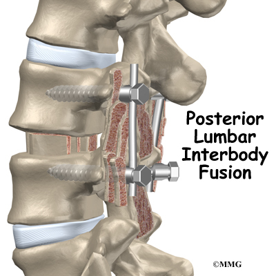
Posterior lumbar interbody fusion (PLIF) is a procedure used to treat problems such as disc degeneration, disc herniation, and spine instability. In this procedure, the surgeon works on the spine from the back (the posterior) and removes a spinal disc in the lower (lumbar) spine. The surgeon inserts bone graft material into the space between the two vertebrae where the disc was removed (the interbody space). The graft may be held in place with a special fusion cage. The goal of the procedure is to stimulate the vertebrae to grow together into one solid bone (known as a fusion). A fusion creates a rigid and immovable column of bone in the problem section of the spine.

This guide will help you understand

  • what surgeons hope to achieve
  • what happens during surgery
  • what to expect as you recover

Posterior Lumbar Interbody Fusion

Anatomy

What parts of the spine and low back are involved?

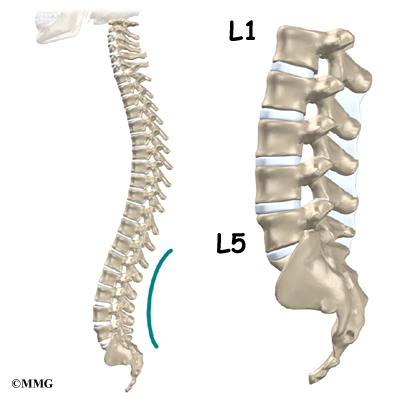
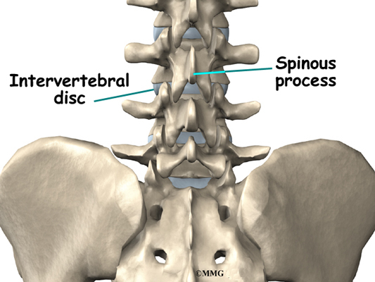
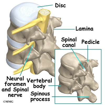
This surgery is done through an incision in the low back. The incision reaches to the spinous processes, the bony projections off the back of the vertebrae. A large block of bone, called the vertebral body makes up the front section of each vertebra. The vertebral bodies are separated by a soft cushion

Posterior Lumbar Interbody Fusion

called an intervertebral disc.

On the back of the vertebral body, the lamina and pedicle bones form a protective ring around the spinal canal. The spinal nerves are enclosed in the spinal canal and exit through small openings on the sides of each vertebral pair, one on the left and one on the right. These passageways are called the neural foramina. (The term neural foramen describes a

Posterior Lumbar Interbody Fusion

single passageway).

Related Document: A Patient’s Guide to Lumbar Spine Anatomy

Rationale

What do surgeons hope to achieve?

Posterior Lumbar Interbody Fusion

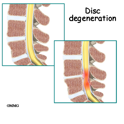
This procedure is often used to stop symptoms from lumbar disc disease. Discs degenerate, or wear out, as a natural part of aging and also from stress and strain on the back. Over time, the disc begins to collapse, and the space decreases between the vertebrae.

Related Document: A Patient’s Guide to Lumbar Degenerative Disc Disease

When this happens, the openings around the spinal nerves (the neural foramina) narrow and may put pressure on the nerves. The long ligaments in the spine slacken due to the collapse in vertebral height. These ligaments may even buckle and put pressure on the spinal nerves.

View animation of degeneration

Pain from disc degeneration can come from a tear in the outer portion of the disc, from chemical inflammation inside the disc, or from a herniated disc that pushes on a nearby spinal nerve. Mechanical pain can also occur from excess movement within the problem part of the spine.

Discectomy is the removal of the disc and any fragments between the vertebrae that are to be fused. Taking out the painful disc is intended to relieve symptoms. It also provides room for placing a graft that will allow the two vertebrae to fuse together.

Posterior Lumbar Interbody Fusion

Once the disc is removed, the surgeon spreads the bones of the spine apart slightly to make room to implant bone graft material. Bone graft is commonly taken from the rim of the pelvis and packed in a special case, called a fusion cage. Bone taken from your own body is called autograft. Bone substitutes are also being used and avoid the need for taking bone from your pelvis.

Another option is to use a wedge of hard, cortical bone taken from preserved human bone. This source of bone graft is called allograft. During the PLIF procedure, the cage or bone wedge is implanted into the interbody space. The PLIF method provides a large surface area for fusion to occur.

Posterior Lumbar Interbody Fusion

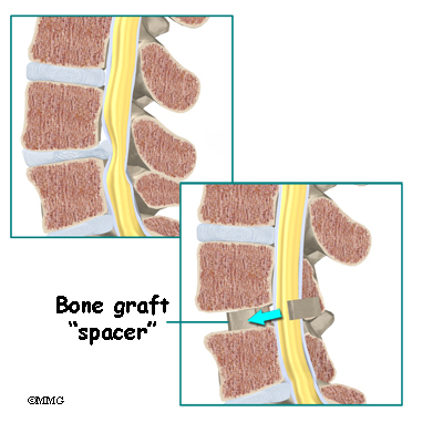
The graft creates a solid spacer to separate and hold the vertebrae apart. Enlarging the space between the vertebrae widens the opening of the neural foramina, taking pressure off the spinal nerves that pass through these openings. Also, the long ligaments that run up and down inside the spinal canal are pulled taut so they don’t buckle into the spinal canal.

View animation of creating a spacer

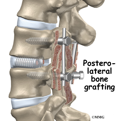
The surgeon also fixes the bones in place using pedicle screws. This instrumentation (or hardware, as it is sometimes called) holds the vertebrae together and prevents them from moving. The less motion there is between two bones trying to heal, the higher the chance they will successfully fuse. The use of instrumentation has increased the success rate of spinal fusions considerably.

Posterior Lumbar Interbody Fusion

During the PLIF procedure, surgeons also commonly add bone graft material along the back sides of the spine. This step is called posterolateral bone grafting. When combined with instrumentation, this approach helps fuse a large surface area on the back (posterior column) of the spine.

In a successful fusion, the vertebrae that are fused together no longer move against one another. The fusion creates one solid bone. No movement happens within the bones that are fused. Instead, they move as one unit. This helps stop the mechanical pain that was coming from the moving parts of the back. Fusion also prevents additional wear and tear on the spinal segment that was fused. By fusing the bones together, surgeons hope to reduce future problems at the spinal segment.

View animation of fusion

Preparations

How will I prepare for surgery?

The decision to proceed with surgery must be made jointly by you and your surgeon. You should understand as much about the procedure as possible. If you have concerns or questions, you should talk to your surgeon.

Once you decide on surgery, your surgeon may suggest a complete physical examination by your regular doctor. This exam helps ensure that you are in the best possible condition to undergo the operation.

On the day of your surgery, you will probably be admitted to the hospital early in the morning. You shouldn’t eat or drink anything after midnight the night before.

Surgical Procedure

What happens during the operation?

Patients are given a general anesthesia to put them to sleep during most spine surgeries. As you sleep, your breathing may be assisted with a ventilator. A ventilator is a device that controls and monitors the flow of air to the lungs.

During surgery the patient usually kneels face down on a special operating table. The special table supports the patient so the abdomen is relaxed and free of pressure. This position reduces blood loss during surgery. It also gives the surgeon more room to work.

Two measurements are made before surgery begins. The first measurement ensures that the surgeon chooses a fusion cage or bone wedge that will fit inside the disc space. To correctly size the fusion cage or bone wedge, the surgeon uses an X-ray image to measure the disc space in a healthy disc, above or below the problem segment.

Second, to size the length of the pedicle screws, a CT scan is used to measure the length of the pedicle bone on the back of the vertebrae to be fused. The CT scan is a special type of X-ray that lets doctors see slices of bone tissue. The machine uses a computer and X-rays to create these slices.

Posterior Lumbar Interbody Fusion

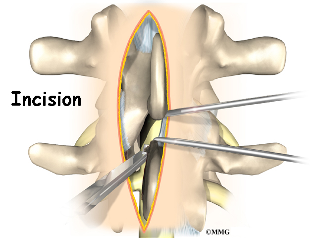
To begin the procedure, an incision is made down the middle of the low back. The tissues just under the skin are separated. Then the small muscles along the sides of the low back are moved aside, exposing the back of the spinal column. Next, the surgeon takes an X-ray to make sure that the procedure is being performed on the correct vertebrae.

The bone graft is prepared. When autograft (bone taken from your body) is used, the same incision made at the beginning of the surgery can be used. The surgeon reaches through the first incision and opens the tissues that cover the back of the pelvis. An osteotome is used to cut the surface of the pelvis bone. An instrument is used to gather a small amount of the pelvis bone. The graft material is prepared and will later be packed into the fusion cages. The tissues covering the pelvis bone are sutured.

Posterior Lumbar Interbody Fusion

Then the surgeon prepares to implant bone graft into the space between the vertebral bodies. The surgeon removes the lamina bones that cover the back of the spinal canal. Next, the surgeon cuts a small opening in the ligamentum flavum, an elastic ligament separating the lamina bones and the spinal nerves. Removing the ligamentum flavum allows the surgeon to see inside the spinal canal. The nerves are checked for tension where they exit the spinal canal. If a nerve root is under tension, the surgeon enlarges the neural foramen, the opening where the nerve travels out of the spinal canal.

The surgeon locates the spot where the pedicle screws are to be placed. A fluoroscope is used to visualize the pedicle bones. A fluoroscope is a special type of X-ray that allows the surgeon to see an X-ray picture continuously on a TV screen. The surgeon uses the fluoroscope to guide one screw through the back of each pedicle, one on the left and one on the right.

The nerve roots inside the spinal canal are then pulled aside with a retractor so the problem disc can be examined. With the nerves held to the side, the surgeon is able to see the disc where it sits just in front of the spinal canal.

Posterior Lumbar Interbody Fusion

A hole is cut into the rim of the back of the disc. Forceps are placed inside the hole in order to clean out disc material within the disc. Reamers and scrapers are used to open up and remove additional disc material.

The surgeon prepares the disc space where the fusion cages or bone wedges are to be inserted. Special spreaders hold the two vertebral bodies apart. A layer of bone is shaved off the flat surfaces of the two vertebrae, causing the surfaces to bleed. Bleeding stimulates the bone graft to heal the bones together.

Adequate room is needed to get the bone graft implants through the spinal column and into the disc space. The nerve roots must be pulled as far to the side as possible to open up enough space.

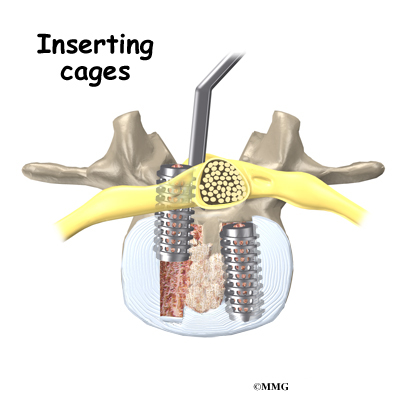
With the disc space held apart by the spreaders, the surgeon has enough room to place the bone graft between the two vertebral bodies. For the fusion cage method, the surgeon packs two cages with bone taken from the pelvis bone or with a suitable bone substitute. Two cages are inserted, one on the left and one on the right. When allograft bone wedges are used, the surgeon inserts the wedges and aligns them within the disc space.

The surgeon uses a fluoroscope to check the position and fit of the graft.

Posterior Lumbar Interbody Fusion

The spreaders used to hold the disc space apart are released. Then the doctor tests the graft by bending and turning the spine to make sure the graft is in the right spot and is locked in place.

Posterior Lumbar Interbody Fusion

Some surgeons add strips of bone graft along the back of the vertebrae to be fused. This is called posterolateral fusion. The bones that project out from each side of the back of the spine are called transverse processes. The back surface of the transverse processes are shaved, causing the surfaces to bleed. Small strips of bone, usually taken from the pelvis bone at the beginning of the surgery, are placed over the transverse processes. The combination of this graft material with the pedicle screws helps hold the spine steady as the interbody fusion heals.

A drainage tube may be placed in the wound. The muscles and soft tissues are then put back in place. The skin is stitched together. The surgeon may place you in a rigid brace that straps across the chest, pelvis, and low back to support the spine while it heals.

Complications

What might go wrong?

As with all major surgical procedures, complications can occur. Some of the most common complications following PLIF include

  • problems with anesthesia
  • thrombophlebitis
  • infection
  • nerve damage
  • problems with the implant or hardware
  • nonunion
  • ongoing pain

This is not intended to be a complete list of possible complications.

Problems with Anesthesia

Problems can arise when the anesthesia given during surgery causes a reaction with other drugs the patient is taking. In rare cases, a patient may have problems with the anesthesia itself. In addition, anesthesia can affect lung function because the lungs don’t expand as well while a person is under anesthesia. Be sure to discuss the risks and your concerns with your anesthesiologist.

Thrombophlebitis (Blood Clots)

View animation of pulmonary embolism

Thrombophlebitis, sometimes called deep venous thrombosis (DVT), can happen after any operation. It occurs when the blood in the large veins of the leg forms blood clots. This may cause the leg to swell and become warm to the touch and painful. If the
blood clots in the veins break apart, they can travel to the lung, where they lodge in the capillaries and cut off the blood supply to a portion of the lung. This is called a pulmonary embolism. (Pulmonary means lung, and embolism refers to a fragment of something traveling through the vascular system.) Most surgeons take preventing DVT very seriously. There are many ways to reduce the risk of DVT, but probably the most effective is getting you moving as soon as possible. Two other commonly used preventative measures include

  • pressure stockings to keep the blood in the legs moving
  • medications that thin the blood and prevent blood clots from forming

Infection

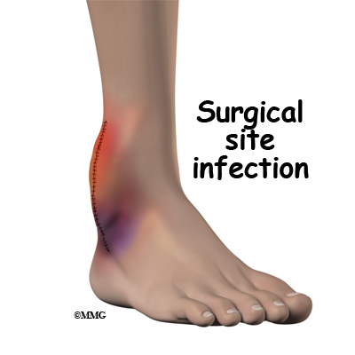
Infection following spine surgery is rare but can be a very serious complication. Some infections may show up early, even before you leave the hospital. Infections on the skin’s surface usually go away with antibiotics. Deeper infections that spread into the bones and soft tissues of the spine are harder to treat. They may require additional
surgery to treat the infected portion of the spine.

Nerve Damage

Posterior Lumbar Interbody Fusion

Any surgery that is done near the spinal canal can potentially cause injury to the spinal cord or spinal nerves. Injury can occur from bumping or cutting the nerve tissue with a surgical instrument, from swelling around the nerve, or from the formation of scar
tissue.

The nerve roots inside the spinal canal are especially at risk during the PLIF procedure. Retractors are used to hold the nerves aside and may cause muscle weakness and a loss of sensation to the areas supplied by an injured nerve. Pressure on the nerves that supply the bowels and bladder can cause incontinence. However, these types of nerve problems after PLIF usually go away soon after surgery.

Problems with the Implant or Hardware

Fusion surgery with cages requires bone grafting. The graft is commonly taken from the top rim of the pelvis (autograft). There is a risk of pain, infection, or weakness in the area where the graft is taken. These risks are avoided when a bone graft substitute is used in place of an autograft.

After the interbody implant is placed, the surgeon checks the position of the fusion cage or bone wedge before completing the surgery. However, the implant may shift slightly soon after surgery to the point that it is no longer able to hold the spine stable. If the implant migrates out of position, it can cause injury to the nearby tissues. A second surgery may be needed to align or replace the implant.

Hardware can also cause problems. Screws or pins may loosen and irritate the nearby soft tissues. Also, the metal plates can sometimes break. If this happens, the surgeon may suggest another surgery either to take out the hardware or to add more hardware to solve the problem.

Nonunion

Posterior Lumbar Interbody Fusion

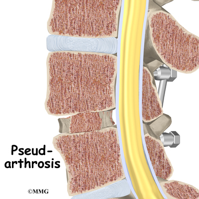
Sometimes the bones do not fuse as planned. This is called a nonunion, or pseudarthrosis. (The term pseudarthrosis means false joint.) When more than one level of the spine is fused at one time, there is a greater chance that nonunion will occur. (Fusion of more than one level means two or more consecutive discs are removed and replaced with bone graft.) If the joint motion from a nonunion continues to cause pain, the patient may need a second operation.

In the second procedure, the surgeon usually adds more bone graft. Additional instrumentation may also be needed to rigidly secure the bones so they will fuse together.

Ongoing Pain

PLIF is a complex surgery. Not all patients get complete pain relief with this procedure. As with any surgery, patients should expect some pain afterward. If the pain continues or becomes unbearable, talk to your surgeon about treatments that can help control your pain.

Afterward

What happens after surgery?

Patients are sometimes placed in a rigid body brace after surgery. The surgical drain is removed within one to two days.

Patients usually stay in the hospital after surgery for three to five days. During this time, patients work daily with a physical therapist. The therapist demonstrates safe ways to move, dress, and do activities without putting extra strain on the back. The therapist
may recommend that the patient use a walker for the first day or two. Before going home, patients are shown ways to help control pain and avoid problems.

Patients are able to return home when their medical condition is stable. However, they are usually required to keep their activities to a minimum in order to give the fusion time to begin healing. Patients are cautioned against bending, lifting, twisting, driving, and prolonged sitting for up to six weeks. Outpatient physical therapy is
usually started a minimum of six weeks after the date of surgery.

Rehabilitation

What should I expect as I recover?

Rehabilitation after PLIF can be a slow process. Many surgeons prescribe outpatient physical therapy beginning a minimum of six weeks after surgery. This delay is needed to make sure the graft has time to begin to fuse. You will probably need to attend therapy sessions for two to three months. You should expect full recovery to take up to eight months.

At first, treatments are used to help control pain and inflammation. Ice and electrical stimulation are commonly used to help with these goals. Your therapist may also use massage and other hands-on treatments to ease muscle spasm and pain.

Active treatments are slowly added. These include exercises for improving heart and lung function. Short, slow walks are generally safe to start with. Swimming and use of a stairclimbing machine are helpful in the later phases of treatment. Therapists also teach
specific exercises to help tone and control the muscles that stabilize the low back.

Posterior Lumbar Interbody Fusion

Your therapist also works with you on how to move and do activities. This form of treatment, called body mechanics, is used to help you develop new movement habits. This training helps you keep your back in safe positions as you go about your work and daily activities. Training includes positions you use when sitting, lying, standing, and walking. You’ll also work on safe body mechanics with lifting, carrying, pushing, and pulling.

As your condition improves, the therapist tailors your program to prepare you to go back to work. Some patients are not able to go back to a job that requires strenuous tasks. Your therapist may suggest changes in job tasks that enable you to go back to your previous job or to do alternate forms of work. You’ll learn to do these tasks in new ways that keep your back safe and free of strain.

Before your therapy sessions end, your therapist will teach you ways to avoid future problems.
Patellar Tendon Graft Reconstruction of the ACL

A Patient’s Guide to Patellar Tendon Graft Reconstruction of the ACL

Introduction

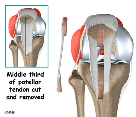
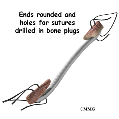
The anterior cruciate ligament (ACL) is a major stabilizer of the knee joint. This key knee ligament is commonly torn during sports activities. The standard operation to fix a torn ACL is with a patellar tendon graft. The surgeon takes out the middle section of the patellar tendon below the kneecap (patella). This new graft includes the strip of tendon, along with attached plugs of bone on each end. For this reason, it is sometimes referred to as a bone-patellar-tendon-bone graft. The surgeon removes the torn ACL and puts the new graft into the knee, making sure to line it up just like the original ligament.

Many types of tissue grafts have been tried. The patellar tendon graft has proven to be one of the strongest for ACL reconstruction. Patients who have this operation generally get back to their usual activities and sports. They often do so faster than people who have their ACL reconstructed with other types of tissue grafts.

This guide will help you understand

  • what parts of the knee are treated during surgery
  • how surgeons perform the operation
  • what to expect before and after the procedure

Patellar Tendon Graft Reconstruction of the ACL

Anatomy

What parts of the knee are involved?

Ligaments are tough bands of tissue that connect the ends of bones together. The ACL is located in the center of the knee joint where it runs from the backside of the femur (thighbone) to the front of the tibia (shinbone).

Patellar Tendon Graft Reconstruction of the ACL

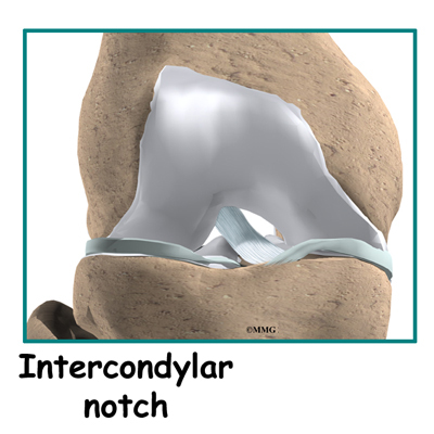
The ACL runs through a special notch in the femur called the intercondylar notch and attaches to a special area of the tibia called the tibial spine.

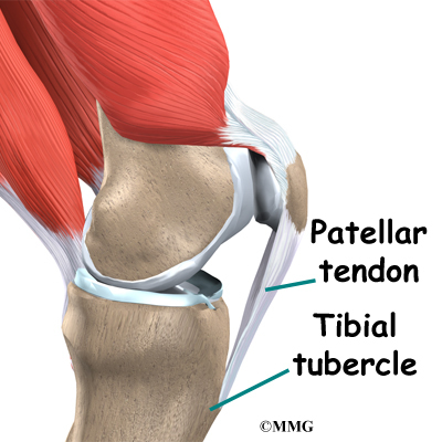
The patellar tendon is a thick and strong band of connective tissue on the front of the knee. It starts at the bottom of the patella and fastens just below the knee to a bony bump on the front of the tibia, called the tibial tubercle. When using the patellar tendon as an ACL graft, surgeons remove a strip from the middle of it. The graft includes the bony attachments from

Patellar Tendon Graft Reconstruction of the ACL

the bottom of the patella and from the tibial tubercle.

Related Document: A Patient’s Guide to Knee Anatomy

Related Document: A Patient’s Guide to Anterior Cruciate Ligament Injuries

Rationale

What does the surgeon hope to accomplish?

The main goal of ACL surgery is to keep the tibia from moving too far forward under the femur bone and to get the knee functioning normally again.

Many surgeons prefer to use the patellar tendon when reconstructing the ACL. The graft is often chosen because it is one of the strongest ACL grafts. It’s easy to get to, holds well in its location, and generally heals fast.

Patellar Tendon Graft Reconstruction of the ACL

The anatomy of the graft helps to speed healing and to create a solid connection. When the surgeon implants the new graft, the bony plugs on each end of the graft fit inside a tunnel of bone. This means there is bone-to-bone contact. The body treats the contact of these two bony surfaces as it would a broken bone. It responds by healing the two surfaces together. Healing at the bone-to-bone surface fixes the patellar tendon graft in place.

Preparation

What do I need to know before surgery?

You and your surgeon should make the decision to proceed with surgery together. You need to understand as much about the procedure as possible. If you have concerns or questions, you should talk to your surgeon.

Once you decide on surgery, you need to take several steps. Your surgeon may suggest a complete physical examination by your regular doctor. This exam helps ensure that you are in the best possible condition to undergo the operation.

You may also need to spend time with the physical therapist who will be managing your rehabilitation after surgery. This allows you to get a head start on your recovery. One purpose of this preoperative visit is to record a baseline of information. Your therapist will check your current pain levels, your ability to do your activities, and the movement and strength of each knee.

A second purpose of the preoperative visit is to prepare you for surgery. Your therapist will teach you how to walk safely using crutches or a walker. And you’ll begin learning some of the exercises you’ll use during your recovery.

On the day of your surgery, you will probably be admitted to the surgery center early in the morning. You shouldn’t eat or drink anything after midnight the night before.

Surgical Procedure

What happens during the operation?

Patellar Tendon Graft Reconstruction of the ACL

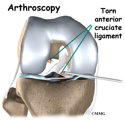
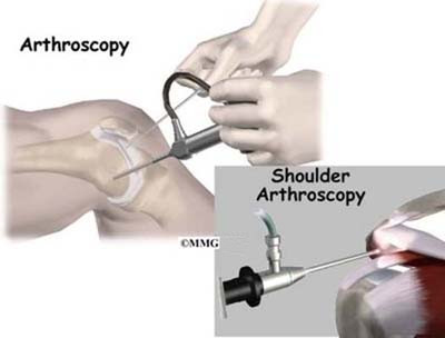
Most surgeons perform this surgery using an arthroscope, a small fiber-optic TV camera that is used to see and operate inside the joint. Only small incisions are needed during arthroscopy for this procedure. The operation doesn’t require the surgeon to open the knee joint.

Before surgery you will be placed under either general anesthesia or a type of spinal anesthesia. The surgeon begins the operation by making two small openings into the knee, called portals. These portals are where the arthroscope and surgical tools are placed into the knee.

Patellar Tendon Graft Reconstruction of the ACL

Care is taken to protect the nearby nerves and blood vessels.

A small incision is also made below the patella. Working through this incision, the surgeon takes out the middle section of the patellar tendon, along with the bone attachments on each end. The bone plugs are rounded and smoothed. Holes are drilled in each bone plug to place sutures (strong stitches) that will pull the graft into place.

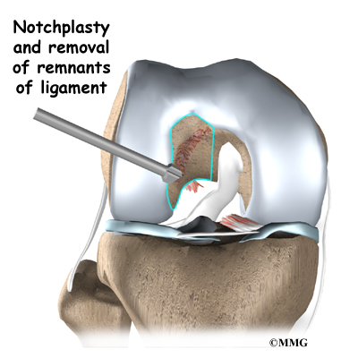
Next, the surgeon prepares the knee to place the graft. The remnants of the original ligament are removed. The intercondylar notch (mentioned earlier) is enlarged so that nothing will rub on the graft. This part of the surgery is referred to as a notchplasty.

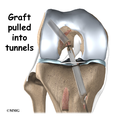
Once this is done, holes are drilled in the tibia and the femur to place the graft. These holes are placed so that the graft will run between the tibia and femur in the same direction as the original ACL.

Patellar Tendon Graft Reconstruction of the ACL

The graft is then pulled into position using sutures placed through the drill holes. Screws are used to hold the bone plugs in the drill holes.

To keep fluid from building up in your knee, the surgeon may place a tube in your knee joint. The portals and skin incision are then stitched together, completing the surgery.

Complications

What problems can happen with this surgery?

As with all major surgical procedures, complications can occur. This document doesn’t provide a complete list of the possible complications, but it does highlight some of the most common problems. Some of the most common complications following patellar tendon graft reconstruction of the ACL are

  • anesthesia complications
  • thrombophlebitis
  • infection
  • problems with the graft
  • problems at the donor site

Anesthesia Complications

Most surgical procedures require that some type of anesthesia be done before surgery. A very small number of patients have problems with anesthesia. These problems can be reactions to the drugs used, problems related to other medical complications, and problems due to the anesthesia. Be sure to discuss the risks and your concerns with your anesthesiologist.

Thrombophlebitis (Blood Clots)

View animation of pulmonary embolism

Thrombophlebitis, sometimes called deep venous thrombosis (DVT), can occur after any operation, but is more likely to occur following surgery on the hip, pelvis, or knee. DVT occurs when blood clots form in the large veins of the leg. This may cause the leg to swell and become warm to the touch and painful. If the blood clots in the veins break apart, they can travel to the lung, where they lodge in the capillaries and cut off the blood supply to a portion of the lung. This is called a pulmonary embolism. (Pulmonary means lung, and embolism refers to a fragment of something traveling through the vascular system.) Most surgeons take preventing DVT very seriously. There are many ways to reduce the risk of DVT, but probably the most effective is getting you moving as soon as possible after surgery. Two other commonly used preventative measures include

  • pressure stockings to keep the blood in the legs moving
  • medications that thin the blood and prevent blood clots from forming

Infection

Following surgery, it is possible that the surgical incision can become infected. This will require antibiotics and possibly another surgical procedure to drain the infection.

Problems with the Graft

After surgery, the body attempts to develop a network of blood vessels in the new graft. This process, called revascularization, takes about 12 weeks. The graft is weakest during this time, which means it has a greater chance of stretching or rupturing. A stretched or torn graft can occur if you push yourself too hard during this period of recovery. When revascularization is complete, strength in the graft gradually builds. A second surgery may be needed to replace the graft if it is stretched or torn.

Problems at the Donor Site

Problems can occur at the donor site (the area below the patella where the graft was taken from the knee). A major drawback of taking out a piece of the patellar tendon to reconstruct the ACL is that most patients end up having difficulty kneeling down long after surgery. Lingering pain in the front of the knee is also common.

A portion of bone is taken from the bottom of the patella during the graft procedure. This can weaken the patella. In rare cases, heavy use of the quadriceps muscle (on the front of the thigh) can cause the patella to fracture. This often requires a second surgery to repair the broken patella.

Taking tissue from the center of the patellar tendon can also cause problems. The body attempts to heal the area but sometimes produces too much scar tissue. The extra scar tissue that forms around the donor site may prevent normal motion in the knee. The patellar tendon is not as strong as it was before surgery. In rare cases, this has been linked to a tear in the patellar tendon. Also, the patellar tendon may become easily inflamed. And problems in this area can keep the quadriceps from regaining normal control and strength.

After Surgery

What should I expect after surgery?

You may use a continuous passive motion (CPM) machine immediately afterward to help the knee begin moving and to alleviate joint stiffness. The machine straps to the leg and continuously bends and straightens the joint. This continuous motion is thought to reduce stiffness, ease pain, and keep extra scar tissue from forming inside the joint. The CPM is often used with a form of cold treatment that circulates cold water through hoses and pads around your knee.

Most ACL surgeries are now done on an outpatient basis. Many patients go home the same day as the surgery. Some patients stay one to two nights in the hospital if necessary. The tube placed in your knee at the end of the surgery is usually removed after 24 hours.

Your surgeon may also have you wear a protective knee brace for a few weeks after surgery. You’ll use crutches for two to four weeks in order to keep your knee safe, but you’ll probably be allowed to put a comfortable amount of weight down while you’re up and walking.

Rehabilitation

What will my recovery be like?

Patients usually take part in formal physical therapy after ACL reconstruction. The first few physical therapy treatments are designed to help control the pain and swelling from the surgery. The goal is to help you regain full knee extension as soon as possible.

The physical therapist will choose treatments to get the quadriceps muscles toned and active again. Muscle stimulation and biofeedback, which involve placing electrodes over the quadriceps muscle, may be needed at first to get the muscle going again and to help retrain it.

As the rehabilitation program evolves, more challenging exercises are chosen to safely advance the knee’s strength and function. Specialized balance exercises are used to help the muscles respond quickly and without thinking. This part of treatment is called neuromuscular training. If you need to stop suddenly, your muscles must react with just the right amount of speed, control, and direction. After ACL surgery, this ability doesn’t come back completely without exercise.

Neuromuscular training includes exercises to improve balance, joint control, muscle strength and power, and agility. Agility makes it possible to change directions quickly, go faster or slower, and improve starting and stopping. These are important skills for walking, running, and jumping, and especially for sports performance.

When you get full knee movement, your knee isn’t swelling, and your strength and muscle control are improving, you’ll be able to gradually go back to your work and sport activities. Some surgeons prescribe a functional brace for athletes who intend to return quickly to their sports.

Ideally, you’ll be able to resume your previous lifestyle activities. However, athletes are usually advised to wait at least six months before returning to their sports. Most patients are encouraged to modify their activity choices.

You will probably be involved in a progressive rehabilitation program for four to six months after surgery to ensure the best result from your ACL reconstruction. In the first six weeks following surgery, expect to see the physical therapist two to three times a week. If your surgery and rehabilitation go as planned, you may only need to do a home program and see your therapist every few weeks over the four to six month period.

Meniscal Surgery

A Patient’s Guide to Meniscal Surgery

Introduction

The meniscus is very important to the long-term health of the knee. In the past, surgeons would simply take out part or all of an injured meniscus. But today’s surgeons know that removing the meniscus can lead to early knee arthritis. Whenever possible, they try to repair the tear. If the damaged area must be removed, care is taken during surgery to protect the surrounding healthy tissue.

This guide will help you understand

  • what parts of the knee are treated during meniscal surgery
  • what operations are used to treat a damaged meniscus
  • what to expect before and after meniscal surgery

Meniscal Surgery

Anatomy

What parts of the knee are involved?

There is one meniscus on each side of the knee joint. The C-shaped medial meniscus is on the inside part of the knee, closest to your other knee. (Medial means closer to the middle of the body.) The U-shaped lateral meniscus is on the outer half of the knee joint. (Lateral means further out from the center of the body.)

Meniscal Surgery

The menisci (plural for meniscus) protect the articular cartilage on the surfaces of the thighbone (femur) and the shinbone (tibia). Articular cartilage is the smooth, slippery material that covers the ends of the bones that make up the knee joint. The articular cartilage allows the joint surfaces to slide against one another without damage to either surface.

Meniscal Surgery

Most of the meniscus is avascular, meaning no blood vessels go to it. Only its outer rim gets a small supply of blood. Doctors call this area the red zone. The ends of a few vessels in the red zone may actually travel inward to the middle section, the red-white zone. The inner portion of the meniscus, closest to the center of the knee, is called the white zone. It has no blood vessels at all. Although a tear in the outer rim has a good chance of healing, damage further in toward the center of the meniscus will not heal on its own.

Related Document: A Patient’s Guide to Knee Anatomy

Related Document: A Patient’s Guide to Meniscal Injuries

Rationale

What does my surgeon hope to accomplish?

Meniscal Surgery

The meniscus is a pad of cartilage that acts like a shock absorber to protect the knee. The meniscus is also vital for knee stability. When the meniscus is damaged or is surgically removed, the knee joint can become loose, or unstable. Without the protection and stability of a healthy meniscus, the surfaces of the knee can suffer wear and tear, leading to a condition called osteoarthritis.

Related Document: A Patient’s Guide to Knee Osteoarthritis

Most tears of the meniscus do not heal on their own. A small tear in the outer rim (the red zone) has a good chance of healing. However, tears in the inner part of the meniscus often require surgery. When tears in this area are causing symptoms, they tend to get bigger. This puts the articular cartilage on the surfaces of the knee joint at risk of injury.

Surgeons aim to save the meniscus. If an injured part must be removed, only the smallest amount of the meniscus is taken out. Preserving the nearby areas of the meniscus is vital for keeping the knee healthy. If a tear can possibly be repaired, surgeons will recommend a meniscal repair.

Meniscal Surgery

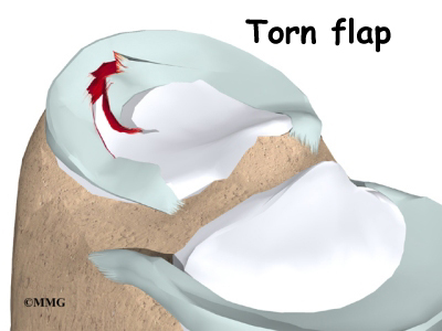
A torn meniscus may cause symptoms of pain and swelling and sometimes catching and locking. The goal of surgery is to take these symptoms away. When the knee locks and you have to tug on it to get it moving, a small flap from a meniscal tear may have developed. The flap may be getting caught in the knee joint as you bend it. Or a small piece of the meniscus could actually be floating around inside the joint. This fragment, called a loose body, can get lodged between the moving parts of the knee, causing the knee to lock. In these cases, surgery may be needed, sometimes right away, to fix the flap or to remove the loose body.

Meniscal Surgery

Only when the majority of the meniscus is damaged beyond repair is the entire meniscus removed. Surgeons are experimenting with solutions to replace the meniscus.

Preparations

What do I need to know before surgery?

You and your surgeon should make the decision to proceed with surgery together. You need to understand as much about the procedure as possible. If you have concerns or questions, be sure and talk to your surgeon.

Once you decide on surgery, you need to take several steps. Your surgeon may suggest a complete physical examination by your regular doctor. This exam helps ensure that you are in the best possible condition to undergo the operation.

You may also need to spend time with the physical therapist who will be managing your rehabilitation after surgery. This allows you to get a head start on your recovery. One purpose of this preoperative visit is to record a baseline of information. The therapist will check your current pain levels, ability to do your activities, and the movement and strength of each knee.

A second purpose of the preoperative visit is to prepare you for surgery. The therapist will teach you how to walk safely using crutches or a walker. And you’ll begin learning some of the exercises you’ll use during your recovery.

On the day of your surgery, you will probably be admitted for surgery early in the morning. You shouldn’t eat or drink anything after midnight the night before.

Surgical Procedure

What happens during meniscal surgery?

Meniscal Surgery

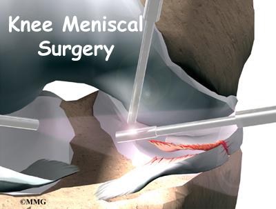
Meniscal surgery is done using an arthroscope, a small fiber-optic TV camera that is used to see and operate inside the joint. Only small incisions are needed during arthroscopy. The surgeon does not need to open the knee joint.

Before surgery you will be placed under either general anesthesia or a type of spinal anesthesia. The surgeon begins the operation by making two or three small openings into the knee, called portals. These portals are where the arthroscope and surgical instruments are placed inside the knee. Care is taken to protect the nearby nerves and blood vessels.

Partial Meniscectomy

Meniscal Surgery

The procedure to carefully remove a damaged portion of the meniscus is called partial meniscectomy. The surgeon starts by inserting the arthroscope into one of the portals. A probe is placed into another portal. The surgeon watches on a screen while probing the meniscus. All parts of the inside of the knee joint are examined. When a meniscal tear is found, the surgeon determines the type and location of the tear. Surgical instruments are placed into another portal and are used to remove the torn portion of meniscus.

When the problem part of the meniscus has been removed, the surgeon checks the knee again with the probe to be sure no other tears are present. A small motorized cutter is used to trim and shape the cut edge of the meniscus. The joint is flushed with sterile saline to wash away debris from the injury or from the surgery. The portals are closed with sutures.

Meniscal Repair

Suture Repair

View animation of sewing the edges of a torn meniscus

Meniscal Surgery

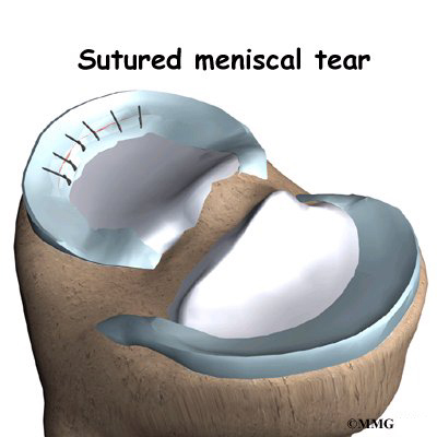
Using the arthroscope and a probe, the surgeon locates the tear. The probe is used to push the torn edges of the meniscus together. A small rasp or shaver is used to roughen the edges of the tear. Then a hollow tube called a cannula is inserted through one of the portals. The surgeon threads a suture through the cannula and into the knee joint. The suture is sewn into the two edges of the tear. The surgeon tugs on the thread to bring the torn edges close together. The suture is secured by tying two to three knots. Additional sutures are placed side by side until the entire tear is fixed.

An alternate method is to pierce the knee joint with one or two curved needles. The needle goes from the outer edge of the meniscus completely through the tear. The surgeon may feed a suture from another portal into the end of the needle. Or the suture can be threaded into the needle from the outside of the knee. Both ways get the suture through the tear and allow the surgeon to sew the torn edges of the meniscus together.

Suture Anchor Repair

View animation of anchoring the edges of a torn meniscus

Meniscal Surgery

Special fasteners, called suture anchors, are sometimes used to anchor the torn edges of the meniscus together. These implants are biodegradable, meaning they eventually break down and are absorbed by the body. Suture anchors have barbed shafts and are pointed like an arrow. They work like a staple or straight pin to hold the healing tissues together.

Repairs using suture anchors work best for younger patients who have a single tear near the outer rim (red zone) of the meniscus. (As described earlier, this part of the meniscus has the richest blood supply.) A probe is often used to line up the torn edges of the meniscus. Then the surgeon uses a small surgical tool to punch an arrow through the damaged part of the meniscus. Usually only two or three arrows are needed. Larger tears may require up to six arrows. The arrows anchor the two torn edges together while the tear heals. It takes about six months before the arrows begin to be absorbed by the body.

Meniscal Transplantation

Meniscal Surgery

If the meniscus cannot be repaired or has been previously removed, a new form of treatment may offer a way to slow the onset of knee arthritis. Meniscal transplantation uses borrowed tissue to take the place of the original meniscus.
Experiments have been tried using various replacement materials. One material that is showing promise is an allograft. An allograft is tissue that is from a donor, usually preserved human meniscus tissue. Because it is so new, this surgery is currently only available for select patients in a limited number of locations.

View animation of removing the meniscus

View animation of preparing the surgical site

View animation of placing the allograft

View animation of suturing the allograft in place

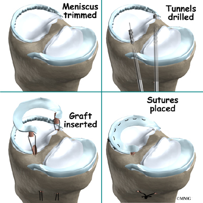
Using the arthroscope, the surgeon removes remnants of the old meniscus. Next, the allograft is prepared. Small sutures are placed around the edges of the allograft. The surgeon slides the allograft with the sutures into the knee through a small incision. The allograft is sewn in place onto the tibia bone. Surgical instruments, including a cannula or needle (described earlier), may be used to secure additional sutures. Some surgeons also use special anchors to firmly fix the allograft in place. A probe is used to make sure the transplanted meniscus holds securely. The arthroscope is removed, and the portals are sewn shut.

Complications

What can go wrong?

As with all major surgical procedures, complications can occur. This document doesn’t provide a complete list of the possible complications, but it does highlight some of the most common problems. Some of the most common complications following meniscal surgery are

  • anesthesia complications
  • thrombophlebitis
  • infection
  • suture anchor problems
  • graft failure
  • slow recovery
  • ongoing pain

Anesthesia Complications

Most surgical procedures require that some type of anesthesia be done before surgery. A very small number of patients have problems with anesthesia. These problems can be reactions to the drugs used, problems related to other medical complications, and problems due to the anesthesia. Be sure to discuss the risks and your concerns with your anesthesiologist.

Thrombophlebitis (Blood Clots)

View animation of pulmonary embolism

Thrombophlebitis, sometimes called deep venous thrombosis (DVT), can occur after any operation, but is more likely to occur following surgery on the hip, pelvis, or knee. DVT occurs when blood clots form in the large veins of the leg. This may cause the leg to swell and become warm to the touch and painful. If the blood clots in the veins break apart, they can travel to the lung, where they lodge in the capillaries and cut off the blood supply to a portion of the lung. This is called a pulmonary embolism. (Pulmonary means lung, and embolism refers to a fragment of something traveling through the vascular system.) Most surgeons take preventing DVT very seriously. There are many ways to reduce the risk of DVT, but probably the most effective is getting you moving as soon as possible after surgery. Two other commonly used preventative measures include

  • pressure stockings to keep the blood in the legs moving
  • medications that thin the blood and prevent blood clots from forming

Infection

Following surgery, it is possible that the skin portals can become infected. This will require antibiotics and possibly another surgical procedure to drain the infection.

Suture Anchor Problems

Suture anchors can cause problems. If one breaks, the free-floating piece may hurt other parts inside the knee joint, particularly the articular cartilage. Also, the end of the anchor may poke too far through the meniscus. If so, the point may rub and irritate nearby tissues. A second surgery may be needed to fix problems with suture anchors.

Graft Failure

Surgeries where tissue is grafted into the body, like bone marrow or kidney transplants, have a high risk that the body will reject the graft. This is not so in meniscal transplant surgery. The preserved graft contains no live cells, so it doesn’t have to be matched up with the person getting the graft. Also, the properties of meniscal tissue makes rejection of a transplanted graft rare. The main reason for graft failure in meniscal transplant surgery occurs when patients try to do too much, too soon after surgery. Doing sports where there are quick starts and stops, sharp pivoting, and jumping can cause the graft to fail. If the graft tears, another transplant surgery will be needed.

Slow Recovery

Not everyone after meniscal surgery gets quickly back to routine activities. Some people feel better and have less swelling, but they still find it hard to do normal activities even several months after surgery. Others with damage in their knee ligaments or in the articular cartilage also tend to have a slower recovery.

Ongoing Pain

Pain relief does not always occur with meniscal surgery. If you have pain that continues or becomes unbearable, talk to your surgeon about treatments that can help control your pain.

After Surgery

What happens after meniscal surgery?

Meniscal surgery is done on an outpatient basis. Patients usually go home the same day as the surgery. The portals are covered with surgical strips, and the knee may be wrapped in an elastic bandage.

Crutches are used after meniscal surgery. They may only be needed for one to two days after a simple meniscectomy. Surgeons specify how much weight can be borne after meniscal repair or allograft transplant. Patients having meniscal repair are usually told not to place any weight on the foot for four to six weeks after surgery. After a transplant procedure, most patients are instructed to touch only the toes of the operated leg on the ground for four to six weeks. Some sugeons allow their patients to place a comfortable amount of weight on the foot four weeks after repair or transplant surgery.

Patients who have had a meniscal repair or transplant usually wear a knee brace for at least four weeks. The brace keeps the knee straight. It is removed often during the day to do easy range-of-motion exercises for the knee.

Follow your surgeon’s instructions about how much weight to place on your foot while standing or walking. Avoid doing too much, too quickly. You may be told to use a cold pack on the knee and to keep your leg elevated and supported.

Rehabilitation

What will my recovery be like?

Your rehabilitation will depend on the type of surgery you had. You probably won’t need formal physical therapy after partial meniscectomy. Most patients can do their exercises as part of a home program. If you require outpatient physical therapy, you will probably need to attend therapy sessions for two to four weeks. You should expect full recovery to take up to three months.

Many surgeons have their patients take part in formal physical therapy after meniscal repair and transplant procedures. The first few physical therapy treatments are designed to help control the pain and swelling from the surgery. Physical therapists will also work with patients to make sure they are putting only a safe amount of weight on the affected leg.

For the first six weeks after a meniscal repair, you should avoid bending the knee more than 90 degrees. Then it is safe to gradually bend the knee fully. However, you should avoid squatting for at least three to four months while the repair fully heals. It is not advisable to run, jump, or twist the knee for at least four to six months. Patients sometimes resume sport activities within four to six months after surgery to repair the meniscus.

Range-of-motion exercises start right away after a transplant. The goal is to get the knee to bend to 90 degrees within four weeks after surgery. As time goes on, more challenging exercises are chosen to safely advance the knee’s range of motion, strength, and function.

Ideally, patients will be able to resume their previous activities. Some patients may be encouraged to modify their activity choices, especially if an allograft was used.

The physical therapist’s goal is to help you keep your pain under control and improve your knee’s range of motion and strength. When you are well under way, regular visits to your therapist’s office will end. The therapist will continue to be a resource, but you will be in charge of doing your exercises as part of an ongoing home program.

Biceps Tendonitis

A Patient’s Guide to Biceps Tendonitis

Introduction

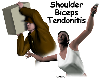
Biceps tendonitis, also called bicipital tendonitis, is inflammation in the main tendon that attaches the top of the biceps muscle to the shoulder. The most common cause is overuse from certain types of work or sports activities. Biceps tendonitis may develop gradually from the effects of wear and tear, or it can happen suddenly from a direct injury. The tendon may also become inflamed in response to other problems in the shoulder, such as rotator cuff tears, impingement, or instability (described below).

This guide will help you understand

  • what parts of the shoulder are affected
  • the causes of biceps tendonitis
  • ways to treat this problem

Anatomy

Biceps Tendonitis

What parts of the shoulder are affected?

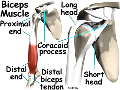
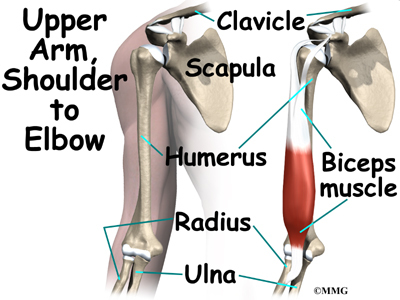
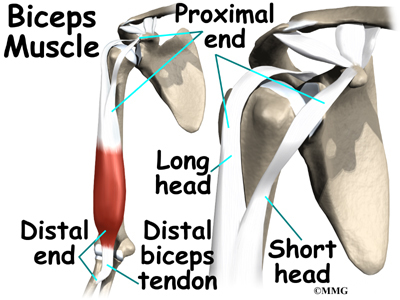
The biceps muscle goes from the shoulder to the elbow on the front of the upper arm. Two separate tendons (tendons attach muscles to bones) connect the upper part of the biceps muscle to the shoulder. The upper two tendons of the biceps are called the proximal biceps tendons, because they are closer to the top of the arm.

Biceps Tendonitis

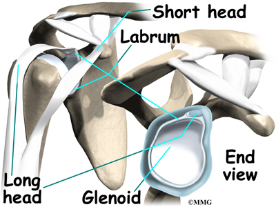
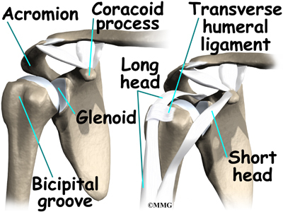
The main proximal tendon is the long head of the biceps. It connects the biceps muscle to the top of the shoulder socket, the glenoid. It also blends with the cartilage rim around the glenoid, the labrum. The labrum is a rim of soft tissue that turns the flat surface of the glenoid into a deeper socket. This arrangement improves the fit of the ball that fits in the socket, the humeral head.

Biceps Tendonitis

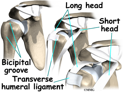
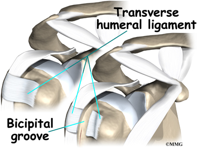
Beginning at the top of the glenoid, the tendon of the long head of the biceps runs in front of the humeral head. The tendon passes within the bicipital groove of the humerus and is held in place by the transverse humeral ligament. This arrangement keeps the humeral head from sliding too far up or forward within the glenoid.

The short head of the biceps connects on the coracoid process of the scapula (shoulder blade). The coracoid process is

Biceps Tendonitis

a small bony knob just in from the front of the shoulder. The lower biceps tendon is called the distal biceps tendon. The word distal means the tendon is further down the arm. The lower part of the biceps muscle connects to the elbow by this tendon. The muscles forming the short and long heads of the biceps stay separate until just above the elbow, where they unite and connect to the distal biceps tendon.

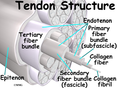
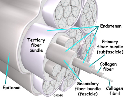
Tendons are made up of strands of a material called collagen.

Biceps Tendonitis

The collagen strands are lined up in bundles next to each other. Because the collagen strands in tendons are lined up, tendons have high tensile strength. This means they can withstand high forces that pull on both ends of the tendon. When muscles work, they pull on one end of the tendon. The other end of the tendon pulls on the bone, causing the bone to move.

Biceps Tendonitis

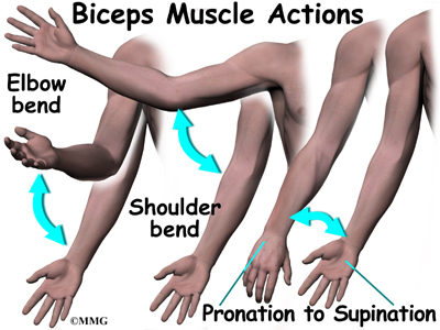
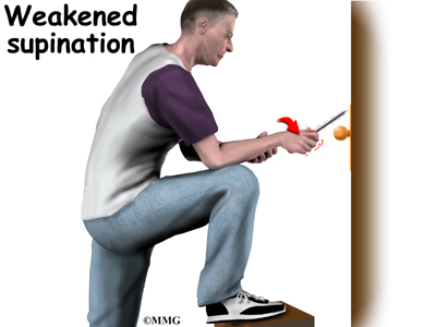
Contracting the biceps muscle can bend the elbow upward. The biceps can also help flex the shoulder, lifting the arm up, a movement called flexion. And the muscle can rotate, or twist, the forearm in a way that points the palm of the hand up. This movement is called supination, which positions the hand as if you were holding a tray.

Related Document: A Patient’s Guide to Shoulder Anatomy

Causes

Why is my biceps tendon inflamed?

Continuous or repetitive shoulder actions can cause overuse of the biceps tendon. Damaged cells within the tendon don’t have time to recuperate. The cells are unable to repair themselves, leading to tendonitis. This is common in sport or work activities that require frequent and repeated use of the arm, especially when the arm motions are performed overhead. Athletes who throw, swim, or swing a racquet or club are at greatest risk.

Years of shoulder wear and tear can cause the biceps tendon to become inflamed. Examination of the tissues in these cases commonly shows signs of degeneration. Degeneration in a tendon causes a loss of the normal arrangement of the collagen fibers that join together to form the tendon. Some of the individual strands of the tendon become jumbled due to the degeneration, other fibers break, and the tendon loses strength. When this happens in the biceps tendon, inflammation, or even a rupture of the biceps tendon, may occur.

Related Document: A Patient’s Guide to Rupture of the Biceps Tendon

Biceps tendonitis can happen from a direct injury, such as a fall onto the top of the shoulder. A torn transverse humeral ligament can also lead to biceps tendonitis. (As mentioned earlier, the transverse humeral ligament holds the biceps tendon within the bicipital groove near the top of the humerus.) If this ligament is torn, the biceps tendon is free to jump or slip out of the groove, irritating and eventually inflaming the biceps tendon.

Biceps tendonitis sometimes occurs in response to other shoulder problems, including

  • rotator cuff tears
  • shoulder impingement
  • shoulder instability

Rotator Cuff Tears

Biceps Tendonitis

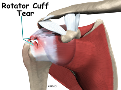
Aging adults with rotator cuff tears also commonly end up with biceps tendonitis. When the rotator cuff is torn, the humeral head is free to move too far up and forward in the shoulder socket and can impact the biceps tendon. The damage may begin to weaken the biceps tendon and cause it to become inflamed.

Related Document: A Patient’s Guide to Rotator Cuff Tears

Shoulder Impingement

Biceps Tendonitis

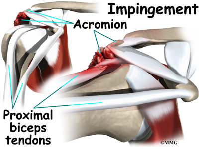
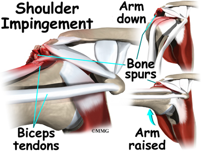
In shoulder impingement, the soft tissues between the humeral head and the top of the shoulder blade (acromion) get pinched or squeezed with certain arm movements.

Related Document: A Patient’s Guide to Shoulder Impingement

Shoulder Instability

Conditions that allow too much movement of the ball within the socket create shoulder instability. When extreme shoulder motions are frequently repeated, such as with throwing or swimming, the soft tissues supporting the ball and socket can eventually get stretched out.

Related Document: A Patient’s Guide to Shoulder Instability

Biceps Tendonitis

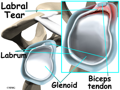
The labrum (the cartilage rim that deepens the glenoid, or shoulder socket) may begin to pull away from its attachment to the glenoid. A shoulder dislocation can also cause the labrum to tear. When the labrum is torn, the humeral head may begin to slip up and forward within the socket. The added movement of the ball within the socket (instability) can cause damage to the nearby biceps tendon, leading to secondary biceps tendonitis.

Related Document: A Patient’s Guide to Labral Tears

Symptoms

What does biceps tendonitis feel like?

Biceps Tendonitis

Patients generally report the feeling of a deep ache directly in the front and top of the shoulder. The ache may spread down into the main part of the biceps muscle. Pain is usually made worse with overhead activities. Resting the shoulder generally eases pain.

The arm may feel weak with attempts to bend the elbow or when twisting the forearm into supination (palm up). A catching or slipping sensation felt near the top of the biceps muscle may suggest a tear of the transverse humeral ligament.

Diagnosis

How can my doctor be sure I have biceps tendonitis?

Your doctor will first take a detailed medical history. You will need to answer questions about your shoulder, if you feel pain or weakness, and how this is affecting your regular activities. You’ll also be asked about past shoulder pain or injuries.

The physical exam is often most helpful in diagnosing biceps tendonitis. Your doctor may position your arm to see which movements are painful or weak. Available arm motion is checked. And by feeling the biceps tendon, the doctor can tell if the tendon is tender.

Special tests are done to see if nearby structures are causing problems, such as a tear in the labrum or in the transverse humeral ligament. The doctor checks the shoulder for impingement, instability, or rotator cuff problems.

X-rays are generally not needed right away. They may be ordered if the shoulder hasn’t gotten better with treatment. An X-ray can show if there are bone spurs or calcium deposits near the tendon. X-rays can also show if there are other problems, such as a fracture. Plain X-rays do not show soft tissues like tendons and will not show a biceps tendonitis.

When the shoulder isn’t responding to treatment, magnetic resonance imaging (MRI) scan may also be ordered. An MRI is a special imaging test that uses magnetic waves to create pictures of the shoulder in slices. This test can tell if there are problems in the rotator cuff or labrum.

Arthroscopy is an invasive way to evaluate shoulder pain that isn’t going away. It is not used to first evaluate biceps tendonitis. It may be used for ongoing shoulder problems that haven’t been found in an X-ray or MRI scan. The surgeon uses an arthroscope to see inside the joint. The arthroscope is a thin instrument that has a tiny camera on the end. It can show if there are problems with the rotator cuff, the labrum, or the portion of the biceps tendon that is inside the shoulder joint.

Treatment

What treatment options are available?

Nonsurgical Treatment

Whenever possible, doctors treat biceps tendonitis without surgery. Treatment usually begins by resting the sore shoulder. The sport or activity that led to the problem is avoided. Resting the shoulder relieves pain and calms inflammation.

Anti-inflammatory medicine may be prescribed to ease pain and to help patients return to normal activity. These medications include common over-the-counter drugs such as ibuprofen.

Doctors may have their patients work with a physical or occupational therapist. Therapists apply treatments to reduce pain and inflammation. When present, conditions causing the biceps tendonitis are also addressed. For example, shoulder impingement may require specialized hands-on joint mobilization, along with strengthening of the rotator cuff and shoulder blade muscles. Treating the main cause will normally get rid of the biceps tendonitis. When needed, therapists also evaluate the way you do your work or sport activities to reduce problems of overuse.

In rare instances, an injection of cortisone may be used to try to control pain. Cortisone is a very powerful steroid. However, cortisone is used very sparingly because it can weaken the biceps tendon, and possibly cause it to rupture.

Surgery

Patients who are improving with conservative treatments do not typically require surgery. Surgery may be recommended if the problem doesn’t go away or when there are other shoulder problems present.

Acromioplasty

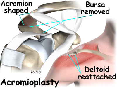
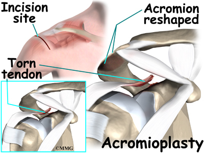
The most common surgery for bicipital tendonitis is acromioplasty, especially when the underlying problem is shoulder impingement. This procedure involves removing the front portion of the acromion, the bony ledge formed where the scapula meets the top of the shoulder joint. By removing a small portion of the acromion, more space is created between the acromion and the humeral head. This takes pressure off the soft tissues in between, including the biceps tendon.

Acromioplasty is usually done through a two-inch incision in the skin over the shoulder joint. In some cases, the surgery can be done using an arthroscope.

Biceps Tendonitis

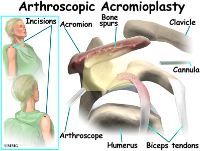
Today, acromioplasty is usually done using an arthroscope. An arthroscope is a slender tool with a tiny TV camera on the end. It lets the surgeon work in the joint through a very small incision. This may result in less damage to the normal tissues surrounding the joint, leading to faster healing and recovery.

To perform the acromioplasty using the arthroscope, several small incisions are made to insert the arthroscope and special instruments needed to complete the procedure. These incisions are small, usually about one-quarter inch long. It may be necessary to make three or four incisions around the shoulder to allow the arthroscope to be moved to different locations to see different areas of the shoulder.

A small plastic, or metal, tube is inserted into the shoulder and connected with sterile plastic tubing to a special pump. Another small tube allows the fluid to be removed from the joint. This pump continuously fills the shoulder joint with sterile saline (salt water) fluid. This constant flow of fluid through the joint inflates the joint and washes any blood and debris from the joint as the surgery is performed.

There are many small instruments that have been specially designed to

Biceps Tendonitis

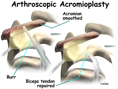
perform surgery in the joint. Some of these instruments are used to remove torn and degenerative tissue. Some of these instruments nibble away bits of tissue and then vacuum them up from out of the joint. Others are designed to burr away bone tissue and vacuum it out of the joint. These instruments are used to remove any bone spurs that are rubbing on the tendons of the shoulder and smooth the under surface of the acromion and AC joint.

If necessary, the acromioplasty can also be performed using the older, open method. The open method requires a small incision in the skin over the shoulder joint.

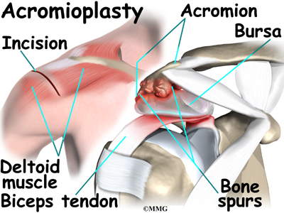
Working through the incision, the surgeon locates the deltoid muscle on

Biceps Tendonitis

the outer part of the shoulder. Splitting the front section of this muscle gives the surgeon a better view of the acromion. Some surgeons also detach the deltoid muscle where it connects on the front of the acromion.

The bursa sac that lies just under the acromion is removed. Next, a surgical tool is used to cut a small portion off the front of the acromion. The ligament arcing from the acromion to the corocoid process (the coracoacromial ligament) may also be removed.

Biceps Tendonitis

The surgeon shaves the undersurface of the acromion to remove any bone spurs. A file is used to smooth the edge of the acromion. Next, a series of small holes is drilled into the remaining acromion. These holes are used to reattach the deltoid muscle to the acromion.

The surgeon inspects the rotator cuff muscle to see if any tears are present. Then the entire area is irrigated to wash away small particles of bone. Finally, the free end of the deltoid muscle is sutured back to the ridge of the acromion using the drill holes made earlier.

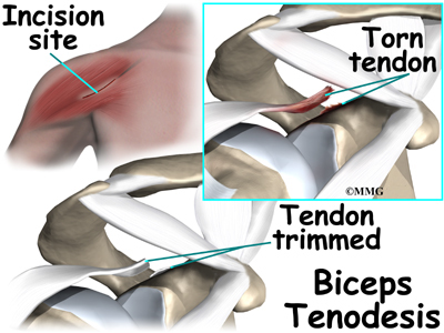
If the biceps tendon is severely degenerated, the surgeon may perform biceps tenodesis (described next). The surgeon completes the procedure by closing the incision with sutures.

Biceps Tenodesis

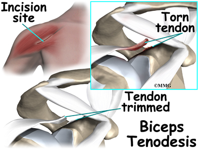
Biceps tenodesis is a method of reattaching the top end of the biceps tendon to a new location. Studies show that the long-term results of this form of surgery are not satisfactory for patients with biceps tendonitis. However, tenodesis may be needed when the biceps tendon is severely degenerated or when shoulder reconstruction for other problems is needed.

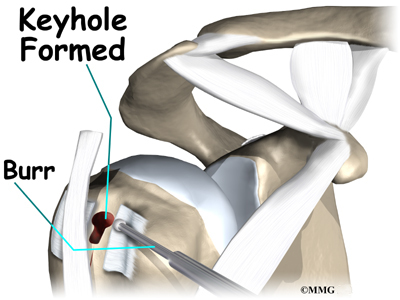
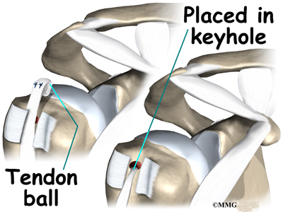
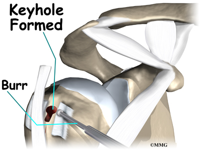
A common way to do this surgery is called the keyhole technique. The keyhole describes the shape of a small hole made by the surgeon in the humerus. The end of the tendon is slid into the top of the keyhole and pulled down to anchor it in place.

This surgery can be done using the arthroscope. The tenodesis procedure is usually combined with other procedures such as those discussed above. If so, the surgeon will simply continue using the arthroscope to do the tenodesis procedure if possible. The advantage of using the arthroscope is that less normal tissue is damaged. This may result in faster healing and recovery.

Biceps Tendonitis

If the procedure is performed using the open method, the surgeon begins by making an incision on the front of the shoulder, just above the axilla (armpit). The overlying muscles are separated so the surgeon can locate the top of the biceps tendon. The end of the biceps tendon is removed from its attachment at the top of the glenoid. The tendon is prepared by cutting away frayed and degenerated tissue.

The transverse humeral ligament is split, exposing the bicipital groove.

Biceps Tendonitis

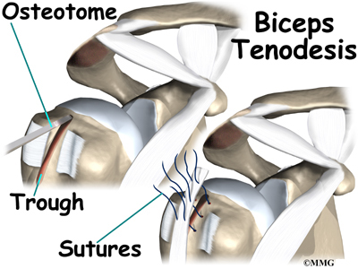
An incision is made along the floor of the bicipital groove. The bleeding from the incision gets scar tissue to form that will help anchor the repaired tendon in place.

Biceps Tendonitis

A burr is used to form a keyhole-shaped cavity within the bicipital groove. The top of the cavity is round. The bottom is the slot of the keyhole. It is made the same width as the biceps tendon.

Biceps Tendonitis

The surgeon rolls the top end of the biceps tendon into a ball. Sutures are used to form and hold the ball.The elbow is bent, taking tension off the biceps muscle and tendon. The surgeon pushes the tendon ball into the top part of the keyhole. As the elbow is gradually straightened, the ball is pulled firmly into the narrow slot in the lower end of the keyhole.

Rehabilitation

What should I expect after treatment?

Nonsurgical Rehabilitation

You will need to avoid heavy arm activity for three to four weeks. As the pain resolves, you should be safe to begin doing more normal activities.

Your doctor may prescribe a carefully progressed rehabilitation program under the supervision of a physical or occupational therapist. This could involve four to six weeks of therapy. At first, treatments are used to calm inflammation and to improve shoulder range of motion. As symptoms ease, specific exercises are used to strengthen the biceps muscle, as well as the rotator cuff and scapular muscles. Overhead athletes are shown ways to safely resume their sport.

After Surgery

Some surgeons prefer to have their patients start a gentle range-of-motion program soon after surgery. When you start therapy, your first few therapy sessions may involve ice and electrical stimulation treatments to help control pain and swelling from the surgery. Your therapist may also use massage and other types of hands-on treatments to ease muscle spasm and pain.

You will gradually start exercises to improve movement in the forearm, elbow, and shoulder. You need to be careful to avoid doing too much, too quickly.

Heavier exercises for the biceps muscle are avoided for two to four weeks after surgery. Your therapist may begin with light isometric strengthening exercises. These exercises work the biceps muscle without straining the healing tendon.

After two to four weeks, you start doing more active strengthening. As you progress, your therapist will teach you exercises to strengthen and stabilize the muscles and joints of the elbow and shoulder. Other exercises will work your arm in ways that are similar to your work tasks and sport activities. Your therapist will help you find ways to do your tasks that don’t put too much stress on your shoulder.

You may require therapy for six to eight weeks. It generally takes three to four months, however, to safely begin doing forceful biceps activity after surgery. Before your therapy sessions end, your therapist will teach you a number of ways to avoid future problems.
Biceps Rupture

A Patient’s Guide to Biceps Rupture

Introduction

Biceps Rupture

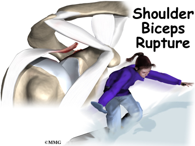
A biceps rupture involves a complete tear of the main tendon that attaches the top of the biceps muscle to the shoulder. It happens most often in middle-aged people and is usually due to years of wear and tear on the shoulder. A torn biceps in younger athletes sometimes occurs during weightlifting or from actions that cause a sudden load on the arm, such as hard fall with the arm outstretched.

This guide will help you understand

  • what parts of the shoulder are affected
  • the causes of a biceps rupture
  • ways to treat this problem

Anatomy

What parts of the shoulder are affected?

Biceps Rupture

The biceps muscle goes from the shoulder to the elbow on the front of the upper arm. Two separate tendons (tendons attach muscles to bones) connect the upper part of the biceps muscle to the shoulder. The upper two tendons of the biceps are called the proximal biceps tendons, because they are closer to the top of the arm.

Biceps Rupture

The main proximal tendon is the long head of the biceps. It connects the biceps muscle to the top of the shoulder socket, the glenoid. Beginning at the glenoid, the tendon of the long head of the biceps travels down the front of the upper arm. The tendon runs within the bicipital groove and is held in place by the transverse humeral ligament.

Biceps Rupture

The short head of the biceps connects on the corocoid process of the scapula. The corocoid process is a small bony knob just in from the front of the shoulder.

The lower biceps tendon is called the distal biceps tendon. The word distal means the tendon is further down the arm. The lower part of the biceps muscle connects to the elbow by this tendon.

The muscles forming the short and long heads of the biceps stay separate until just above the elbow where they unite and connect to the distal biceps tendon.

Biceps Rupture

Tendons are made up of strands of a material called collagen. The collagen strands are lined up in bundles next to each other. Because the collagen strands in tendons are lined up, tendons have high tensile strength. This means they can withstand high forces that pull on both ends of the tendon. When muscles work, they pull on one end of the tendon. The other end of the tendon pulls on the bone, causing the bone to move.

Biceps Rupture

Contracting the biceps muscle can bend the elbow upward. The biceps can also help flex the shoulder, lifting the arm up, a movement called flexion. And the muscle can rotate, or twist, the forearm in a way that points the palm of the hand up. This movement is called supination, which positions the hand as if you were holding a tray.

Related Document: A Patient’s Guide to Shoulder Anatomy

Causes

Why did my biceps rupture?

Biceps Rupture

Biceps ruptures generally occur in people who are between 40 and 60 years old. People in this age group who’ve had shoulder problems for a long time are at most risk. Often the biceps ruptures after a long history of shoulder pain from tendonitis (inflammation of hte tendon) or problems with shoulder impingement. Shoulder impingement is a condition where the soft tissues between the ball of the upper arm and the top of the shoulder blade (acromion) get squeezed with arm motion.

Related Document: A Patient’s Guide to Shoulder Impingement

Years of shoulder wear and tear begin to fray the biceps tendon. Eventually, the long head of the biceps weakens and becomes prone to tears or ruptures. Examination of the tissues within most torn or ruptured biceps tendons commonly shows signs of degeneration. Degeneration in a tendon causes a loss of the normal arrangement of the collagen fibers that join together to form the tendon. Some of the individual strands of the tendon become jumbled due to the degeneration, other fibers break, and the tendon loses strength.

A rupture of the biceps tendon can happen from a seemingly minor injury. When it happens for no apparent reason, the rupture is called nontraumatic.

Aging adults with rotator cuff tears also commonly have a biceps tendon rupture. When the rotator cuff is torn, the ball of the humerus is free to move too far up and forward in the shoulder socket and can impact the biceps tendon. The damage may begin to weaken the biceps tendon and cause it to eventually rupture.

Related Document: A Patient’s Guide to Rotator Cuff Tears

Symptoms

What does a ruptured biceps feel like?

Biceps Rupture

Patients often recall hearing and feeling a snap in the top of the shoulder. Immediate and sharp pain follow. The pain often subsides quickly with a complete rupture because tension is immediately taken off the pain sensors in the tendon. Soon afterward, bruising may develop in the middle of the upper arm and spread down to the elbow. The biceps may appear to have balled up, especially in younger patients who’ve had a traumatic biceps rupture. The arm may feel weak at first with attempts to bend the elbow or lift the shoulder.

The biceps tendon sometimes only tears part of the way. If so, a pop may not be felt or heard. Instead, the front of the shoulder may simply be painful, and the arm may feel weak with the same arm movements that are affected with a complete biceps rupture.

Diagnosis

How can my doctor be sure my biceps ruptured?

Your doctor will first take a detailed medical history. You will need to answer questions about your shoulder, if you feel pain or weakness, and how this is affecting your regular activities. You’ll also be asked about past shoulder pain or injuries.

The physical exam is often most helpful in diagnosing a rupture of the biceps tendon. Your doctor may position your arm to see which movements are painful or weak. By feeling the area of the muscle and tendon, the doctor can often tell if the tendon has ruptured. The muscle may look and feel balled up in the middle of the arm, and a dent can sometimes be felt near the top of the shoulder.

Biceps Rupture

X-rays may be ordered. X-rays show the bones that form the shoulder joint and may show bony changes that have contributed to a ruptured biceps. For example, bone spurs (small projections of bone) may be seen on the X-ray. Spurs that form near the biceps tendon will often puncture the tendon as the arm is used with activity. X-rays can also show if there are other problems, such as a fracture. Plain X-rays do not show soft tissues like tendons and will not show a biceps rupture.

Your doctor may also order a magnetic resonance imaging (MRI) scan. This is the most reliable way to check whether the biceps tendon is only partially torn or if the tendon actually ruptured. An MRI is a special imaging test that uses magnetic waves to create pictures of the shoulder in slices. The MRI can also show if there are other problems in the shoulder.

Treatment

What treatment options are available?

Nonsurgical Treatment

Doctors usually treat a ruptured long head of biceps tendon without surgery. This is especially true for older individuals who can tolerate loss of arm strength or if the injury occurs in the nondominant arm.

Biceps Rupture

Not having surgery usually only results in a moderate loss of strength. The short head of the biceps is still attached and continues to supply strength to raise the arm up. Flexion of the elbow may be affected, but supination (the motion of twisting the forearm such as when you use a screwdriver) is usually affected more. Not repairing a ruptured biceps reduces supination strength by about 20 percent.

Nonsurgical measures could include a sling to rest the shoulder. Patients may be given anti-inflammatory medicine to help ease pain and swelling and to help return people to activity sooner after a biceps tendon rupture. These medications include common over-the-counter drugs such as ibuprofen.

Doctors may have their patients work with a physical or occupational therapist. At first, your therapist will give you tips how to rest your shoulder and how to do your activities without putting extra strain on the sore area.

Your therapist may apply ice and electrical stimulation to ease pain. Exercises are used to gradually strengthen other muscles that help do the work of a normal biceps muscle.

Surgery

Surgery is reserved for patients who need arm strength, are concerned with cosmetics of the balled up biceps, or who have pain that won’t go away.

Biceps Tenodesis

Biceps tenodesis is a surgery to anchor the ruptured end of the biceps tendon. A common method, called the keyhole technique, involves anchoring the ruptured end to the upper end of the humerus. The keyhole describes the shape of a small hole made by the surgeon in the humerus. The end of the tendon is slid into the top of the keyhole

Biceps Rupture

and pulled down to anchor it in place.

The surgeon begins by making an incision on the front of the shoulder, just above the axilla (armpit). The overlying muscles are separated so the surgeon can locate the damaged end of the biceps tendon. The end of the biceps tendon is prepared by cutting away frayed and degenerated tissue.

Biceps Rupture

The transverse humeral ligament is split, exposing the bicipital groove. An incision is made along the floor of the bicipital groove. The bleeding from the incision gets scar tissue to form that will help anchor the repaired tendon in place.

A burr is used to form a keyhole-shaped cavity within the bicipital groove. The top of the cavity is round. The bottom is the slot of the keyhole.

Biceps Rupture

It is made the same width as the biceps tendon.

The surgeon rolls the top end of the biceps tendon into a ball. Sutures are used to form and hold the ball. The elbow is bent, taking tension off the biceps muscle and tendon. The surgeon pushes the tendon ball into the top part of the keyhole. As the elbow is gradually straightened, the ball is pulled firmly into the narrow slot in the lower end of the keyhole.

Biceps Rupture

The surgeon tests the stability of the attachment by bending and straightening the elbow. When the surgeon is satisfied with the repair, the skin incisions are closed, and the shoulder is placed in a protective sling.

Acromioplasty and Direct Tenodesis

This procedure may be used for younger patients who’ve had a recent traumatic biceps rupture, have problems with impingement, and who have an injured rotator cuff.

Biceps Rupture

Acromioplasty involves cutting and reshaping the acromion, the bone that forms the top part of the shoulder. Some surgeons will also sever the corocohumeral ligament, which arches over the top of the shoulder joint. These steps relieve pressure on the tissues between the ball of the humerus and the acromion, including the biceps and rotator cuff tendons. For this reason, this procedure is sometimes called subacromial decompression. The ruptured end of the biceps is then anchored to the upper end of the humerus. This is called direct tenodesis.

The surgeon begins by making an incision across the top of the shoulder. The shoulder muscles are separated to expose the top of the humerus. Bone spurs are removed, along with part of the acromion. The surgeon then smooths the rough ends of the bone.

Biceps Rupture

After the acromioplasty procedure, the surgeon focuses on the biceps tendon. When the bicipital groove is in view, the transverse humeral ligament is cut. Next, an osteotome is used to open the joint capsule and create a trough next to the bicipital groove. Three small holes are drilled along each side of the trough. The surgeon places the loose end of the biceps tendon in the new groove.

Biceps Rupture

Sutures are woven into one drill hole, through the tendon, and out the opposite drill hole. This is repeated for the remaining two sets of drill holes. Next, the top end of the ruptured tendon is cut off. Finally, the three sutures are firmly secured.

When the surgeon is satisfied with the repair, the transverse humeral ligament and joint capsule are sutured, followed by the skin incision. The arm is bent at the elbow and placed in a light splint that is to be worn for four weeks after surgery.

Rehabilitation

What should I expect after treatment?

Nonsurgical Rehabilitation

In cases where the ruptured biceps tendon is treated nonsurgically, you will need to avoid heavy arm activity for three to four weeks. As the pain and swelling resolve, you should be safe to begin doing more normal activities.

If the tendon is only partially torn, however, recovery takes longer. Patients usually need to rest the shoulder using a protective sling. As symptoms ease, a carefully progressed rehabilitation program under the supervision of a physical or occupational therapist usually follows. This often involves four to six weeks of therapy.

After Surgery

Immediately after surgery, you’ll need to wear your shoulder sling for about four weeks. Some surgeons prefer to have their patients start a gentle range-of-motion program soon after surgery. When you start therapy, your first few therapy sessions may involve ice and electrical stimulation treatments to help control pain and swelling from the surgery. Your therapist may also use massage and other types of hands-on treatments to ease muscle spasm and pain.

You will gradually start exercises to improve movement in the forearm, elbow, and shoulder. You need to be careful to avoid doing too much, too quickly.

Heavier exercises for the biceps muscle are avoided until at least four to six weeks after surgery. Your therapist may begin with light isometric strengthening exercises. These exercises work the biceps muscle without straining the healing tendon.

At about six weeks, you start doing more active strengthening. As you progress, your therapist will teach you exercises to strengthen and stabilize the muscles and joints of the elbow and shoulder. Other exercises will work your arm in ways that are similar to your work tasks and sport activities. Your therapist will help you find ways to do your tasks that don’t put too much stress on your shoulder.

You may require therapy for six to eight weeks. It generally takes three to four months, however, to safely begin doing forceful biceps activity after surgery. Before your therapy sessions end, your therapist will teach you a number of ways to avoid future problems.
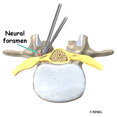
Hip Anatomy

A Patient’s Guide to Hip Anatomy

Introduction

The hip joint is a true ball-and-socket joint. This arrangement gives the hip a large amount of motion needed for daily activities like walking, squatting, and stair-climbing.

Understanding how the different layers of the hip are built and connected can help you understand how the hip works, how it can be injured, and how challenging recovery can be when this joint is injured. The deepest layer of the hip includes the bones and the joints. The next layer is made up of the ligaments of the joint capsule. The tendons and the muscles come next.

In addition to reading this article, be sure to watch our Hip Anatomy Animated Tutorial Video.

This guide will help you understand

  • the parts that make up the hip
  • how these parts work together

Important Structures

The important structures of the hip can be divided into several categories. These include

  • bones and joints
  • ligaments and tendons
  • muscles
  • nerves
  • blood vessels
  • bursae

Hip Anatomy

Bones and Joints

The bones of the hip are the femur (the thighbone) and the pelvis. The top end of the femur is shaped like a ball. This ball is called the femoral head. The femoral head fits into a round socket on the side of the pelvis. This socket is called the acetabulum.

Hip Anatomy

The femoral head is attached to the rest of the femur by a short section of bone called the femoral neck. A large bump juts outward from the top of the femur, next to the femoral neck. This bump, called the greater trochanter, can be felt along the side of your hip. Large and important muscles connect to the greater trochanter. One muscle is the gluteus medius. It is a key muscle for keeping the pelvis level as you walk.

Hip Anatomy

Articular cartilage is the material that covers the ends of the bones of any joint. Articular cartilage is about one-quarter of an inch thick in the large, weight-bearing joints like the hip. Articular cartilage is white and shiny and has a rubbery consistency. It is slippery, which allows the joint surfaces to slide against one another without causing any damage. The function of articular cartilage is to absorb shock and provide an extremely smooth surface to make motion easier. We have articular cartilage essentially everywhere that two bony surfaces move against one another, or articulate.

In the hip, articular cartilage covers the end of the femur and the socket portion of the acetabulum in the pelvis. The cartilage is especially thick in the back part of the socket, as this is where most of the force occurs during walking and running.

Ligaments and Tendons

Hip Anatomy

There are several important ligaments in the hip. Ligaments are soft tissue structures that connect bones to bones. A joint capsule is a watertight sac that surrounds a joint. In the hip, the joint capsule is formed by a group of three strong ligaments that connect the femoral head to the acetabulum. These ligaments are the main source of stability for the hip. They help hold the hip in place.

Hip Anatomy

A small ligament connects the very tip of the femoral head to the acetabulum. This ligament, called the ligamentum teres, doesn’t play a role in controlling hip movement like the main hip ligaments. It does, however, have a small artery within the ligament that brings a very small blood supply to part of the femoral head.

Hip Anatomy

A long tendon band runs alongside the femur from the hip to the knee. This is the iliotibial band. It gives a connecting point for several hip muscles. A tight iliotibial band can cause hip and knee problems.

Hip Anatomy

A special type of ligament forms a unique structure inside the hip called the labrum. The labrum is attached almost completely around the edge of the acetabulum. The shape and the way the labrum is attached create a deeper cup for the acetabulum socket. This small rim of cartilage can be injured and cause pain and clicking in the hip.

Muscles

The hip is surrounded by thick muscles. The gluteals make up the muscles of the buttocks on the back of the hip. The inner thigh is formed by the adductor muscles. The main action of the adductors is to pull the leg inward toward the other leg. The muscles that flex the hip are in front of the hip joint. These include the iliopsoas muscle. This deep muscle begins in the low back and pelvis and connects on the inside edge of the upper femur. Another large hip flexor is the rectus femoris. The rectus femoris is one of the quadriceps muscles, the largest group of muscles on the front of the thigh. Smaller muscles going from the pelvis to the hip help to stabilize and rotate the hip.

Finally, the hamstring muscles that run down the back of the thigh start on the bottom of the pelvis. Because the hamstrings cross the back of the hip joint on their way to the knee, they help to extend the hip, pulling it backwards.

Nerves

Hip Anatomy

All of the nerves that travel down the thigh pass by the hip. The main nerves are the femoral nerve in front and the sciatic nerve in back of the hip. A smaller nerve, called the obturator nerve, also goes to the hip.

These nerves carry the signals from the brain to the muscles that move the hip. The nerves also carry signals back to the brain about sensations such as touch, pain, and temperature.

Blood Vessels

Traveling along with the nerves are the large vessels that supply the lower limb with blood. The large femoral artery begins deep within the pelvis. It passes by the front of the hip area and goes down toward the inner edge of the knee. If you place your hand on the front of your upper thigh you may be able to feel the pulsing of this large artery.

Hip Anatomy

The femoral artery has a deep branch, called the profunda femoris (profunda means deep). The profunda femoris sends two vessels that go through the hip joint capsule. These vessels are the main blood supply for the femoral head. As mentioned earlier, the ligamentum teres contains a small blood vessel that gives a very small supply of blood to the top of the femoral head.

Other small vessels form within the pelvis and supply the back portion of the buttocks and hip.

Bursae

Where friction occurs between muscles, tendons, and bones there is usually a structure called a bursa. A bursa is a thin sac of tissue that contains fluid to lubricate the area and reduce friction. The bursa is a normal structure. The body will even produce a bursa in response to friction.

Think of a bursa like this. If you press your hands together and slide them against one another, you produce some friction. In fact, when your hands are cold you may rub them together briskly to create heat from the friction. Now imagine that you hold in your hands a small plastic sack that contains a few drops of salad oil. This sack would let your hands glide freely against each other without a lot of friction.

Hip Anatomy

A bursa that sometimes causes problems in the hip is sandwiched between the bump on the outer hip (the greater trochanter) and the muscles and tendons that cross over the bump. This bursa, called the greater trochanteric bursa, can get irritated if the iliotibial band (discussed earlier) is tight. Another bursa sits between the iliopsoas muscle where it passes in front of the hip joint. Bursitis here is called iliopsoas bursitis. A third bursa is over the ischial tuberosity, the bump of bone in your buttocks that you sit on.>

Summary

As you can see, the hip is complex with a design that provides a good amount of stability. It allows good mobility and range of motion for doing a wide range of daily activities. Many powerful muscles connect to and cross by the hip joint, making it possible for us to accelerate quickly during actions like running and jumping.

Categories Hip

Open Carpal Tunnel Release

A Patient’s Guide to Open Carpal Tunnel Release

Introduction

Carpal tunnel syndrome (CTS) occurs when the median nerve is squeezed as it courses through the wrist. The passageway through the wrist, called the carpal tunnel, is formed by the small wrist bones (carpals) on one side and a ligament on the other. In an open release for CTS, the surgeon makes an incision on the front of the wrist and hand in order to cut the ligament. The goal is to relieve pressure on the median nerve.

This guide will help you understand

  • what part of the wrist and hand are treated during surgery
  • how surgeons perform the operation
  • what to expect before and after the procedure

Related Document: A Patient’s Guide to Carpal Tunnel Syndrome

Anatomy

What part of the wrist is treated during surgery?

The carpal tunnel is an opening through the wrist into the hand that is formed by the carpal bones of the wrist on the bottom and the transverse carpal ligament on the top. The transverse carpal ligament is at the base of the wrist and crosses from one side of the wrist to the other. (Transverse means across.) It is sometimes referred to as the carpal ligament.

The opening formed by the carpal bones and the carpal ligament is the carpal tunnel. The median nerve passes through the carpal tunnel into the hand. It gives sensation to the thumb, index finger, long finger, and half of the ring finger. It also sends a nerve branch to control the thenar muscles of the thumb.

The median nerve rests on top of the flexor tendons, just below the carpal ligament. Between the skin and the carpal ligament is a thin sheet of connective tissue called the palmar fascia.

Related Document: A Patient’s Guide to Hand Anatomy

Rationale

What does the surgeon hope to achieve?

The surgery releases the carpal ligament, taking pressure off the median nerve. The open procedure for releasing the carpal ligament involves a sizeable wrist incision, usually about two inches long. By creating a large incision, the surgeon is able to clearly see the wrist structures and to carefully do the operation.

Preparation

What should I do to prepare for surgery?

The decision to proceed with surgery must be made jointly by you and your surgeon. You need to understand as much about the procedure as possible. If you have concerns or questions, you should talk to your surgeon.

Once you decide on surgery, your surgeon may suggest a complete physical examination by your regular doctor. This exam helps ensure that you are in the best possible condition to undergo the operation.

On the day of your surgery, you will probably be admitted to the hospital early in the morning. You shouldn’t eat or drink anything after midnight the night before. This surgery can usually be done as an outpatient procedure, meaning you can leave the hospital the same day.

Procedure

Open release for CTS is occasionally done using a general anesthetic (one that puts you to sleep). More often, it is done using a regional anesthetic. A regional anesthetic blocks the nerves going to only a portion of the body. Injections of medications similar to lidocaine are used to block the nerves for several hours. This type of anesthesia could be an axillary block (only the arm is asleep) or a wrist block (only the hand is asleep). The surgery can also be performed by simply injecting lidocaine around the area of the incision.

Once you have anesthesia, your surgeon will make sure the skin of your palm is free of infection by cleaning the skin with a germ-killing solution.

A small incision is made in the palm of the hand, usually about one inch long. In some severe cases, a slightly longer incision is extended into the forearm. The incision makes the palmar fascia visible. This is a sheet of connective tissue in the palm and forearm right under the skin. The surgeon makes an incision through this material and exposes the carpal ligament.

View animation of palmar fascia exposed

View animation of ligament exposed

Once in view, the carpal ligament is released using a scalpel or scissors. Care is taken to make sure that the median nerve and flexor tendons are out of the way and protected. By cutting the carpal ligament, pressure is taken off the median nerve. Upon dividing the carpal ligament, the surgeon stitches just the skin together and leaves the loose ends of the carpal ligament separated. The loose ends are left apart to keep pressure off the median nerve. Eventually, the gap between the two ends of the ligament fills in with scar tissue. After the skin is stitched together, your hand will be wrapped in a bulky dressing. This surgery can usually be done as an outpatient procedure, meaning you can leave the hospital the same day.

Complications

What might go wrong?

As with all major surgical procedures, complications can occur. This document doesn’t provide a complete list of the possible complications, but it does highlight some of the most common problems. Some of the most common complications following open carpal tunnel release are

  • anesthesia
  • infection
  • incision pain
  • scar tissue formation
  • nerve damage
  • hand weakness

Anesthesia

Problems can arise when the anesthesia given during surgery causes a reaction with other drugs the patient is taking. In rare cases, a patient may have problems with the anesthesia itself. In addition, anesthesia can affect lung function because the lungs don’t expand as well while a person is under anesthesia. Be sure to discuss the risks and your concerns with your anesthesiologist.

Infection

Infection is a possible complication after surgery, especially infection of the incision. Therefore, check your incision every day as instructed by your surgeon. If you think you have a fever take your temperature. If you have signs of infection or other complications, call your surgeon right away.

These are warning signs of infection or other complications:

  • pain in your hand that is not relieved by your medicine
  • discharge with an unpleasant odor coming from your incision
  • swelling, heat, and redness along your incision
  • chills or fever over 100.4 degrees Fahrenheit
  • bright red blood coming from your incision

Incision Pain

Some patients continue to have pain along their incision. The area often stays sensitive long after the surgery. However, symptoms of incision sensitivity tend to get better within four to six months after surgery.

Scar Tissue Formation

A common problem after carpal tunnel release is excessive scar tissue buildup. The body attempts to heal the area but goes too far in the process of supplying new cells. Too much scar tissue forms. When this happens the nearby soft tissues can become bound together. The incision may appear raised. The nearby skin may feel tight. You may even feel a bump beneath the incision. Wrist and hand movement may feel restricted. Scar tissue can also bind the flexor tendons and median nerve, preventing them from gliding smoothly within the carpal tunnel. Pain and a loss of range of motion may occur. In severe cases, a second surgery may be needed to remove the extra scar tissue.

Nerve Symptoms

Sometimes people still feel some numbness and tingling after surgery, especially if they had severe pressure on the median nerve prior to surgery. When the thenar muscles (mentioned earlier) are notably shrunken (atrophied) from prolonged pressure on the median nerve, full strength and normal sensation may not fully return even after having the surgery.

Hand Weakness

Muscles that are used to squeeze and grip may seem weak after surgery. During normal gripping, the tendons of the wrist press outward against the carpal ligament. This allows the carpal ligament to work like a pulley to improve grip strength. People used to think that the tendons lose this mechanical advantage after the carpal ligament has been released. However, recent studies indicate that hand weakness is more likely from pain or swelling that occurs in the early weeks after the procedure. With the exception of patients who have severe thenar atrophy at the time of surgery, most people achieve normal hand strength within two to four months of surgery. Those with severe atrophy commonly see improvements in hand strength, but they rarely regain normal size of the thenar muscles.

After Surgery

What happens immediately after surgery?

At first, take time during the day to support your healing arm with your hand elevated above the level of your heart. You may be instructed to put an ice pack on your wrist several times a day to keep swelling down. At various times during the day, move your thumb and fingers five to 10 times. Also, bend and straighten your elbow and lift and lower your shoulder occasionally to keep these joints limber. Keep the dressing on your hand until you return to the surgeon. Avoid getting the stitches wet. Your stitches will be removed 10 to 14 days after surgery.

Heavy gripping and pinching should be avoided for up to six weeks. These actions need to be avoided to keep the tendons from pushing out against the healing carpal ligament After six weeks, you should be safe to resume gripping and pinching without irritating the wrist.

It generally takes longer to recover after open carpal tunnel release. Pain and symptoms usually begin to improve, but you may have tenderness in the area of the incision for several months after surgery.

Patients who wait too long to seek medical advice sometimes have difficulty adjusting after surgery. Poor coping skills in the presence of persistent pain and numbness may result in disappointment or dissatisfaction with the results of surgery. Recovery may take longer than expected when nerve damage is severe. In some cases, symptoms are not entirely alleviated.

Rehabilitation

What should I expect after surgery?

Many surgeons prefer to have their patients attend occupational or physical therapy sessions after the stitches are removed. Patients are treated two to three times each week for four to six weeks. As mentioned, however, it may take several months for the incision pain to go away and for maximum hand strength to return.

At first, therapists attempt to reduce pain and swelling. Common treatments include hot or cold packs, electrical stimulation, and ultrasound. Massage strokes directed from the fingers toward the elbow help move swelling away from the hand and wrist.

Therapists use hands-on stretching and active hand and wrist exercises to encourage range of motion. You’ll be shown how to carefully strengthen your hand by squeezing and stretching special putty. You’ll likely be given home exercises to improve hand and finger movement and strength.

Treatments are used to reduce sensitivity in the incision. The methods are applied gently at first. One method is for the therapist to massage the incision for several minutes. Patients learn the massage technique so they can do it on their own five to six times each day. Another way to desensitize the incision is to grip materials of various textures or to rub them over the incision. These treatments are gradually done with more vigor as the sensitivity of the incision eases.

Another therapy goal is to prevent scar tissue formation. Therapists use scar massage to reduce scar tissue formation in the incision and in the nearby skin and soft tissues. To prevent scar tissue from forming between the flexor tendons and median nerve, therapists instruct their patients in a series of fist positions. These specialized exercises encourage the normal gliding action of the structures within the carpal tunnel.

As you progress, your therapist will also give you exercises to help strengthen and stabilize the muscles and joints in the hand. Other exercises are used to improve fine motor control and dexterity. Some of the exercises you’ll do are designed to get your hand working in ways that are similar to your work tasks and sport activities.

Your therapist will help you find ways to do your tasks that don’t put too much stress on your hand and wrist. Before your therapy sessions end, your therapist will teach you a number of ways to avoid future problems.
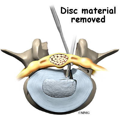
Ankle Anatomy

A Patient’s Guide to Ankle Anatomy

Introduction

Ankle Anatomy

The ankle joint acts like a hinge. But it’s much more than a simple hinge joint. The ankle is actually made up of several important structures. The unique design of the ankle makes it a very stable joint. This joint has to be stable in order to withstand 1.5 times your body weight when you walk and up to eight times your body weight when you run.

Normal ankle function is needed to walk with a smooth and nearly effortless gait. The muscles, tendons, and ligaments that support the ankle joint work together to propel the body. Conditions that disturb the normal way the ankle works can make it difficult to do your activities without pain or problems.

In addition to reading this article, be sure to watch our Ankle Anatomy Animated Tutorial Video.

This guide will help you understand

  • what parts make up the ankle
  • how the ankle works

Important Structures

The important structures of the ankle can be divided into several categories. These include

  • bones and joints
  • ligaments and tendons
  • muscles
  • nerves
  • blood vessels

The top of the foot is referred to as the dorsal surface. The sole of the foot is the plantar surface.

Ankle Anatomy

Bones and Joints

The ankle joint is formed by the connection of three bones. The ankle bone is called the talus. The top of the talus fits inside a socket that is formed by the lower end of the tibia (shinbone) and the fibula (the small bone of the lower leg). The bottom of the talus sits on the heelbone, called the calcaneus.

The talus works like a inside the socket to allow your foot to move up (dorsiflexion) and down (plantarflexion).

Woodworkers and craftsmen are familiar with the design of the ankle joint. They use a similar construction, called a mortise and tenon, to create stable structures. They routinely use it to make strong and sturdy items, such as furniture and buildings.

Inside the joint, the bones are covered with a slick material called articular cartilage. Articular cartilage is the material that allows the bones to move smoothly against one another in the joints of the body.

The cartilage lining is about one-quarter of an inch thick in most joints that carry body weight, such as the ankle, hip, or knee. It is soft enough to allow for shock absorption but tough enough to last a lifetime, as long as it is not injured.

Ligaments and Tendons

Ankle Anatomy

Ligaments are the soft tissues that attach bones to bones. Ligaments are very similar to tendons. The difference is that tendons attach muscles to bones. Both of these structures are made up of small fibers of a material called collagen. The collagen fibers are bundled together to form a rope-like structure. Ligaments and tendons come in many different sizes and like rope, are made up of many smaller fibers. Thickness of the ligament or tendon determines its strength.

Ankle Anatomy

Ligaments on both sides of the ankle joint help hold the bones together. Three ligaments make up the lateral ligament complex on the side of the ankle farthest from the other ankle. (Lateral means further away from the center of the body.) These include the anterior talofibular ligament (ATFL), the calcaneofibular ligament(CFL), and the posterior talofibular ligament (PTFL). A thick ligament, called the deltoid ligament, supports the medial ankle (the side closest to your other ankle).

Ankle Anatomy

Ligaments also support the lower end of the leg where it forms a hinge for the ankle. This series of ligaments supports the ankle syndesmosis, the part of the ankle where the bottom end of the fibula meets the tibia. Three main ligaments support this area. The ligament crossing just above the front of the ankle and connecting the tibia to the fibula is called the anterior inferior tibiofibular ligament (AITFL). The posterior fibular ligaments attach across the back of the tibia and fibula. These ligaments include the posterior inferior tibiofibular ligament (PITFL) and the transverse ligament. The interosseous ligament lies between the tibia and fibula. (Interosseous means between bones.) The interosseus ligament is a long sheet of connective tissue that connects the entire length of the tibia and fibula, from the knee to the ankle.

Ankle Anatomy

The ligaments that surround the ankle joint help form part of the joint capsule. A joint capsule is a watertight sac that forms around all joints. It is made up of the ligaments around the joint and the soft tissues between the ligaments that fill in the gaps and form the sac.

Ankle Anatomy

The ankle joint is also supported by nearby tendons. The large Achilles tendon is the most important tendon for walking, running, and jumping. It attaches the calf muscles to the calcaneus (heelbone) and allows us to raise up on our toes. The posterior tibial tendon attaches one of the smaller muscles of the calf to the underside of the foot. This tendon helps support the arch and allows us to turn the foot inward. The anterior tibial tendon allows us to raise the foot. Two tendons run behind the outer bump of the ankle (the lateral malleolus). These two tendons, called the peroneals, help turn the foot down and out.

Ankle AnatomyAnkle Anatomy

Ankle Anatomy

Muscles

Most of the motion of the ankle is caused by the stronger muscles in the lower leg whose tendons pass by the ankle and connect in the foot. Contraction of the muscles in the leg is the main way that we move our ankle when we walk, run, and jump.

The key ankle muscles have been discussed earlier in the section on ligaments and tendons. These muscles and their actions are also listed here.

  • The peroneals (peroneus longus and peroneus brevis) on the outside edge of the ankle and foot bend the ankle down and out.
  • The calf muscles (gastrocnemius and soleus) connect to the calcaneus by the Achilles tendon. When the calf muscles tighten, they bend the ankle down.
  • The posterior tibialis muscle supports the arch and helps turn the foot inward.
  • The anterior tibialis pulls the ankle upward.

Nerves

The nerve supply of the ankle is from nerves that pass by the ankle on their way into the foot. The tibial nerve runs behind the medial malleolus. Another nerve crosses in front of the ankle on its way to top of the foot. There is also a nerve that passes along the outer edge of the ankle.

Ankle AnatomyAnkle Anatomy

The nerves on the front and outer edge of the ankle control the muscles in this area, and they give sensation to the top and outside edge of the foot.

Blood Vessels

The ankle gets blood from nearby arteries that pass by the ankle on their way to the foot. The dorsalis pedis runs in front of the ankle to the top of the foot. (You can feel your pulse where this artery runs in the middle of the top of the foot.) Another large artery, called the posterior tibial artery, runs behind the medial malleolus. It sends smaller blood vessels to the inside edge of the ankle joint. Other less important arteries entering the foot from other directions also supply blood to the ankle.

Ankle AnatomyAnkle Anatomy

Summary

As you can see, the anatomy of the ankle is very complex. When everything works together, the ankle functions correctly. When one part becomes damaged, it can affect every other part of the ankle and foot, leading to problems.

Wrist Anatomy

A Patient’s Guide to Wrist Anatomy

Introduction

Wrist Anatomy

The anatomy of the wrist joint is extremely complex, probably the most complex of all the joints in the body. The wrist is actually a collection of many bones and joints. These bones and joints let us use our hands in lots of different ways. The wrist must be extremely mobile to give our hands a full range of motion. At the same time, the wrist must provide the strength for heavy gripping.

This guide will help you understand

  • what parts make up the wrist
  • how those parts work together

Important Structures

The important structures of the wrist can be divided into several categories. These include

  • bones and joints
  • ligaments and tendons
  • muscles
  • nerves
  • blood vessels

Bones and Joints

Wrist Anatomy

There are 15 bones that form connections from the end of the forearm to the hand. The wrist itself contains eight small bones, called carpal bones. These bones are grouped in two rows across the wrist. The proximal row is where the wrist creases when you bend it. Beginning with the thumb-side of the wrist, the proximal row of carpal bones is made up of the scaphoid, lunate, and triquetrum. The second row of carpal bones, called the distal row, meets the proximal row a little further toward the fingers. The distal row is made up of the trapezium, trapezoid, capitate, hamate, and pisiform bones.

The proximal row of carpal bones connects the two bones of the forearm, the radius and the ulna, to the bones of the hand. The bones of the hand are called the metacarpal bones. These are the long bones that lie within the palm of the hand. The metacarpals attach to the phalanges, which are the bones in the fingers and thumb.

Wrist Anatomy

One reason that the wrist is so complicated is because every small carpal bone forms a joint with the bone next to it. This means that what we call the wrist joint is actually made up of many small joints.

Wrist Anatomy

Articular cartilage is the material that covers the ends of the bones of any joint. Articular cartilage can be up to one-quarter of an inch thick in the large, weight-bearing joints. It is thinner in joints such as the wrist that don’t support a lot of weight. Articular cartilage is white, shiny, and has a rubbery consistency. It is slippery, which allows the joint surfaces to slide against one another without causing
any damage.

The function of articular cartilage is to absorb shock and provide an extremely smooth surface to make motion easier. We have articular cartilage essentially everywhere that two bony surfaces move against one another, or articulate. In the wrist, articular cartilage covers the sides of all the carpals and the ends of the bones that connect from the forearm to the fingers.

Ligaments and Tendons

Wrist Anatomy

Ligaments are soft tissue structures that connect bones to bones. The ligaments around a joint usually combine to form a joint capsule. A joint capsule is a watertight sac that surrounds a joint and contains lubricating fluid called synovial fluid. In the wrist, the eight carpal bones are surrounded and supported by a joint capsule.

Wrist Anatomy

Two important ligaments support the sides of the wrist. These are the collateral ligaments. There are two collateral ligaments that connect the forearm to the wrist, one on each side of the wrist.

As its name suggests, the ulnar collateral ligament (UCL) is on the ulnar side of the wrist. It crosses the ulnar edge (the side away from the thumb) of the wrist. It starts at the ulnar styloid, the small bump on the edge of the wrist (on the side away from the thumb) where the ulna meets the wrist joint. There are two parts to the cord-shaped UCL. One part connects to the pisiform (one of the small carpal bones) and to the transverse carpal ligament, a thick band of tissue that crosses in front of the wrist. The other goes to the triquetrum (a small carpal bone near the ulnar side of the wrist). The UCL adds support to a small disc of cartilage where the ulna meets the wrist. This structure is called the triangular fibrocartilage complex (TFCC) and is discussed in more detail below. The UCL stabilizes the TFCC and keeps the wrist from bending too far to the side (toward the thumb).

The radial collateral ligament (RCL) is on the thumb side of the wrist. It starts on the outer edge of the radius on a small bump called the radial styloid. It connects to the side of the scaphoid, the carpal bone below the thumb. The RCL prevents the wrist from bending too far to the side (away from the thumb).

Just as there are many bones that form the wrist, there are many ligaments that connect to and support these bones. Injury or problems that cause these ligaments to stretch or tear can eventually lead to arthritis in the wrist.

Wrist Anatomy

At the wrist, the end of the ulna bone of the forearm articulates with two carpal bones, the lunate and the triquetrum. A unique structure mentioned earlier, the triangular fibrocartilage complex (TFCC), sits between the ulna and these two carpal bones. The TFCC is a small cartilage pad that cushions this part of the wrist joint. It also improves the range of motion and gliding action within the wrist joint.

Wrist Anatomy

There are several important tendons that cross the wrist. Tendons connect muscles to bone. The tendons that cross the wrist begin as muscles that start in the forearm. Those that cross the palm side of the wrist are the flexor tendons. They curl the fingers and thumb, and they bend the wrist. The flexor tendons run beneath the transverse carpal ligament (mentioned earlier). This structure lies on the palm side of the wrist. This band of tissue keeps the flexor tendons from bowing outward when you curl your fingers, thumb, or wrist. The tendons that travel over the back of the wrist, the extensor tendons, run through a series of tunnels, called compartments. These compartments are lined with a slick substance called tenosynovium, which prevents friction as the extensor tendons glide inside their compartment.

Muscles

The main muscles that are important at the wrist have been mentioned above in the discussion about tendons. These muscles generally start further up in the forearm. The tendons of these muscles cross the wrist. They control the actions of the fingers, thumb, and wrist.

Nerves

Wrist Anatomy

All of the nerves that travel to the hand cross the wrist. Three main nerves begin together at the shoulder: the radial nerve, the median nerve, and the ulnar nerve. These nerves carry signals from the brain to the muscles that move the arm, hand, fingers, and thumb. The nerves also carry signals back to the brain about sensations such as touch, pain, and temperature.

Wrist Anatomy

The radial nerve runs along the thumb-side edge of the forearm. It wraps around the end of the radius bone toward the back of the hand. It gives sensation to the back of the hand from the thumb to the third finger. It also goes to the back of the thumb and just beyond the main knuckle of the back surface of the ring and middle fingers.

Wrist Anatomy

The median nerve travels through a tunnel within the wrist called the carpal tunnel. The median nerve gives sensation to the palm sides of the thumb, index finger, long finger, and half of the ring finger. It also sends a nerve branch to control the thenar muscles of the thumb. The thenar muscles help move the thumb and let you touch the pad of the thumb to the tips each of each finger on the same hand, a motion called opposition.

Wrist Anatomy

The ulnar nerve travels through a separate tunnel, called Guyon’s canal. This tunnel is formed by two carpal bones (the pisiform and hamate), and the ligament that connects them. After passing through the canal, the ulnar nerve branches out to supply feeling to the little finger and half the ring finger. Branches of this nerve also supply the small muscles in the palm and the muscle that pulls the thumb toward the palm.

The nerves that travel through the wrist are subject to problems. Constant bending and straightening of the wrist and fingers can lead to irritation or pressure on the nerves
within their tunnels and cause problems such as pain, numbness, and weakness in the hand, fingers, and thumb.

Blood Vessels

Wrist Anatomy

Traveling along with the nerves are the large vessels that supply the hand with blood. The largest artery is the radial artery that travels across the front of the wrist, closest to the thumb. The radial artery is where the pulse is taken in the wrist. The ulnar artery runs next to the ulnar nerve through Guyon’s canal (mentioned earlier). The ulnar and radial arteries arch together within the palm of the hand, supplying the front of the hand and fingers. Other arteries travel across the back of the wrist to supply the back of the hand and fingers.

Summary

As you can see, the wrist is a complex area of the body. When you realize all the different ways we use our hands every day and all the different positions we put our hands in, it is easy to understand how hard daily life can be when the wrist doesn’t work well.

Hand Anatomy

A Patient’s Guide to Hand Anatomy

Introduction

Few structures of the human anatomy are as unique as the hand. The hand needs to be mobile in order to position the fingers and thumb. Adequate strength forms the basis for normal hand function. The hand also must be coordinated to perform fine motor tasks with precision. The structures that form and move the hand require proper alignment and control in order for normal hand function to occur.

In addition to reading this article, be sure to watch our Hand Anatomy Animated Tutorial Video.

This guide will help you understand

  • what parts make up the hand
  • how those parts work together

Important Structures

The important structures of the hand can be divided into several categories. These include

  • bones and joints
  • ligaments and tendons
  • muscles
  • nerves
  • blood vessels

The front, or palm-side, of the hand is referred to as the palmar side. The back of the hand is called the dorsal side.

Bones and Joints

There are 27 bones within the wrist and hand. The wrist itself contains eight small bones, called carpals. The carpals join with the two forearm bones, the radius and ulna, forming the wrist joint. Further into the palm, the carpals connect to the metacarpals. There are five metacarpals forming the palm of the hand. One metacarpal connects to each finger and thumb. Small bone shafts called phalanges line up to form each finger and thumb.

The main knuckle joints are formed by the connections of the phalanges to the metacarpals. These joints are called the metacarpophalangeal joints (MCP joints). The MCP joints work like a hinge when you bend and straighten your fingers and thumb.

The three phalanges in each finger are separated by two joints, called interphalangeal joints (IP joints). The one closest to the MCP joint (knuckle) is called the proximal IP joint (PIP joint). The joint near the end of the finger is called the distal IP joint (DIP joint). The thumb only has one IP joint between the two thumb phalanges. The IP joints of the digits also work like hinges when you bend and straighten your fingers and thumb.

The joints of the hand, fingers, and thumb are covered on the ends with articular cartilage. This white, shiny material has a rubbery consistency. The function of articular cartilage is to absorb shock and provide an extremely smooth surface to
facilitate motion. There is articular cartilage essentially everywhere that two bony surfaces move against one another, or articulate.

Ligaments and Tendons

Ligaments are tough bands of tissue that connect bones together. Two important structures, called collateral ligaments, are found on either side of each finger and thumb joint. The function of the collateral ligaments is to prevent abnormal sideways bending of each joint.

In the PIP joint (the middle joint between the main knuckle and the DIP joint), the strongest ligament is the volar plate. This ligament connects the proximal phalanx to the middle phalanx on the palm side of the joint. The ligament tightens as the joint is straightened and keeps the PIP joint from bending back too far (hyperextending). Finger deformities can occur when the volar plate loosens from disease or injury.

The tendons that allow each finger joint to straighten are called the extensor tendons. The extensor tendons of the fingers begin as muscles that arise from the backside of the forearm bones. These muscles travel towards the hand, where they eventually connect to the extensor tendons before crossing over the back of the wrist joint. As they travel into the fingers, the extensor tendons become the extensor hood. The extensor hood flattens out to cover the top of the finger and sends out branches on each side that connect to the bones in the middle and end of the finger.

The place where the extensor tendon attaches to the middle phalanx is called the central slip. When the extensor muscles contract, they tug on the extensor tendon and straighten the finger. Problems occur when the central slip is damaged, as can happen with a tear.

Muscles

Many of the muscles that control the hand start at the elbow or forearm. They run down the forearm and cross the wrist and hand. Some control only the bending or straightening of the wrist. Others influence motion of the fingers or thumb. Many of these muscles help position and hold the wrist and hand while the thumb and fingers grip or perform fine motor actions.

Most of the small muscles that work the thumb and pinky finger start on the carpal bones. These muscles connect in ways that allow the hand to grip and hold. Two muscles allow the thumb to move across the palm of the hand, an important function called thumb opposition.

The smallest muscles that originate in the wrist and hand are called the intrinsic muscles. The intrinsic muscles guide the fine motions of the fingers by getting the fingers positioned and holding them steady during hand activities.

Nerves

All of the nerves that travel to the hand and fingers begin together at the shoulder: the radial nerve, the median nerve, and the ulnar nerve. These nerves carry signals from the brain to the muscles that move the arm, hand, fingers, and thumb. The nerves also carry signals back to the brain about sensations such as touch, pain, and temperature.

The radial nerve runs along the thumb-side edge of the forearm. It wraps around the end of the radius bone toward the back of the hand. It gives sensation to the back of the hand from the thumb to the third finger. It also supplies the back of the thumb and just beyond the main knuckle of the back surface of the ring and middle fingers.

The median nerve travels through a tunnel within the wrist called the carpal tunnel. This nerve gives sensation to the thumb, index finger, long finger, and half of the ring finger. It also sends a nerve branch to control the thenar muscles of the thumb. The thenar muscles help move the thumb and let you touch the pad of the thumb to the tips each of each finger on the same hand, a motion called opposition.

The ulnar nerve travels through a separate tunnel, called Guyon’s canal. This tunnel is formed by two carpal bones, the pisiform and hamate, and the ligament that connects them. After passing through the canal, the ulnar nerve branches out to supply feeling to the little finger and half the ring finger. Branches of this nerve also supply the small muscles in the palm and the muscle that pulls the thumb toward the palm.

The nerves that travel to the hand are subject to problems. Constant bending and straightening of the wrist and fingers can lead to irritation or pressure on the nerves within their tunnels and cause problems such as pain, numbness, and weakness in the hand, fingers, and thumb.

Blood Vessels

Traveling along with the nerves are the large vessels that supply the hand with blood. The largest artery is the radial artery that travels across the front of the wrist, closest to the thumb. The radial artery is where the pulse is taken in the wrist. The ulnar artery runs next to the ulnar nerve through Guyon’s canal (mentioned earlier). The ulnar and radial arteries arch together within the palm of the hand, supplying the front of the hand, fingers, and thumb. Other arteries travel across the back of the wrist to supply the back of the hand, fingers, and thumb.

Summary

The hand is formed of numerous structures that have an important role in normal hand function. Conditions that change the way these structures work can greatly impact whether the hand functions normally. When our hands are free of problems, it’s easy to take the complex anatomy of the hand for granted.

Swan Neck Deformity of the Finger

A Patient’s Guide to Swan Neck Deformity of the Finger

Introduction

Normal finger position and movement occur from the balanced actions of many important structures. Ligaments support the finger joints. Muscles hold and move the fingers. Tendons help control the fine motion of each finger joint. Disease or injury can disturb the balance in these structures, altering normal finger alignment and function. The result may be a crooked finger, such as a swan neck deformity of the finger.

This guide will help you understand

  • what parts of the finger are affected
  • what causes swan neck deformity
  • how the problem is treated
  • what to expect from treatment

Anatomy

What parts of the finger are involved?

Swan Neck Deformity

The fingers are actually made up of three bones, called phalanges. The three phalanges in each finger are separated by two joints, called interphalangeal joints (IP joints). The joint near the end of the finger is called the distal IP joint (DIP joint). (Distal means further away.) The proximal IP joint (PIP joint) is the middle joint between the main knuckle and the DIP joint. (Proximal means closer in.) The IP joints of the fingers work like hinge joints when you bend and straighten your hand.

Swan Neck Deformity

The tendons that allow each finger joint to straighten are called the extensor tendons. The extensor tendons of the fingers begin as muscles that arise from the backside of the forearm bones. These muscles travel toward the hand, where they eventually connect to the extensor tendons before crossing over the back of the wrist joint. As they travel into the fingers, the extensor tendons become the extensor hood. The extensor hood flattens out to cover the top of the finger and sends out branches on each side that connect to the bones in the middle and end of the finger. When the extensor muscles contract, they tug on the extensor tendon and straighten the finger.

Ligaments are tough bands of tissue that connect bones together. Several small ligaments connect the extensor hood with other tendons that travel into the finger to bend the finger. These connections help balance the motion of the finger so that all the joints of the finger work together, giving a smooth bending and straightening action.

Swan Neck Deformity

In the PIP joint (the middle joint between the main knuckle and the DIP joint), the strongest ligament is the volar plate. This ligament connects the proximal phalanx to the middle phalanx on the palm side of the joint. The ligament tightens as the joint is straightened and keeps the PIP joint from bending back too far (hyperextending). Swan neck deformity can occur when the volar plate loosens from disease or injury.

Related Document: A Patient’s Guide to Hand Anatomy

Causes

How does this condition occur?

A swan neck deformity describes a finger with a hyperextended PIP joint and a flexed DIP joint.

Swan Neck Deformity

Conditions that loosen the PIP joint and allow it to hyperextend can produce a swan neck deformity of the finger. Rheumatoid arthritis (RA) is the most common disease affecting the PIP joint. Chronic inflammation of the PIP joint puts a stretch on the volar plate. (As mentioned earlier, the volar plate is a supportive ligament in front of the PIP joint that normally keeps the PIP joint from hyperextending.) As the volar plate becomes weakened and stretched, the PIP joint becomes loose and begins to easily bend back into hyperextension. The extensor tendon gets out of balance, which allows the DIP joint to get pulled downward into flexion. As the DIP joint flexes and the PIP joint hyperextends, the swan neck deformity occurs.

View animation of of PIP joint hyperextension

Related Document: A Patient’s Guide to Rheumatoid Arthritis

Other conditions that weaken the volar plate can produce a swan neck deformity. The small (intrinsic) muscles of the hand and fingers can tighten up from hand trauma, RA, and various nerve disorders, such as cerebral palsy, Parkinson’s disease, or stroke. The muscle imbalance tends to weaken the volar plate and pull the PIP joint into extension. Weakness in the volar plate can also occur from a finger injury that forces the PIP joint into hyperextension, stretching or rupturing the volar plate. As mentioned, looseness (laxity) in the volar plate can lead to a swan neck deformity.

Related Document: A Patient’s Guide to PIP Joint Injuries of the Finger

Swan Neck Deformity

Clearly, PIP joint problems can produce a swan neck deformity. But so can problems that start in the DIP joint at the end of the finger. Injury or disease that disrupts the end of the extensor tendon can cause the DIP joint to droop (flex).

Swan Neck Deformity

An example from sports is a jammed finger that tears or ruptures the extensor tendon at the end of the finger (distal phalanx). Without treatment, the DIP joint droops and won’t straighten out. This condition is called a mallet finger. The extensor tendon may become imbalanced and begin to pull the PIP joint into hyperextension, forming a swan neck deformity.

Related Document: A Patient’s Guide to Mallet Finger Injuries

Chronic inflammation from RA can also disrupt the very end of the extensor tendon. Inflammation and swelling in the DIP joint stretches and weakens the extensor tendon where it passes over the top of the DIP joint. A mallet deformity occurs in the DIP, followed by hyperextension of the PIP joint. Again, the result is a swan neck deformity.

Symptoms

What do swan neck deformities look and feel like?

Swan Neck Deformity

Inflammation from injury or disease (such as RA) may cause pain and swelling of the PIP joint. The PIP joint eventually is free to bend back too far into hyperextension. The DIP joint is bent downward into flexion. Eventually, the imbalance leads to the typical shape of the finger with a swan neck deformity.

Diagnosis

What tests will my doctor do?

Usually the diagnosis is evident just from the physical examination. An X-ray may be ordered so the doctor can check the condition of the joint surfaces, examine joint alignment, and see if a fracture is present (as in a traumatic finger injury). No other tests are required normally.

Treatment

What can be done for a swan neck deformity of the finger?

Treatment for swan neck deformity can be nonsurgical or surgical. The approach your doctor uses depends on whether the PIP joint is flexible or stiff.

Nonsurgical Treatment

Successful nonsurgical treatment is based on restoring balance in the structures of the hand and fingers. The PIP joint must be supple (not stiff). Aligning the PIP joint and preventing hyperextension should help restore DIP extension. If it doesn’t, surgery may be needed.

You may require professional rehabilitation. A physical or occupational therapist addresses the imbalances that have formed the swan neck deformity. Stretching, massage, and joint mobilization are used to try and restore finger alignment and function.

Swan Neck Deformity

A special splint may be used to keep the PIP joint lined up, protect the joint from hyperextending, and still allow the PIP joint to bend. Newer styles are shaped like jewelry rings and are available in stainless steel, sterling silver, or gold. This approach works best for mild cases of swan neck deformity in which the PIP joint is supple.

Splinting and a rigorous therapy program are usually not successful in altering the imbalance responsible for the deformity. However, many hand surgeons will try six weeks with the splint and exercise to improve PIP joint mobility before performing surgery.

Surgery

Soft Tissue Repair

In cases where the balance cannot be restored to a tolerable limit with splinting, surgery may be required to reconstruct and rebalance the structures around the PIP joint. The surgeon releases, aligns, and balances the soft tissues around the PIP joint. The surgery may involve the skin (dermadesis), the tendons (tenodesis), or the ligaments (mobilization or reconstruction).

Surgery to repair the soft tissues that are contributing to a swan neck deformity carries a relatively high risk of failure to achieve completely normal functioning of the finger. All of the repair and reconstruction procedures are dependant on a well designed and rigorous exercise program following the surgery. A physical or occupational therapist will work closely with you during your recovery.

PIP Joint Arthroplasty

Swan Neck Deformity

Swan neck deformity with a stiff PIP joint sometimes requires replacement of the PIP joint, called arthroplasty. The surgeon works from the back surface (dorsum) of the finger joint. Both surfaces of the PIP joint are removed to make room for the new implant. With the new joint in place, the surgeon balances the soft tissues around the joint to ensure that the new joint can easily bend and straighten.

Related Document: A Patient’s Guide to Artificial Joint Replacement of the Finger

Finger Joint Fusion

Swan Neck Deformity

When RA produces a mallet deformity of the DIP joint and the PIP joint is supple, surgeons may consider fusing the DIP joint. Joint fusion is a procedure that binds the two joint surfaces of the finger together, keeping them from moving. Fusing the two joint surfaces together eases pain, makes the joint stable, and helps prevent additional joint deformity.

If past treatments, including surgery, do not stop inflammation or deformity in the PIP joint, fusion of the PIP joint may be recommended. The PIP joint is usually fused in a bent position, between 25 and 45 degrees.

Related Document: A Patient’s Guide to Finger Fusion Surgery

Rehabilitation

What will my recovery be like?

Nonsurgical Rehabilitation

The goal of nonsurgical treatment is to get the finger joints, tendons, and muscles in balance. If nonsurgical treatment is successful, you may see improvement in eight to 12 weeks. Your doctor may have you work with a physical or occupational therapist during this time.

Your therapist will work with you to obtain and use your finger splint. Special forms of stretching may help reduce tightness in the intrinsic muscles of the hand and fingers. Strengthening exercises can help with alignment and function of the hand and fingers.

After Surgery

You’ll wear a splint or brace after surgery. A protective finger splint holds and protects the joint and is used for at least three weeks after surgery. Physical or occupational therapy treatments usually start three to six weeks after surgery.

You will likely need to attend therapy sessions for three to four months, and you should expect full recovery to take up to six months. The first few therapy treatments will focus on controlling the pain and swelling from surgery. Then you’ll begin gentle range-of-motion exercise. Strengthening exercises are started eight to 10 weeks after surgery. You’ll learn ways to grip and support items in order to do your tasks safely and with the least amount of stress on your finger joint. As with any surgery, you need to avoid doing too much, too quickly.

Eventually, you’ll begin doing exercises designed to get your hand and fingers working in ways that are similar to your work tasks and daily activities. Your therapist will help you find ways to do your tasks that don’t put too much stress on your finger joint. Before your therapy sessions end, your therapist will teach you a number of ways to avoid future problems.
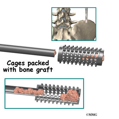
Your therapist’s goal is to help you keep your pain under control, improve your strength and range of motion, and regain fine motor abilities with your hand and finger. When you are well under way, regular visits to your therapist’s office will end. Your therapist will continue to be a resource, but you will be in charge of doing your exercises as part of an ongoing home program.

Postsurgical Infection

A Patient’s Guide to Postsurgical Infection

One of the greatest advances in surgery during the twentieth century has been in understanding how to reduce the risk of infection during and after surgery. Any surgical procedure, no matter how small, carries the risk of infection. Simple procedures, such as placing a needle into a joint to remove fluid or inject medications, have a very low risk. More complex operations that require larger incisions and leave the skin open for longer have a higher risk. Operations done to insert something into the body, such as metal plates or artificial joints, must be done carefully to ensure that the foreign material that is left in the body does not carry infection with it. Preventing infection is one of the most important parts of any operation. All members of the health care team take preventing infection very seriously.

Infection prevention begins before surgery. Your doctor will make sure that no infections exist that may spread to cause problems with your surgery. If you have a skin infection, a bladder infection, or some other type of infection, your surgery may be postponed until the infection is treated and under control.

The morning before surgery, you may be asked to shower with antibacterial soap to reduce the number of bacteria on the skin. Before surgery begins, the skin around the incision site will be cleansed and prepped with a disinfectant, such as iodine. Before surgery, you may be given a dose of antibiotics. This is not true for all types of surgeries. Operations that have a low risk of infection usually do not require antibiotics as a precaution. Many orthopedic operations do require antibiotics, especially those operations where something artificial is left in the body.

During surgery, great care is taken to ensure that the entire operation is done under sterile conditions. Surgery is performed in a sterile room that is designed to prevent the spread of infection. All the items needed for surgery are sterilized.

Your wound dressing will also be applied under sterile conditions before you leave the operating room. The wound dressing is an important barrier against infection until the incision seals itself. This usually occurs in the first few days. If an incision is still seeping any type of drainage, this can be an avenue for germs to enter the wound. The dressing will be kept in place until all drainage stops and the incision seals itself.

After surgery, you may continue on antibiotics for 24 to 72 hours. This is to help reduce the chance of infection.

While you are in the hospital or surgery center, your health care team will make sure your bandage stays dry and clean. A bandage that has soaked through can create an entryway for germs to enter the wound. Alert your nurse if this occurs. Before leaving the hospital or surgery center, you will be taught how to take care of your incision. The same instructions apply if you see extra drainage after your return home. Change the dressing, and call to let your nurse know.

Notice that the health care team always washes their hands before changing the bandage. You should do the same. If you develop an infection anywhere else in your body after surgery, let your doctor know. Some infections can spread and cause problems with the surgery.

Follow these guidelines:

  • Wash your hands before changing your bandage.
  • Change your bandage as instructed.
  • Don’t get the incision wet unless your doctor tells you to.
  • Don’t put any medication on the incision unless your doctor tells you to.
  • Don’t scratch or pick at the incision.

If you develop any of the following warning signs of infection, contact your doctor:

  • fever
  • increased redness around the incision
  • increased swelling around the incision
  • drainage from the wound for more than five days after surgery
  • drainage from the wound that is cloudy, yellow, or foul smelling
  • pain that increases and becomes constant.

Cumulative Trauma Disorder

A Patient’s Guide to Cumulative Trauma Disorder

Introduction

Cumulative trauma disorder (CTD) is a broad category that includes many common diseases that affect the soft tissues of the body. CTD in itself is not a disease. Doctors use the concept to understand and explain what may have caused, or contributed to, certain conditions. Examples of the conditions that may be caused or aggravated by cumulative trauma include carpal tunnel syndrome, tennis elbow, and low back pain.

Other terms are often used to describe the concept of CTD. These include repetitive stress injury (RSI), overuse strain (OS), and occupational overuse syndrome (OOS). This document will refer to these categories generally as CTD.

This guide will help you understand

  • what factors may contribute to CTD
  • how doctors diagnose conditions related to CTD
  • what treatment options are available
  • how to prevent CTD

Causes

What causes CTD?

Opinions abound as to what may cause CTD, but there is very little agreement. Some of the theories about how CTD starts are described below. The theories include

  • overuse
  • muscle tension
  • nerve tension
  • psychosocial factors
  • mind-body interaction
  • contributing factors

Overuse

Using muscles and joints after they have become fatigued, or overly tired, increases the likelihood of injury. Overloaded muscles and soft tissues without proper rest have no chance to recover fully. This problem often hampers athletes who have to throw, jump, or run repeatedly. It can also affect people who work in jobs where they keep doing the same action again and again, such as typing, gripping, and lifting.

All body tissues are in a constant state of change. Minor damage occurs continuously, which the body must repair in the normal course of a day. But the damage can occur faster than the repair mechanisms can keep up with it. When this happens, the tissues become weaker. They may begin to hurt. The weaker the tissues become, the more likely they will suffer even more damage. A cycle begins that looks like a spiral–constantly downward.

Muscle Tension

Some doctors think muscle tension causes CTD. To function, or work properly, the body and each of its parts needs a steady supply of blood, rich in oxygen and nutrients. Nutrients are the body’s fuel–glucose, for example. Cutting off or slowing the blood supply harms the tissues of the body.

Tense muscles are believed by some to actually squeeze off their own flow of energy and fuel. Muscles can get energy without oxygen, but the process produces a chemical called lactic acid. This chemical can be a potent pain-causing chemical. Lactic acid is a chemical that can produce a burning feeling when muscles are overexercised. Some physicians believe that lactic acid produced by tense muscles may cause some of the symptoms of CTD.

As pain develops, muscles tighten even further because they attempt to guard the surrounding area. Guarding is a term that is used to describe a reflex that all muscles in the body share. When pain occurs anywhere in the body, muscles around the painful area go into spasm (they tighten uncontrollably) to try to limit the movement in the area. As a result, blood flow is slowed down even more. The muscles begin to ache more. The nerves that have their blood supply reduced and squeezed by muscles begin to tingle or go numb.

Nerve Tension

This theory suggests that nerves become extra sensitive when they’ve become shortened and irritable. It is thought that poor postures used over long periods causes muscles to bulk up and interfere with blood flow. The nerves that course through the body then become shortened and may begin to stick to the nearby tissues. Moving the arm or leg puts tension on the nerve and can cause pain to radiate along the limb. The problem is thought to get worse from stress because the muscles and nerves tense up and become even tighter. Also, when the same activities are done over and over again, the tight nerve is pulled and strained to the point that it can’t heal and eventually becomes a chronic source of symptoms.

Psychosocial Factors

Problems with CTD tend to be more common among people who suffer from boredom, who have poor working relations, who aren’t satisfied with their jobs, and who have unhappy social circumstances. Reasons why this is so are unclear. The number of CTD cases reported may also be influenced by state worker’s compensation rules. States where claims are processed quickly and with greater benefits tend to have higher volumes of CTD cases. Both of these findings suggest that many cases of CTD may be highly influenced by the patient’s perception of the overall situation. Some patients may subconsciously, or consciously, rationalize their symptoms due to many factors that are not medical but have to do with their overall job and social situation.

Mind-Body Interaction

A newer theory suggests that there isn’t really an injury going on in the soft tissues where symptoms are felt. Instead, the problem is said to be coming from influences within the mind. It is theorized that the brain starts producing pain signals as a cover-up for deep-rooted feelings of past emotional pain or problems. Though the idea sounds hard to believe, practitioners using this approach claim they have had success rates as high as 95 percent. Their patients are reported to have gotten swift relief from treatments aimed at the underlying and unconscious emotional triggers.

Contributing Factors

The way people do their tasks can put them at risk for CTD. Some risk factors include

  • force
  • awkward or static postures
  • poor tool and equipment design
  • fatigue
  • repetition
  • temperature
  • vibration

One of these risk factors alone may not cause a problem. But doing a task where several factors are present may pose a greater risk. And the longer a person is exposed to one or more risks, the greater the possibility of developing CTD. Many different symptoms can arise from the accumulation of small injuries or stresses to the body. CTD is not so much a disease as it is a response to excessive demands these factors can place on our bodies without giving them adequate time to recover between.

Symptoms

What does CTD feel like?

The symptoms of CTD usually start gradually. Patients usually don’t recall a single event that started their symptoms. They may report feelings of muscle tightness and fatigue at first. People commonly report feeling numbness, tingling, and vague pain. Others say they feel a sensation of swelling in the sore limb. Some patients with arm symptoms sense a loss of strength and may drop items because of problems with coordination. Symptoms often worsen with activity and ease with rest.

Diagnosis

How do doctors diagnose CTD?

Your doctor begins the evaluation by taking a history of the problem. You’ll probably be asked questions about your job, such as the type of work you do and how you do your job tasks. Other questions give your doctor information about your work conditions, such as the postures you use, the weights you have to lift or push, and whether you have to do repetitive tasks. You may be asked about how you like your job and whether you get along with your supervisors and coworkers.

Your doctor will do a thorough physical examination. Your description of the symptoms and the physical examination are the most important parts in the diagnosis of CTD. Your doctor will first try to determine what conditions are affecting you. For example you may have symptoms of carpal tunnel syndrome or tennis elbow that need to be treated. Second, your doctor will try and determine if cumulative trauma is playing a role in your condition. If so, part of the treatment will be to try and eliminate the source of the cumulative trauma.

There are no specific tests that can diagnose CTD. There are many tests that may be ordered as your doctor looks for specific conditions.

Treatment Options

What treatments are available?

Nonsurgical Treatments

Many nonsurgical treatment approaches are used by physicians, physical and occupational therapists, and other health providers to reduce the symptoms of CTD-related conditions. A splint may be suggested initially to protect and rest the sore area. Anti-inflammatory drugs are often used together with therapy treatments such as ultrasound, cold packs, or electrical stimulation. A series of exercises may be suggested to help tissues move safely while healing.

Surgical Treatment

Surgery is rarely indicated for CTD. Specific conditions that can occur as a result of CTD may require surgery. Unless the doctor is quite sure there is a structural problem, such as a pinched nerve or severely inflamed tendon, then surgery is not usually suggested.

Prevention

How can I help prevent problems of CTD?

The best medicine for treating CTD is to prevent the problem from occurring in the first place. Key items to consider when attempting to prevent problems with CTD are listed below.

Use healthy work postures and body alignment. Posture can have a significant role in CTD. Faulty alignment of the spine or limbs can be a source of symptoms. Using healthy posture and body alignment in all activities decreases the possibility that CTD will strike. Incorrect posture may lead to muscle imbalances or nerve and soft tissue pressure, leading to pain or other symptoms. Most people spend many hours at their work place, and using unhealthy posture during these long hours increases the likelihood that CTD will develop.

Ergonomics

Assessing where and how a person does work is called ergonomics. Even subtle changes in the way a work station is designed or how a job is done can lead to pain or injury.

Rest and Relax

Rest and relaxation (R and R) have recently become front-line defenses in the prevention of CTD. Methods can be as simple as deep breathing, walking, napping, or exercising.

This strategy is useful during work and off hours. Whether at home or work, our bodies need time to recover, which simply means giving them a chance to heal. Rest and relaxation allow the body to recover and provide a way of repairing these injured tissues along the way, keeping them healthy.

The following ideas may be used to foster rest and relaxation at work:

  • Be relaxed. Try to work with your muscles relaxed by pacing your work schedule, staying well ahead of deadlines, and taking frequent breaks.
  • Stop to exercise. Gentle exercise performed routinely through the day helps keep soft tissues flexible and can ease tension.
  • Change positions. Plan ways to change positions during work tasks. This could include using a chair rather than standing or simply readjusting your approach to your job activity.
  • Rotate jobs or share work duties. This can be fun by offering a new work setting, and it allows the body to recover from the demands of the previous job task.
  • Avoid caffeine and tobacco. These can heighten stress, reduce blood flow, and elevate your perception of pain.

Rehabilitation

What can I expect with treatment?

Getting treated right away for symptoms of CTD can shorten the time it takes to heal. Symptoms can sometimes go away within two to four weeks when steps are taken quickly to address the factors that may be causing your symptoms. However, people who keep doing activities when they have symptoms and don’t seek help right away may be headed for a long and frustrating recovery time, perhaps as long as a year or more.

Your doctor may have you work with a physical or occupational therapist to speed your recovery. Your therapist will want to gather more information and will further evaluate your condition. The answers you give and the results of the examination will guide the therapist in tailoring a treatment program that is right for you.

Therapists often teach patients ways to relax. Relaxation ideas often include helping you learn to breathe deeply by using your diaphragm muscle. Taking the time to relax and breathe deeply eases tense muscles and speeds nutrients and oxygen to sore tissues.

Your therapist may apply treatments such as heat, ice, ultrasound, or gentle hands-on stretches to reduce pain or other symptoms. Muscle stretching may be used to restore muscle balance and to improve your posture and alignment. Therapists sometimes apply stretches that are designed to help nerves glide where they course from the spine to the arms or legs.

Strengthening exercises are used to restore muscle balance and to improve your ability to use healthy postures throughout the day.

Therapists pay close attention to your posture and movement patterns. You may receive verbal instruction and hands-on guidance to improve your alignment and movement habits. Helping you see and feel normal alignment improves your awareness about healthy postures and movements, allowing you to release tension and perform your activities with greater ease.

Your therapist will spend time helping you understand more about CTD and why you are feeling symptoms. You may be given tips on how to combat symptoms at work using rest and relaxation. You may also be given specific stretches and exercises to do at work. Your therapist may visit your work place to analyze your job site and to watch how you do your job tasks. Afterward, your therapist can recommend changes to help you do your job with less strain and less chance of injury. These changes are usually inexpensive and can make a big difference in helping you be more productive with less risk of pain or injury.

Your therapist’s goal is to help you understand your condition, to look for and change factors that may be causing your symptoms, and to help you learn how to avoid future problems. When patients are well underway, regular visits to the therapist’s office will end. Your therapist will continue to be a resource, but you will be in charge of practicing the strategies and exercises you’ve learned as part of an ongoing program.
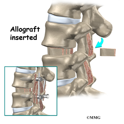
Cast Care

Taking Care of Your New Cast

Cast Care Instructions

Unless you have a waterproof cast, you should keep your cast dry. However, even waterproof casts should be dried out thoroughly after getting them wet. If you don’t dry your cast out after getting it wet, the skin underneath stays damp and can become moldy and smelly. Also, don’t swim with your cast on unless you have the waterproof kind.

To keep your cast dry in the shower, you can enclose it in a plastic garbage bag. Tape the open end of the bag so that water can’t get in. You can also buy a special cast protector.

Casts don’t completely harden for about two days. Be especially careful with your cast during this time. Don’t rest the full weight of the cast on a hard surface during these first two days. Doing so can dent the cast and can cause pressure sores on the skin under the cast. If the cast involves your foot, don’t walk on it for forty-eight hours, even if you have what is known as a walking cast. Walking on a soft cast may cause it to crack or dent.

Keep the casted or splinted limb elevated (propped up) above the level of your heart when you’re able to do so. This will reduce the swelling and help to keep the cast from becoming too tight.

Avoid too much activity and situations that may re-injure you or damage your cast. Remember your cast is there to help your arm or leg heal. It can’t do its job without your cooperation.

Warning Signs

If you have any of the following warning signs after your cast is placed or changed, you should consult your doctor.

Cast Care

Severe Pain

Your injury will probably cause some pain, but if the pain becomes steadily worse after the cast has been placed or changed, that may be a sign that the cast is too tight.

Bluish Nailbeds

Cast Care

The area under the fingernails and toenails is called the nailbed. Normally the nailbeds are pink. When the nailbed is pinched and released, it turns white for a few seconds and then pink again. This is because small blood vessels under the nail are squeezed shut. When you release the pressure, the blood vessels quickly open back up, turning the nail bed pink once again. This is called blanching. If the nailbed has a blue color and doesn’t turn pink again after being pinched and released, this may be a warning sign that the cast is too tight.

Numbness or Tingling

Cast Care

You may feel some numbness after a broken bone has been straightened or fixed if any type of anesthesia (such as a regional block or local anesthesia) was used. This should wear off in a few hours. But if you feel constant numbness or tingling in the fingers or toes of the casted arm or leg, this may indicate that the cast is too tight or that a nerve has been injured.

Immobility of Fingers or Toes

Cast Care

Because of your injury, you may have pain that makes it difficult for you to move your fingers or toes. But if you have no ability to move the fingers or toes of the casted arm or leg, it may be a signal that the cast is too tight and the muscles or nerves aren’t working properly.

Severe Coolness of Fingers or Toes

Many people notice a difference in the temperature of the casted arm or leg. Usually, this difference is minor. But if you have any other problems mentioned here that suggest that the circulation is not good, and the hand or foot is cool, it may be a sign that the cast is too tight.

Severe Swelling

Cast Care

If you have severe swelling above or below your cast, try keeping the casted limb propped up above the level of your heart. Most injuries cause at least some swelling. But if the swelling continues to get worse after the cast is put on, it may be either a sign that the cast is too tight or that something is going on under the cast that needs to be checked.

Dented or Cracked Cast

Cast Care

If your cast is dented or cracked, or starts falling apart before you are due for a checkup, this could lead to problems with healing. Small cracks are probably alright, but if the cast isn’t doing what it is supposed to, it could affect the healing of the injury. You should also check with your doctor if you think the cast isn’t holding the arm or leg still enough.

Arthroscopy

A Patient’s Guide to Arthroscopy

Introduction

Until recently, surgery on the inside of any joint meant making a large incision and opening the joint to do even the most minor procedure. Twenty years ago, fiber optics began changing all that and is continuing to change how orthopedic surgeons operate on joints in the body.

  • What is arthroscopy?
  • How is it used?
  • Why is it better?
  • What joints are being scoped?
  • What goes on during an arthroscopy?
  • What are the risks of arthroscopy?
  • What should I do after my arthroscopy?

What is it?

The term arthroscopy basically means to look into the joint. (Arthro means joint, and scopy means look.) So the common phrase scope the joint means to insert an arthroscope into the joint and have a look. In the early days before the development of miniature video cameras, about all the surgeon could do was take a look.

Over the past several years, the development of very small video cameras and specialized instruments have allowed surgeons to do more than simply take a look into the joint. The arthroscope is now used more and more for actual surgical procedures.

How is it used?

Using the arthroscope to assist with joint surgery usually involves making smaller incisions into the joint than those made in a regular open-incision surgery. Once the arthroscope is inserted into the joint, it is used first to try to see the problem. In this way, the problem can be confirmed before making any large incisions and causing any damage unnecessarily. Using the arthroscope as his eyes the surgeon can then use small specialized instruments inserted into the joint through other small incisions to perform the operation. As surgeons have become familiar with this type of surgery, more surgical procedures that were once done with large incisions are now being done arthroscopically.

Why is it better?

All surgical procedures that are done result in damage to tissues that are otherwise normal, because an incision must be made to see the problem. This is particularly bothersome for joints because to enter a joint, the joint capsule and ligaments must be incised (cut into). For minor surgical procedures inside the joint, it is not unusual for the recovery time to be much longer. This is because the normal tissues that were cut must also heal. Also, large incisions into the joint to perform surgical procedures increase the chances for infection. Long procedures where the joint is open to the air can lead to injury to the articular cartilage (the smooth surface of all joints) because it dries the cartilage out.

Arthroscopy causes less damage to normal structures by requiring much smaller incisions through the joint capsule and ligaments around the joint. Arthroscopy also allows the joint to remain closed and reduces the risk of infection and drying out of the articular cartilage. Because of this, the healing time for most arthroscopic procedures is greatly reduced. Rehabilitation is faster, and unnecessary damage to normal structures is avoided.

Arthroscopy has also greatly expanded orthopedic surgeon’s understanding of problems around the joints. In many cases, conditions which were completely unknown before the invention of the arthroscope have been discovered and are now being treated very effectively with arthroscopic surgery.

What joints are being scoped?

Just about every joint in the body has been scoped, but the vast majority of surgical procedures done with the arthroscope are done on the knee and the shoulder. The knee was the primary site of arthroscopic procedures in the early days of arthroscopy and continues to be a common target of the scope.

Probably the most common use of the arthroscope initially was to remove loose bodies from the knee joint or to remove a torn cartilage or torn meniscus. A loose body is a fragment of bone or cartilage that moves around inside the joint and can get caught between the two bones of the joint and cause pain. The menisci (or cartilages) of the knee are two small pieces of tissue that sit between the two bones of the knee joint and act similar to a gasket. A torn meniscus is a common problem that involves these structures and can get caught between the knee and cause pain.

Very complex surgical procedures are now done inside the knee with the aid of the arthroscope. For example, the anterior and posterior cruciate ligaments inside the knee are now almost always reconstructed without opening the knee joint. The procedure is done by using the arthroscope to help see where to drill holes in the bone and to place the ligament graft. Even fractures that involve the joint surface of the lower leg bone (the tibia) are being fixed by watching the fracture through the arthroscope while screws are inserted through small incisions in the skin–a procedure that once required a large incision into the joint.

The arthroscope is continuing to change the way orthopaedic surgeons deal with shoulder surgery as well. The arthroscope is now being used to repair torn tendons around the shoulder, to reconstruct the shoulder in patients who dislocate their shoulder and have instability, and to identify problems in the shoulder that were unknown prior to shoulder arthroscopy.

Other common joints that may be treated with arthroscopy include the ankle, wrist, elbow, and hip.

What goes on during an arthroscopy?

Preoperative

Your doctor will meet with you to explain what the surgery is intending to accomplish and what you can expect. You will be asked to sign a surgical consent form that describes the operation and the potential risks. It is a record that you understand the procedure. If you have any questions, now is the time to ask.

You will also need to review your other medical problems with the doctor in case you have a medical problem that will require evaluation before surgery. If you have serious lung or heart problems, you may need to see your family physician to get medical clearance before the procedure.

Admission

Most arthroscopic surgical procedures are done on an outpatient basis, which means you will probably be able to go home the same day. (Some arthroscopically assisted procedures still require larger incisions to be made around the joint, and these larger procedures may require a night or two stay in the hospital.) Generally, you are asked to not eat or drink anything after midnight on the evening before surgery. If you commonly take medications in the morning, ask your doctor whether you should take them or not.

Anesthesia

There are many different types of anesthesia that can be done, depending on your wishes, your doctor’s recommendations, and, of course, which joint is involved. In general, anesthesia may be local, regional, or general. Each of these types of anesthesia has advantages and disadvantages, but all are quite safe.

Local Anesthesia

Local anesthesia involves injecting lidocaine (or a similar medication) into the joint and in the areas where the incisions are made. This is the same type of anesthesia that is typically used to sew up a laceration, for example. The benefits of this type of anesthesia are that it is relatively safe, has less effect on the rest of your body, and returns to normal quickly after the procedure. The disadvantages are that you may still feel some discomfort during the procedure. This type of anesthesia is not generally used for shoulders or hips.

Regional Anesthesia

Regional anesthesia is probably the most common type used for arthroscopy. It is often referred to as a block (for example, a spinal block). The block is done by injecting lidocaine around the nerves that go to an extremity (in a spinal block, these are the nerves that go to the legs). The advantages to this type of anesthesia are that it does not affect the function of the lungs (a concern if you have lung problems). It does not cause as much nausea as general anesthesia, and it is relatively safer than general anesthesia. You are also awake and can watch the procedure on the TV monitor. The disadvantages are that it takes longer to take affect than going to sleep, and the actual block involves an injection–which may be uncomfortable for a short period of time.

General Anesthesia

General anesthesia is commonly referred to as going to sleep. It is commonly used for arthroscopic procedures–especially procedures which may be long and complicated. The advantages of general anesthesia are that you are not aware of anything that occurs during surgery. The disadvantages are that you may have a hangover with nausea and vomiting due to the drugs used, and you may have a higher risk of lung problems after surgery.

Surgery

Once the anesthesia is working, the operating room nurses and your doctor will prepare the equipment for arthroscopy. There are lots of electronics and equipment that need to be assembled. The surgical limb will be positioned to make it easier for the surgeon to do the surgery. If the procedure is going to be done on the knee, ankle, elbow, or wrist, a tourniquet may be used to stop the blood flow temporarily during the procedure. This makes it easier to see inside the joint.

When the surgery starts, several small incisions are made into the joint. These are usually about one-quarter of an inch long. The number varies from one to about six depending on what will be done. The joint is then filled with clear fluid (usually similar to the salt solution used in intravenous fluids). This expands the joint and allows the camera to work better. The fluid flows through the joint continuously to wash away blood and other material that is present during the procedure.

Once the surgery is under way, various instruments are used to perform the procedure. The camera is used to view the inside of the joint while the other instruments are used to either remove or repair the problem. If you are awake, you can watch the TV monitor and see exactly what your doctor is seeing. Still pictures or video can be taken through the arthroscope to record the condition of the joint. Your doctor performs the procedure while watching the TV monitor and guiding the instruments by what he or she sees on the TV. That’s why arthroscopy is like a very sophisticated video game.

Recovery

After surgery you will be taken from the operating room into the recovery room. There you will be closely monitored by the nurses until they are sure you are not having any problems from either the surgery or the anesthesia. You will probably be able to have something to drink if you are not nauseated. Normally, you will remain in the recovery room thirty to sixty minutes.

Discharge

Once you are ready to be released from the recovery room, you will probably return to where you started–the outpatient surgery department. Here you can wait with your family or friends until you are ready to be released to go home. You can probably have something to eat at this point and prepare to go home. You will be given some instructions at this point about what you should expect and what you should, and should not, be doing at home.

What are the risks of arthroscopy?

Like all surgical procedures, arthroscopy has potential complications. (Some of the more complicated procedures have specific risks.)

In general, arthroscopy requires anesthesia, and there are risks associated with anesthesia. Different types of drugs are given during the procedure that may cause a reaction–even death in very rare instances.

The arthroscopy procedure itself can result in damage to the joint structures or damage to nerves and blood vessels around the joint. Infection of the joint or soft tissues around the joint can occur after arthroscopy and may require additional operations to drain or treat the infection. Infection rates are generally less after arthroscopic procedures due to smaller incisions, shorter operations, and the fact the joint is constantly being flushed out during the procedure.

View animation of pulmonary embolism

Thrombophlebitis (blood clots), sometimes called deep venous thrombosis (DVT), can occur after arthroscopy (usually after arthroscopy of the hip or lower extremity joints). DVT occurs when the blood in the large veins of the leg forms blood clots within the veins. This may cause the leg to swell and become warm to the touch and painful. If the blood clots in the veins break apart, they can travel to the lung, where they get lodged in the small blood vessels of the lung and cut off the blood supply to a portion of the lung. This is called a pulmonary embolism. (Pulmonary means lung, and embolism refers to a fragment of something traveling through the vascular system.) Most surgeons take preventing DVT very seriously. There are many ways to reduce the risk of DVT, but probably the most effective is getting you moving as soon as possible.

What should I do after my arthroscopy?

You will be given specific instructions about what to do after your arthroscopy depending on what type of procedure you had performed. In general, you should take it easy for several days following surgery. You should watch for any signs of infection such as fever or chills, redness around the incisions, or increasing swelling or fluid build-up around the joint. If you have nausea and vomiting that persists for more that twenty-four hours after returning home you may want to discuss a medication change with your doctor. For specific instructions for your procedure you should consult your doctor.

Osteoporosis

A Patient’s Guide to Osteoporosis

Introduction

Osteoporosis is a very common disorder affecting the skeleton. In a patient with osteoporosis, the bones begin losing their minerals and support beams, leaving the skeleton brittle and prone to fractures.

In the U.S., 10 million individuals are estimated to already have the disease and almost 34 million more have low bone mass, placing them at increased risk for osteoporosis. Of the 10 million Americans affected by osteoporosis, eight million are women and two million are men. Most of them over age 65.

Bone fractures caused by osteoporosis have become very costly. Half of all bone fractures are related to osteoporosis. More than 300,000 hip fractures occur in the United States every year. A person with a hip fracture has a 20 percent chance of dying within six months as a result of the fracture. Many people who have a fracture related to osteoporosis spend considerable time in the hospital and in rehabilitation. Often, they need to spend some time in a nursing home.

This guide will help you understand

  • what happens to your bones when you have osteoporosis
  • how doctors diagnose the condition
  • what you can do to slow or stop bone loss

Anatomy

What happens to bones with osteoporosis?

Osteoporosis

Most people think of their bones as completely solid and unchanging. This is not true. Your bones are constantly changing as they respond to the way you use your body. As muscles get stronger, the bones underneath them get stronger, too. As muscles lose strength, the bones underneath them weaken. Changes in hormone levels or the immune system can also change the way the bones degenerate and rebuild themselves.

As a child, your bones are constantly growing and getting denser. At about age 25, you hit your peak bone mass. As an adult, you can help maintain this peak bone mass by staying active and eating a diet with enough calories, calcium, and vitamin D. But maintaining this bone mass gets more difficult as we get older. Age makes building bone mass more difficult. In women, the loss of estrogen at menopause can cause the bones to lose density very rapidly.

The bone cells responsible for building new bone are called osteoblasts. Stimulating the creation of osteoblasts helps your body build bone and improve bone density. The bone cells involved in degeneration of the bones are called osteoclasts. Interfering with the action of the osteoclasts can slow down bone loss.

In high-turnover osteoporosis, the osteoclasts reabsorb bone cells very quickly. The osteoblasts can’t produce bone cells fast enough to keep up with the osteoclasts. The result is a loss of bone mass, particularly trabecular bone–the spongy bone inside vertebral bones and at the end of long bones. Postmenopausal women tend to have high-turnover osteoporosis (also known as primary type one osteoporosis). This relates to their sudden decrease in production of estrogen after menopause. Bones weakened by this type of osteoporosis are most prone to spine and wrist fractures.

In low-turnover osteoporosis, osteoclasts are working at their normal rate, but the osteoblasts aren’t forming enough new bone. Aging adults tend to have low-turnover osteoporosis (also known as primary type two osteoporosis). Hip fractures are most common in people with this type of osteoporosis.

Secondary osteoporosis describes bone loss that is caused by, or secondary to, another medical problem. These other problems interfere with cell function of osteoblasts and from overactivity of osteoclasts. Examples include medical conditions that cause inactivity, imbalances in hormones, and certain bone diseases and cancers. Some medications, especially long term use of corticosteroids, are known to cause secondary osteoporosis due to their impact on bone turnover.

Osteoporosis creates weak bones. When these weak bones are stressed or injured, they often fracture. Fractures most often occur in the hip or the bones of the spine (the vertebrae). They can also occur in the upper arm, wrist, knee, and ankle.

Causes

What causes osteoporosis?

Aging is one of the main risk factors for osteoporosis and osteoporotic fractures. If you are lucky enough to live a long life, you are much more likely to develop weakened bones from osteoporosis. In women, the loss of estrogen in menopause causes bone loss of up to two percent per year. White women over age 50 have a lifetime risk of fracture of about 50 percent. This figure increases with increasing age.

A number of factors contribute to osteoporosis:

  • advanced age
  • female gender
  • low body weight or a thin and slender build
  • recent weight loss
  • history of fractures
  • family history of fractures
  • tobacco use
  • alcohol abuse
  • lack of exercise
  • extended use of certain medications (e.g., corticosteroids, anticonvulsants, and thyroid medicine)
  • Asian or Caucasian race

These risk factors are just as important as a measurement of low bone mass in determining how likely you are to have a fracture. People with low bone mass but no additional risk factors often don’t develop fractures. People with small amounts of bone loss but many risk factors are more likely to eventually develop fractures.

Symptoms

What does osteoporosis feel like?

Fractures caused by osteoporosis are often painful. But osteoporosis itself has no symptoms. That is why it is especially important to get tested if you are a woman past menopause and have any of the above risk factors. Women over 65 should be tested whether or not they have other risk factors. People with other bone problems or who take drugs that weaken the bones should also be tested. An initial screening for osteoporosis is painless and easy.

Diagnosis

How do doctors diagnose osteoporosis?

Free osteoporosis screenings are available in many drug stores and malls. Most of these screenings use a machine that scans the bone in the heel of your foot. It is a fast and simple way to get an idea of your bone density. However, this test is not entirely accurate. Because the heel bears a lot of weight, the test may show normal bone in the heel, even though the hipbones or spine may have low bone density. If the foot scan shows a low bone mass, you should talk to your doctor.

Your doctor will take a detailed medical history to help weigh your risk factors for osteoporosis. Information about your lifestyle and diet will also help your doctor develop a plan to help you build or maintain bone density.

Your doctor may also recommend more precise testing. Dual-energy X-ray absorptiometry (DEXA) is the most common method of measuring bone mass. A DEXA test uses special X-rays of the bones of your hip and spine to show your bone mass in these areas. The bone mass is then compared to that of a healthy thirty-year-old, called a T score. If you are within one standard deviation (SD) for bone density, you have normal bone. (SD is a statistic to measure variations in how a group is distributed.) If you are between one and 2.5 SDs below ideal levels, you are considered to be osteopenic. This means you have a mild form of osteoporosis. If the bone mass is more than 2.5 SDs below ideal levels, you have osteoporosis.

Be aware that DEXA scans are not perfect. Different equipment or different technicians can get somewhat different readings. If you need to have more precise data, your doctor may recommend additional types of bone scans or ultrasound tests.

A single DEXA scan also can’t show your doctor whether your bone mass is stable, increasing, or decreasing. Your doctor may have you take certain medications that create markers in the blood or urine to show what is happening in your bones. These tests will tell your doctor if you have high-turnover or low-turnover osteoporosis.

If bone density tests show that you have weakened bones, your doctor will need to rule out other causes. In some cases, problems with bone marrow or hormone levels can cause bone loss. Blood tests can show these conditions.

In other cases the bone weakening is actually from a condition called osteomalacia. Osteomalacia involves a softening of the bones caused by a lack of vitamin D. Vitamin D in your body comes from food and sunlight. Due to a lack of sunlight, almost 10 percent of people with hip fractures in the northern parts of the world have osteomalacia rather than osteoporosis. Urine and blood tests can help rule out osteomalacia.

In some cases, your primary care physician may refer you to a specialist. If you still have significant bone loss while on medication to prevent bone resorption, you may need to see a specialist. Referral is also advised for patients who have recurring fractures during therapy or repeated, unexplained fractures. Your doctor will help you find the right specialist for your situation.

Treatment Options

What can be done for osteoporosis?

The goal of your treatment plan will be to prevent fractures. This is especially important if you’ve already suffered a fracture from osteoporosis. To prevent fractures, you need to increase your bone mass. If you have high-turnover osteoporosis, you also need to prevent rapid bone reabsorption.

You need to take several steps to increase your bone mass

  • Make sure you get enough calcium and vitamin D. (Vitamin D helps your body absorb calcium.) Researchers think that increased calcium intake alone could reduce the number of fractures by 10 percent. More and more of us don’t get enough calcium and vitamin D, especially as we age. It is difficult to get recommended levels from the food we eat, so supplements are probably necessary. Talk to your doctor about what kind to buy. Calcium comes in many forms–for example, calcium carbonate, calcium citrate, calcium phosphate, and calcium from bone meal. Some forms of calcium need to be taken with food, and others need to be taken with certain types of food. Taking extra calcium and vitamin D improves the effectiveness of all other treatments for osteoporosis.
  • Eat enough calories to maintain a healthy weight. Being too thin increases your risk of osteoporotic fractures. Weight loss can be a cause of bone loss.
  • Exercise. Your bones are constantly adjusting to the demands you put on them. Even low levels of exercise can help you maintain better bone mass. Low-impact exercises like fast walking, stair climbing, and safe forms of dance help stimulate osteoblasts, slowing down reabsorption. Muscle-strengthening exercises, using light weights, can help keep the bones underneath the muscles strong. Balance training can help you prevent the falls that can cause fractures. Your doctor may recommend seeing a physical therapist to help you develop an exercise program with all three kinds of exercises. (See below.)
  • Premenopausal women should avoid overtraining and certain eating disorders, which can cause missed periods (amenorrhea).
  • If you smoke, quit immediately.
  • If you drink alcohol, do so moderately.

Medication

If you follow these recommendations and still have significant bone loss, your doctor may prescribe medications to slow down your body’s reabsorption of bone.

Many drugs are now available for the prevention and/or treatment of osteoporosis. Finding the right drug for each patient takes into consideration benefits and risks of the drug. These are matched against specific patient characteristics and risk factors. Ultimately, the best drug is the one most likely to be taken consistently and/or correctly by the patient. Osteoporosis management is most effective when drugs are taken in such a way that they have their full benefit.

If you are past menopause, hormone replacement therapy can be very effective. Bisphosphonates and calcitonin can also slow your body’s reabsorption of bone.

Studies have shown that 80 percent of women actually build bone mass up to two percent per year while on estrogen replacement therapy. Estrogen has been shown to decrease the occurrence of fractures in the vertebrae by 50 percent and fractures in the hip by 25 percent. Studies have also shown that hormone replacement therapy can also lower rates of coronary artery disease, relieve some symptoms of menopause, and maybe even prevent or postpone Alzheimer’s disease.

Hormone replacement therapy worries many women. Studies have shown that it may increase the risk of breast cancer. For women with a family history of breast cancer or who have had a stroke or thrombophlebitis (blood clots), hormone replacement therapy is probably not appropriate. Other women should at least consider taking estrogen. Its effects on osteoporosis are dramatic. Researchers estimate that, if estrogen were widely used, it could reduce all osteoporotic fractures by 50 to 75 percent.

Hormone replacement therapy must be continued to be effective, however. When a woman stops taking estrogen, she’ll start to lose bone at a very fast rate again. Within seven years, her bone density will be as low as that of a woman who never took estrogen.

Doctors often prescribe calcitonin to patients with fractures. Calcitonin is a non-sex, non-steroid hormone. Calcitonin binds to osteoclasts (the bone cells that reabsorb bone) and decreases their numbers and activity levels. Calcitonin used to be given only by injection, but now it is available in a nasal spray and a rectal suppository. Nasal calcitonin is used most often for women with osteoporosis who are five years or more past menopause and unable to take other approved agents. For unknown reasons, calcitonin seems to relieve pain. Calcitonin from salmon is much more effective than calcitonin from humans.

You and your doctor need to work together to monitor the effects of calcitonin. It is a new drug, and its long-term effects and benefits are still not fully known. More than 20 percent of patients develop a resistance to calcitonin over time, and it stops working for them.

Bisphosphonates also slow reabsorption by affecting the osteoclasts. The FDA has approved a variety of bisphosphonates for the treatment of osteoporosis. You may have heard the names of some of these such as Alendronate (Fosamax), Risedronate (Actonel), or Ibandronate (Boniva).

Some bisphosphonates are taken orally (pill form) on a daily basis. Others are available in weekly or monthly doses. A new injectable bisphosphonate (Zoledronate) can be given annually (once a year). Boniva comes in pill form and can also be injected once every three months. The injectable forms of this drug are used in the management of postmenopausal osteoporosis.

Studies have shown that bisphosphonates increase bone mass and prevent fractures. No one is sure how well bisphosphonates work when used for a long time. But stopping the drug doesn’t seem to cause the rapid bone loss that happens when someone stops taking estrogen. Because there can be some side effects with these medications, you need to work closely with your doctor if you take them.

The FDA is currently studying several drugs that may be used to treat osteoporosis. Some of these drugs, such as sodium fluoride, can be helpful in low-turnover osteoporosis. These drugs affect your osteoblasts in ways that cause them to create more bone. Sodium fluoride may be available in the near future. Other FDA-approved drugs are now available for the treatment of osteoporosis. For example, Raloxifene (Evista) is an anti-estrogen. Anti-estrogens are also called selective estrogen-receptor modifiers (SERMs). SERMs improve bone density and prevent fractures similar to estrogen, yet without increasing the chances of hormone-related cancer. Their main benefit over hormone replacement therapy is that they do not increase the risk of breast cancer.

Raloxifene is used most often for postmenopausal women younger than 65. They must not be at risk for blood clots or have cardiovascular disease. Men may be prescribed the only anabolic agent (Teriparatide/Forteo) approved for the management of osteoporosis. Anabolic usually refers to hormones that build up muscle or bone mass. Forteo is a form of parathyroid hormone used for patients at high risk of fracture. It is usually followed by an agent with antiresorptive effects such as a bisphosphonate.

Lifestyle changes, hormone replacement therapy, exercise prescription, and recent advances in drug therapy can help you take control of your osteoporosis. You and your doctor should be able to find ways to help you prevent the debilitating fractures of osteoporosis.

Physical Therapy

Many patients benefit from working with a physical therapist. People learn safe ways of moving, lifting, and exercising. Treatments also help people gain muscle strength and improved posture.

The physical therapist relies on your test results and the information received by you and your doctor. The therapist also looks at your body height, posture, body movements, strength, flexibility, balance, and your risk for having a fall.

Accurately measuring and recording your body height is a key part of the evaluation. It can give your therapist an idea of how osteoporosis is affecting your bones and posture, and comparing the recordings over a period of time can help track your success with treatments.

Posture exercises are used to help you “be tall”, regaining body height commonly lost with osteoporosis. This training can help patients who have stooped posture, called kyphosis, in the upper part of the spine. In healthy spine posture, the head is balanced on top of the spine rather than jutted forward.

In posture exercises, the goal is to get your body lined up from head to toe, with weight going through your hips. In people with advanced osteoporosis, the upper body is commonly bent forward at the hips. This prevents the hip bones from getting the right amount of stress and weight on them. As a result, the bones weaken and become more prone to fracture.

Your therapist will explain ways you can put good posture into practice. This is called body mechanics–the way you align your body when you do your activities. Remember that healthy posture is balanced with the body aligned from the head to toes. The same posture should be used when you bend forward to pick things up. Instead of rounding out your shoulders and upper back, keep the back in its healthy alignment as you bend forward at the hip joint. This keeps your back in a safe position and prevents the vertebrae from pinching forward. When bones are weakened from osteoporosis, rounding the spine forward when bending and lifting increases the risk of a spine fracture. As the back rounds forward, it pinches the front section of the vertebrae and can cause a fracture.

Your therapist will work with you in designing a safe program of exercise. Weight-bearing exercise strengthens existing bone and the muscles around joints. These types of exercises include walking outdoors or on a treadmill, doing safe forms of dance, and performing resistance training.

Some of the keys to safe exercise for osteoporosis include using good body alignment, avoiding bending or heavy twisting of the trunk, building up the amount of weight and number of repetitions gradually, and being consistent with your exercise program. Avoid exercises that curl your trunk forward such as stationary bike riding, sit-ups, toe-touches, and knee-to-chest exercises. Don’t exercise using abdominal crunch machines or rowing machines. Emphasize exercises that promote upright posture of the spine, such as walking. And do upper body exercises with your back supported in optimal alignment.

Your physical therapist will also check to make sure you have good balance. Poor balance can lead to a hazardous fall. When people with osteoporosis fall, they often end up fracturing a bone–a potentially life-threatening situation. Exercises to improve balance can be as simple as standing on one foot. As your balance gets better, more challenging types of exercises may be given.

People with balance problems can also benefit from practicing tai chi, an exercise form originating in China. In addition to gaining better balance, people who use the exercise movements show improved posture, flexibility, and strength.

Your therapist will continue to compare your test results of body height, posture, balance, and strength to see how well you are improving. The therapist’s goal is to help you become proficient and safe with your exercises, to improve you stature, strength, and flexibility, and to give you tips on how to avoid future problems.
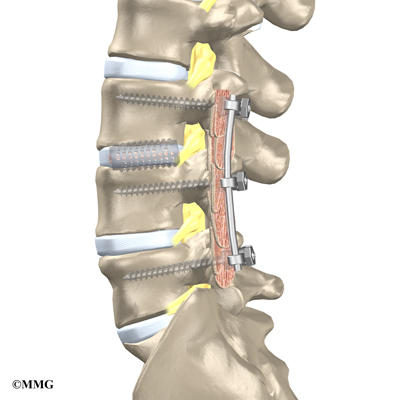
When patients are well underway, regular visits to the therapist’s office will end. The therapist will continue to be a resource, but patients will be in charge of doing their exercises as part of an ongoing home program.

Viral Arthritis

A Patient’s Guide to Viral Arthritis

Introduction

We know that some viruses cause joint pain and inflammation (swelling, redness, and heat). Researchers have wondered for a long time whether some kinds of arthritis with unexplained causes may be the result of a virus. So far no one knows, partly because the virus itself may be long gone before a patient ever develops the pain and inflammation of arthritis.

This guide will help you understand

  • how viral arthritis develops
  • how doctors diagnose viral arthritis
  • what can be done for the condition

Anatomy

Where does viral arthritis develop?

Most viral infections in the body cause a limited illness. Then the body’s immune system destroys the virus, and the symptoms of the illness go away.

In viral arthritis, the immune system’s response to the virus causes inflammation in the joints. Even after the virus is eliminated from the body, the changes in the joint can continue to cause pain and swelling. The joint may even become permanently damaged.

Causes and Symptoms

Which viruses cause viral arthritis?

Several viruses are known to cause problems with joint inflammation and pain. Some of the most common are listed below.

Parvovirus B-19

Parvovirus B-19 is a common virus. About 60 percent of adults have been infected with it at some point in their lives. Parvovirus B-19 causes the illness called fifth disease, which causes a rash on the face and body. People usually get fifth disease as children. About 15 percent of children with fifth disease have some kind of joint pain, which usually goes away quickly. Almost 80 percent of adults who get fifth disease report sore joints within three weeks of the infection. Any joint can be involved, but usually it affects the foot, hand, knee, wrist, and ankle joints on both sides of the body. Most of the time the joint pain clears up in about two weeks, but it has been known to come and go for as long as ten years.

Hepatitis B

Hepatitis B infection, which causes severe inflammation of the liver, can cause a severe and sudden form of arthritis that affects many joints on both sides of the body. The hands and knees are the most common sites, but the wrists, ankles, elbows, shoulders, and other large joints are also affected. The arthritis often starts before the jaundice (yellowness of the skin) of hepatitis, and it may last for several weeks after the jaundice is gone. For patients with chronic (meaning long-lasting) hepatitis, joint pain may come and go.

Rubella

Rubella, a mild but highly infectious viral disease, causes joint pain in many adults, especially women. Joint symptoms tend to appear within a week of the rash common with this disease. The joints are usually not inflamed, but they are stiff and painful. The hands, knees, wrists, ankles, and elbows are most commonly affected. The joint pain of rubella usually goes away within two weeks, but in some cases it can last for several years.

The rubella vaccine also causes joint pain in about 15 percent of people. Joint stiffness occurs about two weeks after the shot and lasts for about a week. The vaccine has been known to cause more severe joint stiffness in some people, however, which can last for more than a year.

HIV

Human immunodeficiency virus (HIV) is the virus that causes AIDS. It is connected to several different forms of arthritis. When people are newly infected with HIV, they often have flu-like symptoms and joint pain. About 10 percent of HIV patients have severe joint pain that comes and goes, mostly in the shoulders, elbows, and knees. Patients with HIV are much more likely to develop reactive arthritis, Reiter’s syndrome, and psoriatic arthritis. In these cases, doctors aren’t sure whether HIV actually causes these forms of arthritis, or whether the arthritis occurs separately. Up to 30 percent of HIV patients also suffer from fibromyalgia.

In some cases, viruses that cause arthritis type symptoms can be carried by insects. Alphaviruses, one such family of viruses, are carried by mosquitoes in Africa, Australia, Europe, and Latin America. All can cause arthritis symptoms.

Diagnosis

How do doctors identify viral arthritis?

There is no specific test for any type of viral arthritis. Your doctor will make the diagnosis based on other symptoms. Usually blood work and X-rays don’t help. In many cases the diagnosis of viral arthritis is made after a search for other causes turns up negative and your symptoms or history suggest a virus as the cause.

Treatment

What can be done for the condition?

In most cases, viral arthritis runs its course fairly quickly. Your doctor may recommend a medication to help with the discomfort. Most of the time this will be an over-the-counter pain reducer like acetaminophen (Tylenol) or a nonsteroidal anti-inflammatory drug (NSAID), such as aspirin or ibuprofen. Your doctor may also recommend use of hot or cold packs on the inflamed joints and rest.

Tendonitis

A Patient’s Guide to Tendonitis

Introduction

Chronic, or long-term, tendon problems are common. Tendon problems are especially common in people who play certain types of sports. Tendon problems account for almost 30 percent of all running injuries and 40 percent of all tennis injuries.

We use the term tendonitis, which means inflammation of the tendon, to refer to these chronic tendon problems. Doctors now know that the tendon does not always become inflamed when it is injured. Other changes in the tendon can cause tendon pain. However, tendonitis is still the most commonly used term.
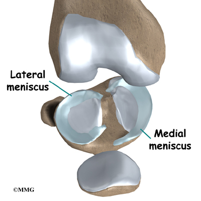
This guide will help you understand

  • how tendonitis develops
  • how doctors diagnose the condition
  • what can be done for tendonitis

Anatomy

Where does tendonitis develop?

Tendonitis

Tendons connect muscle to bone. A tendon is made of material called collagen. Collagen is a key building block of the body. Collagen is considered a connective tissue because it forms tough strands that are like the strands of a nylon rope. Like the strands in a rope, the strands of collagen line up. The more strands, and the better they line up, the stronger they are. The tendon is wrapped in a thin, slippery covering called the tendon sheath. The tendon sheath allows the tendon to slide easily against the tissues around it.

Tendonitis

Many parts of the tendon can be injured. Tendon problems can involve the area where the tendon attaches to the bone, the tissue that surrounds the tendon (the tendon sheath), or the main tissues of the tendon. Doctors use different terms to refer to injuries of different parts of the tendon.

Tendon injuries can show up anywhere in your body. Doctors see tendonitis most often in certain sites.

Achilles Tendonitis

Tendonitis

The tendon sheath, the tissues of the tendon, and the attachment to the bone can all become injured in the Achilles tendon, found in the lower leg. Damaged Achilles tendons carry a higher risk of rupturing because of the weight they bear while standing and walking.

Related Document: A Patient’s Guide to Achilles Tendon Problems

Posterior Tibial Tendonitis

Tendonitis

Tendonitis along the inside edge of the ankle and into the instep of the foot is called posterior tibial tendonitis. It is usually caused by age-related degeneration. If this tendon breaks, it can cause the arch of the foot to become flat and painful.

Related Document: A Patient’s Guide to Posterior Tibial Tendon Problems

Patellar and Quadriceps Tendonitis

Tendonitis

Problems in the tendons of the knee occur mostly in people whose exercise involves running or jumping. Patellar tendonitis is also called jumper’s knee.

De Quervain’s Disease and Trigger Finger

Tendon problems are common in the hand and wrist. De Quervain’s disease causes pain in the wrist just above the thumb. Trigger finger generally causes pain in the palm just below the knuckles, but it eventually causes problems with movement.

Related Document: A Patient’s Guide to De Quervain’s Tenosynovitis

Related Document: A Patient’s Guide to Trigger Finger and Trigger Thumb

Lateral Epicondylitis

Lateral epicondylitis, also called tennis elbow, affects the area where the tendons of the elbow attach to bone on the outside of the elbow. It causes pain when using the wrist and hand.

Tendonitis

Related Document: A Patient’s Guide to Lateral Epicondylitis

Tendonitis

Medial Epicondylitis

Medial epicondylitis, also called Golfer’s elbow, affects the area where the tendons of the elbow attach to bone on the inside of the elbow. It causes pain when using the wrist and hand.

Related Document: A Patient’s Guide to Medial Epicondylitis

Rotator Cuff Tendonitis

Tendonitis

Rotator cuff problems of the shoulder range from mild damage to complete tears. They can cause pain even when resting.

Related Document: A Patient’s Guide to Rotator Cuff Tears

Often, the muscles or other tissues of the joints become tight, misaligned, or weak around the area of tendon injury. Some of the pain and swelling of tendonitis may actually be in the surrounding tissues.

Causes

Why do I have this problem?

Doctors don’t know exactly what causes most tendon problems. They think that repetitive stress on the tendon is the most common cause. The tendon can be injured by the repetitive pounding of running and jumping, or by the stress caused by lifting heavy loads over and over again. Tendonitis usually builds up over weeks or months.
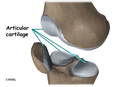
If the tendon is too damaged, or if it doesn’t get time to heal, the problem becomes chronic (long-lasting). In general, the heavier the load or the more often the stress is repeated, the more likely you are to develop tendonitis.

Too much stress on the tendons can be made worse by other factors. Lack of flexibility or weakness in your muscles can make tendonitis more likely. Shoes that don’t fit right, poor equipment, or incorrect technique can also increase your risk of tendon injury. Don’t underestimate the benefit of upgrading your equipment. The improved design of athletic shoes over the past few decades seems to have decreased tendon injuries.

Aging seems to cause tendon damage in some cases. As we age, the tissues of the tendon can break down, or degenerate. Age-related tendon problems do not seem to cause inflammation. The tendon material itself is more affected in these conditions and some surgeon’s refer to this type of tendon problem as tendonosis.

Tendonitis

Some researchers think that a decreased blood supply to the tendons can cause the tendon damage in tendonosis. The decreased blood supply does not allow the tendon to get enough oxygen from the blood. This leads to a condition where the tendon degenerates. The collagen material that makes up the tendon actually becomes weaker and loses its nylon rope appearance. This type of degeneration has been noted in the rotator cuff around the shoulder, in the Achilles tendon in the heel and in the tendons of the elbow.

Many factors can work together to cause tendonitis. For instance, a woman in her forties who takes up running may have tendonitis caused jointly by the degeneration of aging and the mechanical stress of running.

Symptoms

What does tendonitis feel like?

Tendonitis causes pain. This is the primary symptom of tendonitis and tendonosis. The affected tendons are sometimes swollen. In some cases this swelling occurs from actual thickening of the tendon itself. In other cases the swelling comes from thickening or swelling of the tendon sheath. Tendon problems often cause pain after resting, such as when you first get up in the morning. This pain usually goes away within minutes, or even seconds.

Tendonitis

The pain or swelling in your tendon may make your joint hard to move. Some types of tendon problems cause crepitus, a crackling feeling when the joint moves. In rare instances the weakened tendon may actually rupture, or break, with a sudden force. This may require surgery to repair.

Diagnosis

How do doctors identify tendonitis?

Your doctor will take a detailed medical history, including many questions about your activities, your job, and your symptoms. Your doctor will also physically examine the sore area. The probing and movement may cause pain, but it is important for your doctor to know exactly where it hurts.

X-rays do not usually show tendon damage. Your doctor may still ask you to get an X-ray or another imaging test to rule out other problems. Sometimes tendon injuries and other joint or muscle problems occur together. In some cases your doctor may recommend a magnetic resonance imaging (MRI) scan to look at the tendons. The MRI scan is a test that uses magnetic waves instead of X-rays. This test shows the tendons and other soft tissues of the body. It can show the damage in the material that makes up the tendon.
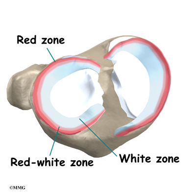
In rare cases it is difficult to find the exact source of your pain. In these cases, your doctor may ask you to go through more sophisticated imaging tests. Your doctor may also inject a local anesthetic into the tendon suspected of causing the pain. If the pain goes away, you have found the right tendon.

Treatment

What can be done for the problem?

Tendon problems can be difficult to treat effectively. They can last for many months to several years, even with treatment. You should expect your treatment to take from six to nine months. Even if treatment is effective, your pain may come back. The exact treatment your doctor recommends depends on which tendon is affected.

Your doctor will probably recommend nonsteroidal anti-inflammatory drugs (NSAIDs), such as aspirin and ibuprofen, to help control the inflammation and pain. NSAIDs are usually used for a short time with tendon problems. Your doctor may also suggest ice or heat treatments.
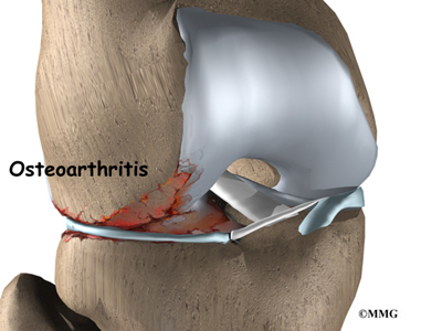
If nothing helps relieve the pain, corticosteroid injections around the tendon are sometimes used. Doctors disagree on whether to use corticosteroid injections in tendons. Even if your doctor does use injections, they are not safe in all cases. Injections into tendons can cause more injury–in some cases, the tendons actually tear. It is generally recommended to give no more than three corticosteroid injections, at least three months apart. Patients need to avoid heavy activity for a few weeks after the injection.

You will need to rest to give your body time to heal. If playing tennis injured your tendon, stop playing tennis until it is completely healed. You should only take part in activities that don’t strain the injured tendon.

Your doctor may refer you to a physical or occupational therapist. A therapist will teach you stretches and exercises to help your tendon heal and regain its strength. A therapist can also assess your work site and athletic equipment and recommend changes to reduce the strain on your tendon. Depending on your type of injury, you may be asked to try such special equipment as arch supports, heel lifts, and splints.

Most people with chronic tendon problems can find ways to relieve the pain and take part in their normal activities, even if the problem doesn’t completely go away. In a few cases, patients can’t find ways to manage the pain even after six months. For these patients, surgery may be necessary. Surgery can be very successful in relieving the pain of chronic tendonitis.

Septic Arthritis

A Patient’s Guide to Septic Arthritis

Introduction

Septic arthritis is an infection inside a joint that is caused by bacteria. (Septic describes an infection in the body caused by bacteria.) It comes on suddenly and causes very noticeable pain and swelling in the affected joint. It can affect anyone, including infants and children. But it is most likely to affect people over 60 and people with certain health or joint problems.

This guide will help you understand

  • how septic arthritis develops
  • how doctors diagnose the condition
  • what can be done for septic arthritis

Anatomy

Where does septic arthritis develop?

In septic arthritis, the synovial fluid–the lubricating fluid inside a joint–becomes contaminated, or infected, with bacteria. In response to the infection, the body produces white blood cells that try to fight and kill the bacteria. This creates pus that mixes with the synovial fluid inside the joint. The white blood cells that fight infection produce chemicals that may not only kill the bacteria but may also damage the joint surfaces. Once this process begins, the breakdown of the joint can continue even after the infection goes away and the bacteria are all dead.

About 80 percent of cases of septic arthritis involve only one joint. Roughly half of the time a knee is affected. The other most common sites are the hip, shoulder, wrist, ankle, elbow, and small joints in the hands and feet. When more than one joint is affected, the knees are usually involved.

Causes

Why do I have this problem?

The cause of septic arthritis is known. It is caused by a bacterial infection in a joint. Bacteria can get into a joint in many ways. The bacteria may be introduced into the joint as a result of injury, such as a laceration that goes into the joint. Surgery on the joint can introduce bacteria into the joint. There is always a small chance of causing an infection if a needle is placed in the joint to either remove synovial fluid for testing or to inject medications into the joint. The joint infection can also come from infections in the skin around the joint or infections in the bones around the joint. The bacteria can also be carried by the blood from an area of infection somewhere else in the body. Once in the joint, synovial fluid provides a good place for the infection to lodge and grow.

Although they do not cause septic arthritis, several risk factors make it more likely to develop. These risk factors include a suppressed immune system, drug abuse, and other diseases. Other diseases that go along with septic arthritis include rheumatoid arthritis, diabetes, cancer, sickle cell disease, anemia, lupus, liver disease, skin infections, and hemophilia.

Artificial joints and recent joint surgery or arthrocentesis (inserting needles into the joint) are also risk factors for septic arthritis. However, the risk from these sources is small. The rate of septic arthritis from artificial joints is less than one percent. The rate following corticosteroid injections is less than 0.01 percent. Rates of septic arthritis following arthroscopy (surgery conducted using a small TV camera inserted into a joint) range from 0.04 percent to four percent. Septic arthritis caused by arthroscopy usually begins within two weeks of the procedure.

The biggest risk factor in septic arthritis is advanced age. Roughly half the cases of septic arthritis occur in people over age 60. In these patients, 75 percent of the infections occur in joints that have already been affected by arthritis, especially the hips, knees, and shoulders.

Symptoms

What does septic arthritis feel like?

Septic arthritis causes moderate to severe joint pain that comes on from a few hours to a few days. (Doctors refer to this as acute onset.) The pain is much worse when you move or touch the joint. The joint is usually warm and red, and it can’t move easily. You may have other evidence of infection, including a fever, chills, flu-like symptoms, and high white blood cell counts. However, 20 percent of patients with septic arthritis run no fever. Even fewer people over 60 run a fever or have high white blood cell counts when they have septic arthritis.

Diagnosis

How do doctors identify the condition?

The sooner a diagnosis of septic arthritis is made, the better. The more time the infection goes unchecked, the more damage it can do to the joint.

If you have acute onset in one joint and a risk factor of any kind, your doctor will probably test for septic arthritis. A needle will be inserted into the joint and synovial fluid removed and sent to the lab. Your doctor will perform several tests on the synovial fluid. The most important test is probably the measuring the white blood cell count in the fluid. An infection in the joint causes the white blood cell count to be extremely high. Other tests are also useful especially making sure there are no crystals in the fluid that could indicate the presence of gout. A gout attack can easily be mistaken for an infection, and vice versa.

The fluid will be examined under a microscope to look for bacteria. Your doctor will also have the lab perform a culture of the synovial fluid to try and grow the bacteria. In this test a small sample of the fluid is placed in a special container and allowed to incubate in a warm cabinet. The test usually takes from 24 to 36 hours to see if any bacteria grow. This test can also help determine the specific type of bacteria that is causing the infection. If bacteria grow in culture the lab can also test different antibiotics on the bacteria to see which antibiotic will work the best.

Your doctor may ask you to have an X-ray. The X-ray will be used to look for any problems in the joint. It will also provide a baseline of information so your doctor can see the changes in your joint over the course of the infection.

Treatment

What can be done for septic arthritis?

Your doctor will prescribe an antibiotic right away, taking into account your medical history, other diseases or infections you have, and the lab test results. The antibiotic may be changed as your doctor gets information from the testing of the synovial fluid. You will probably be started on intravenous (IV) antibiotics. You will need to continue on oral antibiotics for up to six weeks.

Antibiotics alone may not get rid of septic arthritis. The fluid in the joint may need to be drained at least once a day to remove the pus. This can be done using a large needle. In larger joints such as the knee, arthroscopy may also be used to wash the joint out and remove dead or damaged tissue. Surgery may be necessary to drain and clean joints such as the hip and shoulder. Surgery may also be necessary in joints with bone infections and in joints where the infection can’t be easily controlled.

As your infection begins to clear up, your doctor may suggest that you begin strengthening and range of motion exercises. They will help your joint regain its strength and flexibility. A physical or occupational therapist may oversee your exercise program.

How well your septic arthritis responds to treatment will depend on a number of factors. Most important is how soon you got treatment. Only about 25 percent of patients who wait a week to see a doctor can hope for a complete recovery. So you may feel as though your doctor is rushing you through tests and treatment, but it is for a very good reason.

Rheumatoid Arthritis

A Patient’s Guide to Rheumatoid Arthritis

Introduction

Rheumatoid arthritis (RA) is a chronic, or long-term, inflammatory form of arthritis. RA is considered an autoimmune disease, in which your immune system attacks the tissues of your own body. In RA, the immune system mostly attacks tissues in the joints, but it can also affect other organs of your body. In some people, RA seems to run its course more or less by itself. In others, RA gets progressively worse and leads to the destruction of joints. RA can greatly affect your ability to move and do normal tasks. RA can appear at any age, but most patients are between the ages of 30 and 50. About two million Americans have RA, and most of them are women.

This guide will help you understand

  • how RA develops
  • how doctors diagnose the condition
  • what can be done for RA

Anatomy

Where does RA develop?

In RA, two things are happening in the joints. First, the immune system causes inflammation in the synovial membrane, called synovitis. The synovial membrane is the thin tissue that lines the inside of all joints. At first this causes extra fluid, swelling, and oozing clots in the joint. The pain and swelling of synovitis can be reversed.

Second, the synovitis itself causes other problems in the joint. The blood cells and the swollen membranes release chemicals into the synovial fluid (the lubricating fluid of the joint) that can break down or damage the tissues of the joint. This breakdown can cause permanent damage to the cartilage, bone, ligaments, and tendons inside and around the joint. The structural damage usually happens in the first to third year of the disease. The synovitis can come and go, but the structural damage progresses. As a result, the joint becomes painful and very difficult to move.

RA usually affects many corresponding joints on both sides of body. (For example, both knees, both ankles, both wrists, and the same joints in both hands may be affected.) Research indicates that almost all the joints that will be affected show symptoms of RA in the first year of the disease. This means that each joint may continue to get worse, but you probably won’t have many more joints that will develop the symptoms of RA.

Most RA patients have inflammation in the tendons around the joint. (Tendons connect muscle to bone.) Nodules, or bumps, may form on the tendons, or the tendon sheath (the membrane that surrounds the tendon) may become inflamed. Inflammation can also occur in other parts of the body, like the lungs. In general, inflammation refers to symptoms of swelling, redness, heat, and pain.

Most people think of RA as a disease of the joints, but it is actually a systemic disease–it affects the whole body. That means that RA can show up in other organs, too, such as the heart, blood vessels, lungs, and eyes. Sometimes RA occurs in joints and other organs, and sometimes it occurs only in other organs. RA works somewhat differently outside the joints, but the underlying problems are still damage to the tissue and loss of function.

Causes

Why do I have this problem?

No one knows exactly what causes RA. There are probably different causes in different people. Many doctors and researchers think that a virus or bacteria might cause RA. So far studies haven’t proved this. However, researchers do know that bacteria can cause swelling in the synovial membrane.

Heredity–your genes–plays a part in RA. The disease tends to run in families. If a close relative has RA, you are 16 times more likely to develop the disease yourself.

Symptoms

What does RA feel like?

The primary symptom of RA is pain in corresponding joints (both elbows, both knees, and so on). In rare cases the pain is only in one joint. Most often the pain develops over several weeks. But the pain can come on suddenly. As the pain spreads to other joints, it becomes more symmetrical, meaning that it shows up in the same places on both sides of your body. The pain is directly related to the amount of swelling in the synovial membranes. When the swelling is at its worst, your joints themselves will feel warm and swollen. The pain can come and go with the swelling.

RA patients also describe severe morning stiffness that can last up to two hours. The stiffness can be so bad that it makes it hard for you to get dressed, make breakfast, or even get out of bed. This stiffness also corresponds to the synovitis. When the synovitis goes away for a time, so does the stiffness.

About half of RA patients have rheumatoid nodules. The nodules are hard knots, from the size of a pea to the size of a golf ball, that grow under the skin in three distinct layers. They are usually found on the outside of the elbow, the Achilles tendon on your heel, the underside of your fingers, the lower abdomen, and certain toe joints. They look like the kind of bump that grows around a splinter. They don’t usually hurt. Over time they tend to shrink or disappear.

Because RA is a systemic disease, most patients feel tired and weak during flare-ups. In patients who test positive for rheumatoid factor (RF) in their blood, other organ systems can also become inflamed when the joints do. About 50 percent of RA patients have systemic inflammation during joint outbreaks of RA.

Conjunctivitis, or inflammation of the eye, is common. It may be related to a disease of the eye called Sjogren’s syndrome, which often occurs along with RA. The main symptom is eye dryness, but patients often can’t even feel it.

RA can affect the lungs. Occasionally it can cause an inflammation of the membrane that surrounds the lungs (called the pleura), which causes pain in the side and sometimes coughing and problems breathing deeply.

RA commonly affects the nervous system, but it can be hard to tell from other symptoms of RA. Damage to the joints in the cervical spine (the neck) can eventually lead to weakness and instability between the cervical vertebrae. This damage can cause problems with the spinal cord as it travels through the neck.

Some symptoms depend on the affected joints:

  • Cervical spine (the neck): Symptoms include neck stiffness, weakness, and loss of motion. Other symptoms often can’t be felt or seen in exams. Ligaments are often inflamed, and there may be problems with the spinal cord or nervous system. Neck pain alone tends to get better, even when the joints are damaged. Damage to the nervous system does not usually improve.
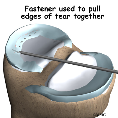
  • Shoulders: The main symptom is loss of motion. Your body’s unconscious reaction to shoulder pain is simply not to use your shoulders. Since daily life doesn’t require much shoulder use, frozen shoulder syndrome can set in quickly.
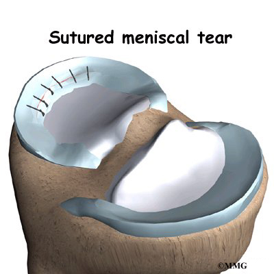
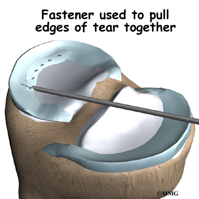
  • Hands and wrists: Almost everyone with RA has affected wrists. Joints in the middle of your hand and fingers are usually affected. The knuckles at the ends of your fingers usually are not. RA can cause joint deformities that freeze your fingers in unusual positions. Rheumatoid nodules and tendon inflammation can make it hard to bend the fingers. Nodules can cause a locking and catching action as your fingers bend.
  • Knees: It is easy to feel the swelling in the knees. A fluid-filled lump called a Baker’s cyst often appears behind the knee. It can burst and leak fluid into the calf.
  • Feet and ankles: RA commonly affects the joints in the middle of the toes and the ankle joints. The deformities and pain in the toes can cause problems with walking. The sole of the foot can feel tingly or numb.

The progression of RA is hard to predict. The swelling of RA flares up and dies down, and milder forms of the disease often don’t require much treatment. Mild RA may even go undiagnosed.

Diagnosis

How do doctors identify RA?

No single test can confirm a diagnosis of RA. Many findings over a period of time lead to the diagnosis. In fact, your doctor can’t even positively diagnose RA until you’ve had symptoms for at least several weeks. Early on, many characteristics of RA haven’t developed yet, such as the pattern of joints that are affected, X-ray findings, and blood test changes. And RA in its early stages can look a lot like other forms of arthritis, such as lupus, psoriatic arthritis, and diseases of the spine. Your doctor will need to consider each of these diagnoses and perhaps do tests to rule them out.

Your doctor will start with a detailed health history. You will need to describe your pain and be very specific about where your pain is located and when it came on. You will also need to tell your doctor about any other medical conditions you have had and drugs you are taking. Even if these other conditions are not related to your joint pain, your doctor will need to know these things to help you find effective treatment. Your doctor will also examine your joints closely. Your doctor will be looking for bone-on-bone crepitus, a high-pitched screech that you can feel or hear in the joint. It is the sound of bone rubbing on bone, and nothing else makes this sound.

Every patient with RA has inflammation of the synovial membranes. Your doctor can confirm this by checking the count of white blood cells (WBC) in your synovial fluid. This involves inserting a thin needle into your joint and drawing out a small amount of the fluid for testing. The fluid can also be tested for other things. The WBC alone doesn’t prove that you have RA. Your doctor will need to rule out other causes of synovitis.

Your doctor will also ask you to undergo a blood test. RF, or rheumatoid factor, is found in the blood of about 85 percent of RA patients. But this test alone can’t confirm RA either. Some patients with RA do not have RF, and people with RF can have other forms of arthritis.

Another blood test is the erythrocyte sedimentation rate (ESR, or sed rate), which measures how fast red blood cells settle in the test tube. Red blood cells that settle faster than normal indicate inflammation in the body. But the ESR varies greatly between people. It is even possible for a patient with RA to have a normal ESR. The ESR may be more useful in monitoring the progress of your disease than in diagnosing it. A higher ESR usually means that the inflammation is more severe.

The C-reactive protein test can also monitor inflammation. It is a newer test that may be more accurate than the ESR. This test measures the amount of a certain protein that is produced by the body due to inflammation. When inflammation is very active the amount of C-reactive protein is high, and when inflammation is brought under control the level of protein decreases.

At some point your doctor will probably ask you to get X-rays of your affected joints and organs. X-rays and other imaging techniques can show damage to the cartilage and bone and the swelling in the soft tissues of the joint.

In some cases your doctor may want to biopsy the rheumatoid nodules. A small amount of the nodule is removed and examined in a laboratory.

Treatment

What can be done for the condition?

Doctors have learned much about RA in recent years, but they still don’t know much more about how to truly cure the disease. They do have many strategies for treating the symptoms of RA. If you start treatment within a few months after your symptoms appear, the better you will probably do in the long-term. Early detection and treatment can help avoid the worst joint damage. Sudden remission does occur, but it’s unclear how often, and it appears to be more likely within the first two years of the disease. Patients who develop RA at a young age, are RF positive, have close relatives with RA, and have RA nodules tend to have a more difficult time managing the disease.

Your doctor will prescribe one or more medications. Nonsteroidal anti-inflammatory drugs (NSAIDs), such as aspirin and ibuprofen, can help decrease the pain and swelling. Corticosteroids taken by mouth can also help with inflammation. Because steroid use can cause other problems in your body, they are usually not be used over long periods of time, if at all possible. Corticosteroid injections into the affected joints can ease the swelling and give you immediate, short-term relief. And your doctor may prescribe eye medications, even if you have no eye symptoms. Because eye inflammation is so common with RA and is hard to diagnose, the eye drops can help prevent it from developing or becoming severe.

Disease-modifying antirheumatic drugs (DMARDs) are important in treating RA. No one is sure exactly how well DMARDs actually slow or prevent the structural damage from RA. However, tests have shown all DMARDs to be effective for at least one year of treatment.

DMARDs can be very hard on your body and can interact with other drugs, so it is not always easy to find the best medication for you. Often more than one drug is taken at the same time. Several DMARDs are frequently prescribed:

  • Hydroxychloroquine is a relatively nontoxic drug that was made to treat malaria. It can be safely used with other DMARDs. It is most useful in early, mild RA. You should get regular eye check-ups while taking this drug.
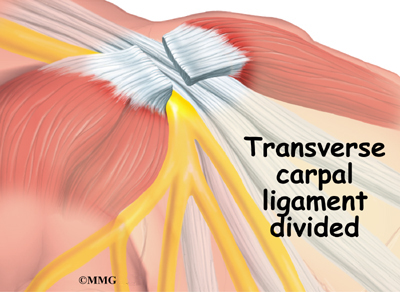
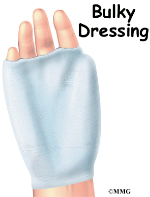
  • Sulfasalazine is much like hydroxychloroquine. This drug requires regular blood monitoring.
  • Gold salts can cause short-term remissions. Over the long-term, however, the RA does progress. Blood and urine monitoring is required.
  • Methotrexate can help manage RA, but it is unclear how much it actually changes the course of the disease. Methotrexate can be very useful over the long-term, but there are problems with flare-ups when patients stop taking it.
  • Azathioprine is used with moderate and severe RA.
  • Penicillamine is only used in patients who have systemic disease that doesn’t respond well to other medications.
  • Cyclosporine is expensive and hard on the kidneys, so it is most often used in severe RA.
  • Cyclophosphamide is very effective but very toxic, so it is only used in specific cases.
  • Certain antibiotics are somewhat effective in mild cases.

Combining methotrexate with another drug to get the desired results (decreased joint pain, swelling, and stiffness) is a popular and effective strategy for many patients. One group of disease modifying medications used along with methotrexate is called anti-tumor necrosis factor (TNF) agents.

Another term for the anti-TNF agents is TNF inhibitors. These are a special type of antibody also referred to as human monoclonal antibodies. They specifically target (and inhibit) tumor necrosis factor. Tumor necrosis factor (TNF) promotes the inflammatory response, which in turn causes many of the clinical problems associated with autoimmune disorders such as rheumatoid arthritis.

Anyone taking tumor necrosis inhibitors must be careful to report any signs of infection to the physician right away. There have been reports of deaths possibly associated with unrecognized or untreated infections in patients taking these immune suppressing drugs.

Symptoms of infection anywhere but commonly affecting skin, lungs, or urinary tract include fever, chills, fatigue, enlarged lymph nodes, skin rash or red streaks, cough, and/or sore throat. Upset stomach, painful urination or blood in the urine or stools are additional important symptoms that could be a signal of infection and should be reported to the physician as well.

In general, your doctor may recommend some of the following treatments that will require some effort and lifestyle changes from you:

  • Patient education. RA is a frustrating and complex disease. The more you understand it, the better you can help treat your own symptoms and prevent flare-ups.
  • Range-of-motion and strengthening exercises. These will most often be designed and monitored by a physical or occupational therapist.
  • Equipment and gadgets, such as canes and jar openers that can help you go about your daily business without putting too much stress on affected joints.
  • Gentle aerobic exercise.
  • Support groups.

At least half of RA patients don’t find much relief from treatment and eventually need surgery on the affected joints. Surgery, including total joint replacement, can be a very effective way to help you overcome the pain and loss of movement of RA.

For most patients, RA is a disease that comes and goes throughout their lives. But it doesn’t have to be crippling. With your doctor’s help, you should be able to find treatment that works for you.