Anterior Cruciate Ligament Injuries

A Patient’s Guide to Anterior Cruciate Ligament Injuries

Introduction

The anterior cruciate ligament (ACL) is probably the most commonly injured ligament of the knee. In most cases, the ligament is injured by people participating in athletic activity. As sports have become an increasingly important part of day-to-day life over the past few decades, the number of ACL injuries has steadily increased. This injury has received a great deal of attention from orthopedic surgeons over the past 15 years, and very successful operations to reconstruct the torn ACL have been invented.

This guide will help you understand

  • where in the knee the ACL is located
  • how an ACL injury causes problems
  • how doctors treat the condition

Anterior Cruciate Ligament Injuries

Anatomy

Where is the ACL, and what does it do?

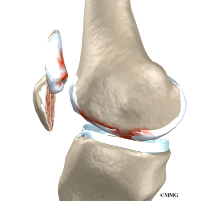
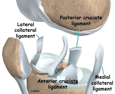
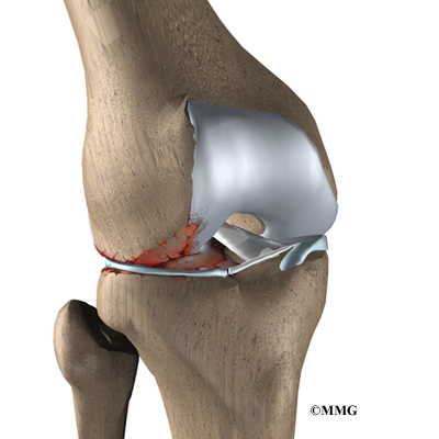
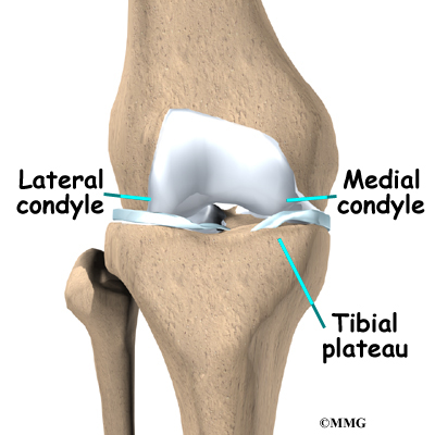
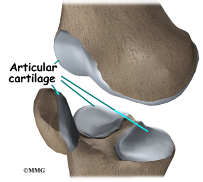
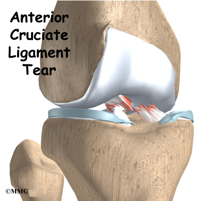
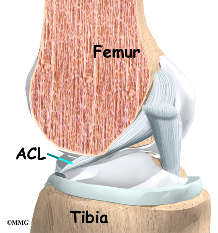
Ligaments are tough bands of tissue that connect the ends of bones together. The ACL is located in the center of the knee joint where it runs from the backside of the femur (thighbone) to connect to the front of the tibia (shinbone).

Anterior Cruciate Ligament Injuries

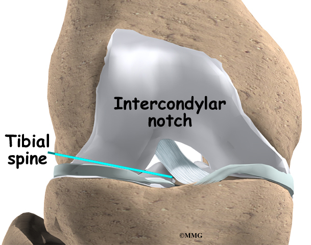
The ACL runs through a special notch in the femur called the intercondylar notch and attaches to a special area of the tibia called the tibial spine.

Anterior Cruciate Ligament Injuries

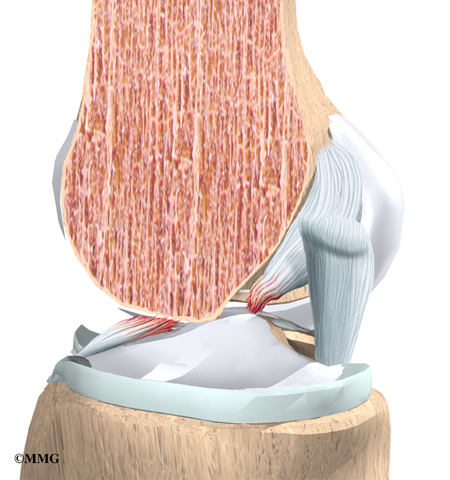
The ACL is the main controller of how far forward the tibia moves under the femur. This is called anterior translation of the tibia. If the tibia moves too far, the ACL can rupture. The ACL is also the first ligament that becomes tight when the knee is straightened. If the knee is forced past this point, or hyperextended, the ACL can also be torn.

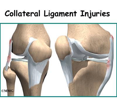
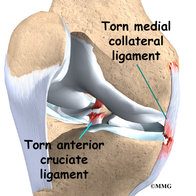
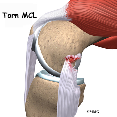
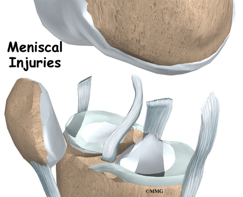
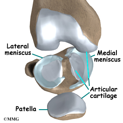
Other parts of the knee may be injured when the knee is twisted violently, as in a clipping injury in football. It is not uncommon to also see a tear of the medial collateral ligament (MCL) on the inside edge of the knee, and the lateral meniscus, which is the U-shaped cushion between the outer half of the tibia and femur bones.

Related Document: A Patient’s Guide to Knee Anatomy

Causes

How do ACL injuries occur?

The mechanism of injury for many ACL ruptures is a sudden deceleration (slowing down or stop), hyperextension, or pivoting in place. Sports-related injuries are the most common.

The types of sports that have been associated with ACL tears are numerous. Those sports requiring the foot to be planted and the body to change direction rapidly (such as basketball) carry a high incidence of injury. In this way, most ACL injuries are considered noncontact. However, contact-related injuries can result in ACL tears. For example, a blow to the outside of the knee when the foot is planted is the most likely contact-related injury.

Football is also frequently the source of an ACL tear. Football combines the activity of planting the foot and rapidly changing direction and the threat of bodily contact. Downhill skiing is another frequent source of injury, especially since the introduction of ski boots that come higher up the calf. These boots move the impact of a fall to the knee rather than the ankle or lower leg. An ACL injury usually occurs when the knee is forcefully twisted or hyperextended while the foot remains in contact with the ground. Many patients recall hearing a loud pop when the ligament is torn, and they feel the knee give way.

The number of women suffering ACL tears has dramatically increased. This is due in part to the rise in women’s athletics. But studies have shown that female athletes are two to four times more likely to suffer ACL tears than male athletes in the same sports.

Recent research has shown several factors that contribute to women’s higher risk of ACL tears. Women athletes seem less able to tighten their thigh muscles to the same degree as men. This means women don’t get their knees to hold as steady, which may give them less knee protection during heavy physical activity. Also, tests show that women’s quadriceps and hamstring muscles work differently than men’s. Women’s quadriceps muscles (on the front of the thigh) work extra hard during knee-bending activities. This pulls the tibia forward, placing the ACL at risk for a tear.

Meanwhile, women’s hamstring muscles (on the back of the thigh) respond more slowly than in men. The hamstring muscles normally protect the tibia from sliding too far forward. Women’s sluggish hamstring response may allow the tibia to slip forward, straining the ACL. Other studies suggest that women’s ACLs may be weakened by the effects of the female hormone estrogen. Taken together, these factors may explain why female athletes have a higher risk of ACL tears.

Symptoms

What does a torn ACL feel like?

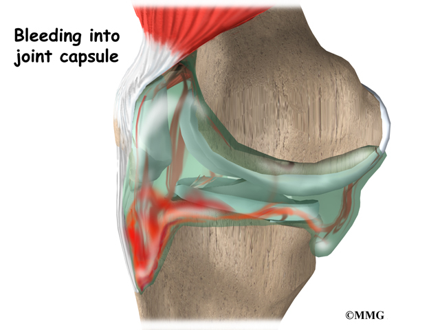
The symptoms following a tear of the ACL can vary. Some patients report hearing and/or feeling a pop. Usually, the knee joint swells within a short time following the injury. This is due to bleeding into the knee joint from torn blood vessels in the damaged ligament. The instability caused by the torn ligament leads to a feeling of insecurity and giving way of the knee, especially when trying to change direction on the knee. The knee may feel like it wants to slip backwards. There may be activity-related pain and/or swelling. Walking downhill or on ice is especially difficult. And you may have trouble coming to a quick stop.

The pain and swelling from the initial injury will usually be gone after two to four weeks, but the knee may still feel unstable. The symptom of instability and the inability to trust the knee for support are what require treatment. Also important in the decision about treatment is the growing realization by orthopedic surgeons that long-term instability leads to early arthritis of the knee.

Related Document: A Patient’s Guide to Osteoarthritis of the Knee

Diagnosis

How do doctors identify ACL injuries?

The history and physical examination are probably the most important ways to diagnose a ruptured or deficient ACL. In the acute (sudden) injury, the swelling is a good indicator. A good rule of thumb that orthopedic surgeons use is that any tense swelling that occurs within two hours of a knee injury usually represents blood in the joint, or a hemarthrosis. If the swelling occurs the next day, the fluid is probably from the inflammatory response.

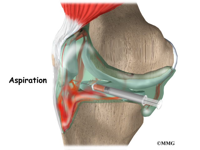
Placing a needle in the swollen joint and aspirating (or draining as much fluid as possible) gives relief from the swelling and provides useful information to your doctor. If blood is found when draining the knee, there is about a 70 percent chance it represents a torn ACL. This fluid can also show if the cartilage on the surface of the knee joint was injured.

During the physical examination, special stress tests are performed on the knee. Three of the most commonly used tests are the Lachman test, the pivot-shift test, and the anterior drawer test. The doctor will place your knee and leg in various positions and then apply a load or force to the joint. Any excess motion or unexpected movement of the tibia relative to the femur may be a sign of ligament damage and insufficiency.

Another way to check for anterior tibial translation is with the KT-1000 and KT-2000 arthrometers. The patient’s leg is bent and supported on a wedge with the knee in 30 degrees of flexion. The arthrometer is placed against the knee to be tested and strapped to the lower leg. Usually, the normal knee is tested first. The arthrometer applies an anterior force of 15 pounds against the tibia. The amount of anterior tibial translation is measured. The test is repeated with a force of 20 pounds. A third test applies a manual maximal force to the posterior (back) of the tibia. This is similar to the Lachman test.

The results of these tests will help your doctor determine how badly the ACL was injured. Other tests may be combined with tests of ACL integrity to determine whether other knee ligaments, joint capsule, or joint cartilage have also been injured.

Anterior Cruciate Ligament Injuries

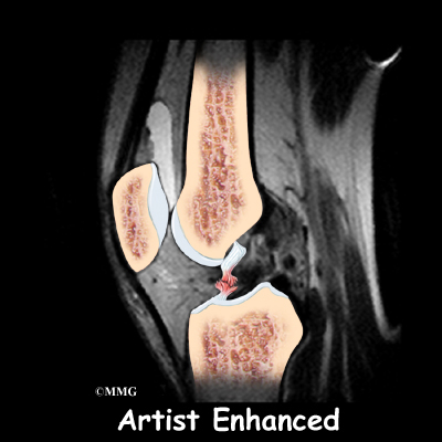
Your doctor may order X-rays of the knee to rule out a fracture. Ligaments and tendons do not show up on X-rays, but bleeding into the joint can result from a fracture of the knee joint, or when portions of the joint surface are chipped off. Magnetic resonance imaging (MRI) is probably the most accurate test for diagnosing a torn ACL without actually looking into the knee. The MRI machine uses magnetic waves rather than X-rays to show the soft tissues of the body. This machine creates pictures that look like slices of the knee. The pictures show the anatomy, and any injuries, very clearly. This test does not require any needles or special dye and is painless.

Anterior Cruciate Ligament Injuries

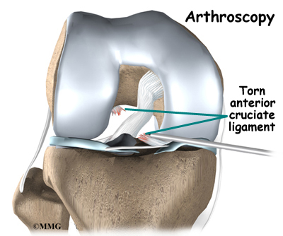
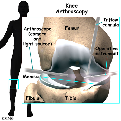
In some cases, arthroscopy may be used to make the definitive diagnosis if there is a question about what is causing your knee problem.

Arthroscopy is an operation that involves inserting a small fiber-optic TV camera into the knee joint, allowing the orthopedic surgeon to look at the structures inside the joint directly. The vast majority of ACL tears are diagnosed without resorting to this type of surgery, though arthroscopy is sometimes used to repair a torn ACL.

Treatment

How do doctors treat an ACL injury?

Nonsurgical Treatment

Initial treatment for an ACL injury focuses on decreasing pain and swelling in the knee. Rest and mild pain medications, such as acetaminophen (Tylenol®), can help decrease these symptoms. You may need to use crutches until you can walk without a limp. Most patients are instructed to put a normal amount of weight down while walking. The knee joint may need to be drained with a needle (mentioned earlier) to remove any blood in the joint.

Most patients receive physical therapy after having an ACL injury. Therapists treat swelling and pain with the use of ice, electrical stimulation, and rest periods with your leg supported in elevation.

Exercises are used to help you regain normal movement of joints and muscles. Range-of-motion exercises should be started right away with the goal of helping you swiftly regain full movement in your knee. This includes the use of a stationary bike, gentle stretching, and careful pressure applied to the knee by the therapist. Exercises are also given to improve the strength of the hamstring and quadriceps muscles. As your symptoms ease and strength improves, you will be guided in specialized exercises to improve knee stability.

An ACL brace may be suggested. This type of brace is usually custom-made and not the type you can buy at the drugstore. It is designed to improve knee stability when the ACL doesn’t function properly. An ACL brace is often recommended when the knee is unstable and surgery is not planned. As mentioned, a torn ACL that isn’t corrected often leads to early knee arthritis. There is no evidence that an ACL brace will prevent further damage to the knee due to wear and tear arthritis. The ACL brace may help keep the knee from giving way during moderate activity. However, it can give a false sense of security and won’t always protect the knee during sports that require heavy cutting, jumping, or pivoting. Many orthopedists will also recommend wearing a brace for at least one year after a surgical reconstruction, so even if you decide to have ACL surgery, a brace is probably a good investment.

Surgery

If the symptoms of instability are not controlled by a brace and rehabilitation program, then surgery may be suggested. The main goal of surgery is to keep the tibia from moving too far forward under the femur bone and to get the knee functioning normally again.

Even when surgery is needed, most surgeons will have their patients attend physical therapy for several visits before the surgery. This is done to reduce swelling and to make sure you can straighten your knee completely. This practice also reduces the chances of scarring inside the joint and can speed recovery after surgery.
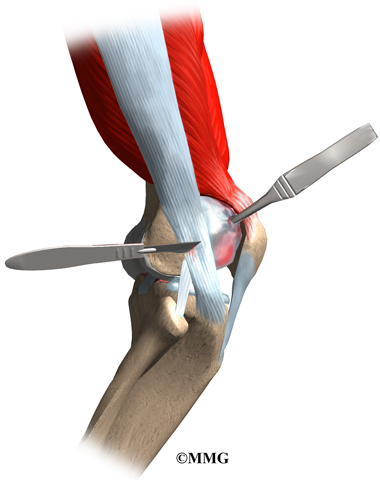
Arthroscopic Method

Most surgeons now favor reconstruction of the ACL using a piece of tendon or ligament to replace the torn ACL. This surgery is most often done with the aid of the arthroscope. Incisions are usually still required around the knee, but the surgery doesn’t require the surgeon to open the joint. The arthroscope is used to view the inside of the knee joint as the surgeon performs the work. Most ACL surgeries are now done on an outpatient basis, and many patients go home the same day as the surgery. Some patients stay one or two nights in the hospital if necessary.

Patellar Tendon Graft

Anterior Cruciate Ligament Injuries

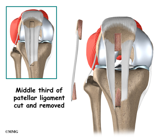
One type of graft used to replace the torn ACL is the patellar tendon. This tendon connects the kneecap (patella) to the tibia. The surgeon removes a strip from the center of the ligament to use as a replacement for the torn ACL.

Related Document: A Patient’s Guide to Patellar Tendon Graft Reconstruction of the ACL

Hamstring Tendon Graft

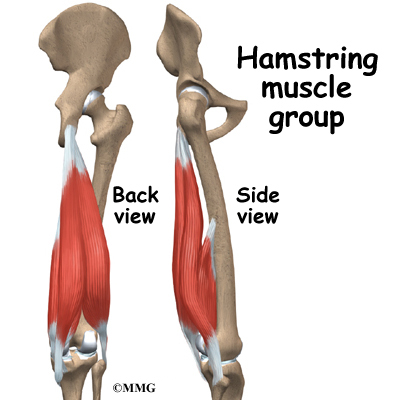
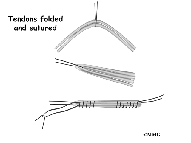
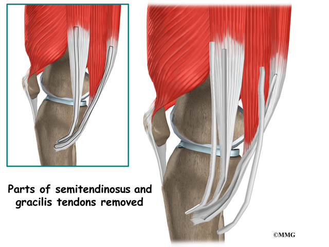
Surgeons also commonly use a hamstring graft to reconstruct a torn ACL. This graft is taken from one of the hamstring tendons that attaches to the tibia just below the knee joint. The hamstring muscles run down the back of the thigh. Their tendons cross the knee joint and connect on each side of the tibia. The graft used in ACL reconstruction is taken from the hamstring tendon, called the semitendinosus. This tendon runs along the inside part of the thigh and knee. Surgeons also commonly include as part of the hamstring graft a tendon just next to the semitendinousus, called the gracilis. When arranged into three or four strips, the hamstring graft has nearly the same strength as a patellar tendon graft.

Related Document: A Patient’s Guide to Hamstring Tendon Graft Reconstruction of the ACL

Allograft Reconstruction

Other materials are also used to replace the torn ACL. In some cases, an allograft is used. An allograft is tissue that comes from someone else. This tissue is harvested from tissue and organ donors at the time of death and sent to a tissue bank. The tissue is checked for any type of infection, sterilized, and stored in a freezer. When needed, the tissue is ordered by the surgeon and used to replace the torn ACL. The allograft (your surgeon’s choice of graft) can be from the tibialis tendon, patellar tendon, hamstring tendon, or Achilles tendon (the tendon that connects the calf muscles to the heel).

Many surgeons use patellar tendon allograft tissue because the tendon comes with the original bone still attached on each end of the graft (from the patella and from the tibia). This makes it easier to fix the allograft in place.

The advantage of using an allograft is that the surgeon does not have to disturb or remove any of the normal tissue from your knee to use as a graft. The operation also usually takes less time because the graft does not need to be harvested from your knee.

Rehabilitation

What should I expect after treatment?

Nonsurgical Rehabilitation

Nonsurgical rehabilitation for a torn ACL will typically last six to eight weeks. Therapists apply treatments such as electrical stimulation and ice to reduce pain and swelling. Exercises to improve knee range of motion and strength are added gradually. If your doctor prescribes a brace, your therapist will work with you to obtain and use the brace.

You can return to your sporting activities when your quadriceps and hamstring muscles are back to nearly their full strength and control, you are not having swelling that comes and goes, and you aren’t having problems with the knee giving way.

After Surgery

Most doctors have their patients take part in formal physical therapy after ACL reconstruction. You will probably be involved in a progressive rehabilitation program for four to six months after surgery to ensure the best result from your ACL reconstruction. At first, expect to see the physical therapist two to three times a week. If your surgery and rehabilitation go as planned during the first six weeks, you may only need to do a home program and see your therapist every few weeks over the four to six month period.

Artificial Joint Replacement of the Knee

A Patient’s Guide to Artificial Joint Replacement of the Knee

Introduction

A painful knee can severely affect your ability to lead a full, active life. Over the last 25 years, major advancements in artificial knee replacement have improved the outcome of the surgery greatly. Artificial knee replacement surgery (also called knee arthroplasty) is becoming increasingly common as the population of the world begins to age.

In addition to reading this article, be sure to watch our Artificial Knee Replacement (Knee Arthroplasty) Animated Tutorial Video.

This guide will help you understand

  • what your surgeon hopes to achieve with knee replacement surgery
  • what happens during the procedure
  • what to expect after your operation

Anatomy

What is the normal anatomy of the knee?

Knee joint replacement

The knee joint is formed where the thighbone (femur) meets the shinbone (tibia). A smooth cushion of articular cartilage covers the end surfaces of both of these bones so that they slide against one another smoothly. The articular cartilage is kept slippery by joint fluid made by the joint lining (synovial membrane). The fluid is contained in a soft tissue enclosure around the knee joint called the joint capsule.

Knee joint replacement

The patella, or kneecap, is the moveable bone on the front of the knee. It is wrapped inside a tendon that connects the large muscles on the front of the thigh, the quadriceps muscles, to the lower leg bone. The surface on the back of the patella is covered with articular cartilage. It glides within a groove on the front of the femur.

Related Document: A Patient’s Guide to Knee Anatomy

Rationale

What does the surgeon hope to achieve?

The main reason for replacing any arthritic joint with an artificial joint is to stop the bones from rubbing against each other. This rubbing causes pain. Replacing the painful and arthritic joint with an artificial joint gives the joint a new surface, which moves smoothly and without causing pain. The goal is to help people return to many of their activities with less pain and with greater freedom of movement.

Preparation

How should I prepare for surgery?

The decision to proceed with surgery should be made jointly by you and your surgeon. The decision should only be made after you feel that you understand as much about the procedure as possible.

Once you decide to proceed with surgery, several things may need to be done. Your orthopedic surgeon may suggest a complete physical examination by your regular doctor. This is to ensure that you are in the best possible condition to undergo the operation. You may also need to spend time with the physical therapist who will be managing your rehabilitation after the surgery. The therapist will begin the teaching process before surgery to ensure that you are ready for rehabilitation afterwards.

One purpose of the preoperative visit is to record a baseline of information. This includes measurements of your current pain levels, functional abilities, the presence of swelling, and the available movement and strength of each knee.

A second purpose of the preoperative therapy visit is to prepare you for your upcoming surgery. You will practice some of the exercises used just after surgery. You will also be trained in the use of either a walker or crutches. (Whether the surgeon uses a cemented or noncemented artificial knee will determine how much weight you will apply through your foot at first while walking.) Finally, an assessment will be made of any needs you will have at home once you’re released from the hospital.

You may be asked to donate some of your own blood before the operation. This blood can be donated three to five weeks before the operation, and your body will make new blood cells to replace the loss. At the time of the operation, if you need to have a blood transfusion you will receive your own blood back from the blood bank.

Surgical Procedure

What happens during the operation?

Before we describe the procedure, let’s look first at the artificial knee itself.

The Artificial Knee

There are two major types of artificial knee replacements:

  • cemented prosthesis
  • uncemented prosthesis

Both are still widely used. In many cases a combination of the two types is used. The patellar (kneecap) portion of the prosthesis is commonly cemented into place. The decision to use a cemented or uncemented artificial knee is usually made by the surgeon based on your age, your lifestyle, and the surgeon’s experience.

Each prosthesis is made up of three main parts.

Knee joint replacement

The tibial component (bottom portion) replaces the top surface of the lower bone, the tibia. The femoral component (top portion) replaces the bottom surface of the upper bone (the femur) and the groove where the patella fits. The patellar component (kneecap portion) replaces the surface of the patella where it glides in the groove on the femur.

The femoral component is made of metal. The tibial component is usually made of two parts: a metal tray that is attached directly to the bone, and a plastic spacer that provides the slick surface. The plastic used is so tough and slick that you could ice skate on a sheet of it without damaging the material much. The patellar component is usually made of plastic as well. In some types of knee implants, the patellar component is made of a combination of metal and plastic.

A cemented prosthesis is held in place by a type of epoxy cement that attaches the metal to the bone. An uncemented prosthesis has a fine mesh of holes on the surface that allows bone to grow into the mesh and attach the prosthesis to the bone.

The Operation

To begin the procedure, the surgeon makes an incision on the front of the knee to allow access to the joint. Several different approaches can be used to make the incision. The choice is usually based on the surgeon’s training and preferences.

Knee joint replacement

Once the knee joint is opened, a special positioning device (a cutting guide) is placed on the end of the femur. This cutting guide is used to ensure that the bone is cut in the proper alignment to the leg’s original angles, even if the arthritis has made you bowlegged or knock-kneed. With the help of the cutting guide, the surgeon cuts several pieces of bone from the end of the femur. The artificial knee will replace these worn surfaces with a metal surface.

Knee joint replacement

Next, the surface of the tibia is prepared. Another type of cutting guide is used to cut the tibia in the correct alignment.

Knee joint replacement

Then the articular surface of the patella is removed.

Knee joint replacement

The metal femoral component is then placed on the femur. In the uncemented prosthesis, the metal piece is held snugly onto the femur because the femur is tapered to accurately match the shape of the prosthesis. The metal component is pushed onto the end of the femur and held in place by friction. In the cemented variety, an epoxy cement is used to attach the metal prosthesis to the bone.

Knee joint replacement

The metal tray that holds the plastic spacer is then attached to the top of the tibia. This metal tray is either cemented into place, or held with screws if the component is of the uncemented variety. The screws are primarily used to hold the tibial tray in place until bone grows into the porous coating. (The screws remain in place and are not removed.)

The plastic spacer is then attached to the metal tray of the tibial component. If this component should wear out while the rest of the artificial knee is sound, it can be replaced. The replacement procedure is called a retread.

Knee joint replacement

The surgeon then sizes the patellar component and puts it into place behind the patella. This piece is usually cemented in place.

Finally, the soft tissues are sewn back together, and staples are used to hold the skin incision together.

View animation of removing the joint surfaces

View animation of inserting the femoral component

View animation of inserting the tibial component

View animation of removing the patella and inserting the patellar component

View animation of the completed artificial knee

Complications

What might go wrong?

As with all major surgical procedures, complications can occur. This document doesn’t provide a complete list of the possible complications, but it does highlight some of the most common problems. Some of the most common complications following artificial knee replacement are

  • anesthesia complications
  • thrombophlebitis
  • infection
  • stiffness
  • loosening

Anesthesia Complications

Most surgical procedures require that some type of anesthesia be done before surgery. A very small number of patients have problems with anesthesia. These problems can be reactions to the drugs used, problems related to other medical complications, and problems due to the anesthesia. Be sure to discuss the risks and your concerns with your anesthesiologist.

Thrombophlebitis (Blood Clots)

View animation of pulmonary embolism

Thrombophlebitis, sometimes called deep venous thrombosis (DVT), can occur after any operation, but is more likely to occur following surgery on the hip, pelvis, or knee. DVT occurs when blood clots form in the large veins of the leg. This may cause the leg to swell and become warm to the touch and painful. If the blood clots in the veins break apart, they can travel to the lung, where they lodge in the capillaries and cut off the blood supply to a portion of the lung. This is called a pulmonary embolism. (Pulmonary means lung, and embolism refers to a fragment of something traveling through the vascular system.) Most surgeons take preventing DVT very seriously. There are many ways to reduce the risk of DVT, but probably the most effective is getting you moving as soon as possible after surgery. Two other commonly used preventative measures include

  • pressure stockings to keep the blood in the legs moving
  • medications that thin the blood and prevent blood clots from forming

Infection

Infection can be a very serious complication following an artificial joint surgery. The chance of getting an infection following artificial knee replacement is probably around one percent. Some infections may show up very early, even before you leave the hospital. Others may not become apparent for months, or even years, after the operation. Infection can spread into the artificial joint from other infected areas. Your surgeon may want you to take antibiotics when you have dental work or surgical procedures on your bladder and colon to reduce the risk of spreading germs to the joint.

Stiffness

In some cases, the ability to bend the knee does not return to normal after knee replacement surgery. To be able to use the leg effectively to rise from a chair, the knee must bend at least to 90 degrees. A desirable range of motion is greater than 110 degrees.

The most important factor in determining range of motion after surgery is whether the ligaments and soft tissues were balanced during surgery. The surgeon tries to get the knee in the best alignment so there is equal tension on all the ligaments and soft tissues.

Sometimes extra scar tissue develops after surgery and can lead to an increasingly stiff knee. If this occurs, your surgeon may recommend taking you back to the operating room, placing you under anesthesia once again, and manipulating the knee to regain motion. Basically, this allows the surgeon to break up and stretch the scar tissue without you feeling it. The goal is to increase the motion in the knee without injuring the joint.

Loosening

The major reason that artificial joints eventually fail continues to be a process of loosening where the metal or cement meets the bone. Great advances have been made in extending how long an artificial joint will last, but most will eventually loosen and require a revision. Hopefully, you can expect 12 to 15 years of service from an artificial knee, but in some cases the knee will loosen earlier than that. A loose prosthesis is a problem because it usually causes pain. Once the pain becomes unbearable, another operation will probably be required to revise the knee replacement.

Related Document: A Patient’s Guide to Revision Arthroplasty of the Knee

After Surgery

What happens after surgery?

Some orthopedic surgeons recommend a device known as a continuous passive motion (CPM) machine immediately after surgery. The unit is thought to help prevent blood clots and speed healing of the wound. It may help patients get by with less need for medication. The unit may help improve knee mobility after knee replacement surgery. However, patients seem to do equally well in regaining knee motion by doing their exercises.

You may also have physical therapy treatments once or twice each day as long as you are in the hospital. Therapy treatments will address the range of motion in the knee. Gentle movement will be used to help you bend and straighten the knee. If you are using a CPM device, it will be checked for alignment and settings. Your leg may be elevated to help drain extra fluid in the leg.

Your therapist will also go over exercises to help improve knee mobility and to start exercising the thigh and hip muscles. Ankle movements are used to help pump swelling out of the leg and to prevent the possibility of a blood clot.

When you are stabilized, your therapist will help you up for a short outing using your crutches or your walker.

Most patients are able to go home after spending two to four days in the hospital. You’ll be on your way home when you can demonstrate a safe ability to get in and out of bed, walk up to 75 feet with your crutches or walker, go up and down stairs safely, and access the bathroom. It is also important that you regain a good muscle contraction of the quadriceps muscle and that you gain improved knee range of motion. Patients who still need extra care may be sent to a different unit until they are safe and ready to go home.

Most orthopedic surgeons recommend regular checkups after your artificial joint replacement. How often you need to be seen varies from every six months to every five years, according to your situation and what your surgeon recommends. You should always consult your orthopedic surgeon if you begin to have pain in your artificial joint, or if you begin to suspect something is not working correctly.

Knee joint replacement

Most patients who have an artificial joint will have episodes of pain, but when you have a period that lasts longer than a couple of weeks you should consult your surgeon. The surgeon will examine your knee in search of reasons for the pain. X-rays may be taken of your knee to compare with X-rays taken earlier to see whether the artificial joint shows any evidence of loosening.

Rehabilitation

What should I expect during my rehabilitation?

Once discharged from the hospital, you may see your therapist for one to six in-home treatments. This is to ensure you are safe in and about the home and getting in and out of a car. Your therapist will make recommendations about your safety, review your exercise program, and continue working with you on knee range of motion. In some cases you may require additional visits at home before beginning outpatient physical therapy. Home therapy visits end when you can safely leave the house.

Visits to the physical therapist’s office come next. Your therapist may use heat, ice, or electrical stimulation to reduce any remaining swelling or pain.

You should continue to use your walker or crutches as instructed. If you had a cemented procedure, you’ll advance the weight you place through your sore leg as much as you feel comfortable. If yours was a noncemented procedure, place only the toes down until you’ve had a follow-up X-ray and your surgeon or therapist directs you to put more weight through your leg (usually by the fifth or sixth week postoperatively).

Your therapist may use hands-on stretches for improving range of motion. Strength exercises address key muscle groups including the buttock, hip, thigh, and calf muscles. Endurance can be achieved through stationary biking, lap swimming, and using an upper body ergometer (upper cycle).

Therapists sometimes treat their patients in a pool. Exercising in a swimming pool puts less stress on the knee joint, and the buoyancy lets you move and exercise easier. Once you’ve gotten your pool exercises down and the other parts of your rehab program advance, you may be instructed in an independent program.

When you are safe in putting full weight through the leg, several types of balance exercises can be chosen to further stabilize and control the knee.

Finally, a select group of exercises can be used to simulate day-to-day activities, such as going up and down steps, squatting, rising on your toes, and bending down. Specific exercises may then be chosen to simulate work or hobby demands.

Many patients have less pain and better mobility after having knee replacement surgery. Your therapist will work with you to help keep your knee joint healthy for as long as possible. This may require that you adjust your activity choices to keep from putting too much strain on your new knee joint. Heavy sports that require running, jumping, quick stopping or starting, and cutting are discouraged. Cycling, swimming, and level walking are encouraged, as are low impact sports like golfing or bowling.

The therapist’s goal is to help you improve knee range of motion, maximize strength, and improve your ability to do your activities. When you are well under way, regular visits to the therapist’s office will end. The therapist will continue to be a resource, but you will be in charge of doing your exercises as part of an ongoing home program.

Articular Cartilage Problems of the Knee

A Patient’s Guide to Articular Cartilage Problems of the Knee

Introduction

Articular cartilage problems in the knee joint are common. Injured areas, called lesions, often show up as tears or pot holes in the surface of the cartilage. If a tear goes all the way through the cartilage, surgeons call it a full-thickness lesion. When this happens, surgery is usually recommended. However, these operations are challenging. Repair and rehabilitation are difficult. Your surgeon will consider many factors when determining the procedure that’s best for you.

This guide will help you understand

  • what your surgeon hopes to achieve
  • what happens during the procedure
  • what to expect after surgery

Anatomy

Where is the articular cartilage, and what does it do?

Knee Articular Cartilage Problems

Articular cartilage covers the ends of bones. It has a smooth, slippery surface, which allows the bones of the knee joint to slide over each other without rubbing. This slick surface is designed to minimize pressure and friction as you move.

When the surface of the cartilage is injured, it is usually not painful at first. This is because cartilage tissues are not supplied with nerves. However, any holes or rough spots in the cartilage can throw off the intricate design of the joint. If this happens, the joint can become inflamed and painful. If the injury, or lesion, is large enough, the bone below the cartilage loses protection, and pressure and strain on this unprotected portion of the bone can also become a source of pain. Finally, if the cartilage injury isn’t treated, it may eventually cause other problems in the joint.

Surgeons classify defects in the knee cartilage using a grading scale from I (one) to IV (four). In a grade I tear, the cartilage has a soft spot. Grade II lesions show minor tears in the surface of the cartilage. Grade III lesions have deep crevices. In grade IV lesions, the tear goes all the way to the underlying bone.

The following images show each type of defect:

A grade IV lesion goes completely through all layers of the cartilage. It is diagnosed as a full-thickness lesion. Sometimes part of the torn cartilage will break off inside the joint. Since it is no longer attached to the bone, it can begin to move around within the joint, causing even more damage to the surface of the cartilage. Some doctors refer to this unattached piece as a loose body.

Knee Articular Cartilage Problems

Cartilage lacks a supply of blood or lymph vessels, which normally nourish other parts of the body. Without a direct supply of nourishment, cartilage is not able to heal itself if it gets injured. If the cartilage is torn all the way down to the bone, however, the blood supply from inside the bone is sometimes enough to start some healing inside the lesion. In cases like this, the body will form a scar in the area using a special type of cartilage called fibrocartilage. Fibrocartilage is a tough, dense, fibrous material that helps fill in the torn part of the cartilage. Yet it’s not an ideal replacement for the smooth, glassy articular cartilage that normally covers the surface of the knee joint.

Related Document: A Patient’s Guide to Knee Anatomy

Rationale

What does the surgeon hope to accomplish?

Articular cartilage lesions do not always cause symptoms. In fact, surgeons many times happen upon lesions in the knee joint cartilage while doing knee surgery for a completely different problem. Just because there isn’t any pain does not mean the lesion is not causing problems. In general, partially torn lesions do not heal by themselves. And they often get worse over time, not better.

Likewise, full-thickness lesions may not cause any symptoms at first. The fibrocartilage that fills in the injured space often doesn’t match the shape of the joint surface. The body may have problems adapting to the altered shape of the joint, which can eventually even change the way the joint works.

When the lesion causes pain, surgery will most likely be recommended. If the lesion is not causing symptoms, there is less certainty about what to do. Will surgery help? Or could it make the situation worse? In these cases, surgeons will weigh many factors before recommending surgery, such as the patient’s age and lifestyle, the overall condition of the knee, and how bad the lesion actually is.

Even if patients have pain, they may not have surgery right away. Doctors may start by recommending ways to manage the symptoms. This could be as simple as applying heat or ice and taking prescription medication. Often, doctors will recommend patients work with a physical therapist. A knee brace or shoe orthotic may be issued to improve knee alignment to ease pressure on the sore knee.

Preparation

What should I expect before surgery?

Before surgery, your surgeon will need to find out as much as possible about your knee. In addition to your physical exam, you will need more X-rays and possibly other imaging tests, such as magnetic resonance imaging (MRI) and bone scans. Your surgeon may also need to use an arthroscope (discussed later) to check the lesion’s location, size, and depth.

Surgical Procedure

What happens during surgery?

Many types of surgery have been developed for fixing articular cartilage injuries in the knee. When the decision is made to go ahead with surgery, the surgeon will consider whether to do a procedure to restore or to repair the cartilage. A reparative surgery can help fill in the lesion, but it doesn’t completely restore the actual makeup and function of the original cartilage. (Sometimes that simply isn’t possible given the amount of damage in the knee.) Reparative procedures may provide pain relief and improve knee motion and function.

Your surgeon would ideally like to help your knee return to its natural state, with full function and no pain. This requires restorative surgery, meaning that the end result is a lesion filled to the full depth by tissue identical to the original. Surgeons rely on some fairly new procedures to substitute or replace the original cartilage. One method is to transplant cartilage and underlying bone from a nearby area in the knee joint. Another method is to take some chondrocytes (the primary cells of cartilage) from your knee cartilage, grow them in a laboratory, and then use the newly grown tissue to fill in the lesion at a later date.

The final decision about which surgery to use will be based on your specific injury, age, activity level, and the overall condition of your knee.

Reparative Surgery: Cell Stimulation Methods

These procedures are used to stimulate the body to begin healing the injury. They are considered reparative surgeries because the lesion mainly fills in with fibrocartilage.

Arthroscopic Debridement

Knee Articular Cartilage Problems

Surgeons use an arthroscope, a tiny camera inserted into the knee during surgery, to see into the joint and clean up the joint by trimming rough edges of cartilage and removing loose fragments. Sometimes this procedure is referred to as chondroplasty. It is only intended to be a short-term solution, but it is often successful in relieving symptoms for a few years. This procedure is usually used when the lesion is too large for a grafting type procedure or the patient is older and an artificial knee is planned for the future.

Abrasion Arthroplasty

Knee Articular Cartilage Problems

When osteoarthritis affects a joint, the articular cartilage can wear away, leaving bone rubbing on bone. This causes the bone to become hard and polished. During arthroscopy the surgeon can use a special instrument known as a burr to perform an abrasion arthroplasty. In this procedure, the surgeon carefully scrapes off the hard, polished bone tissue from the surface of the joint. The scraping action causes a healing response in the bone. In time new blood vessels enter the area and fill it with scar tissue (fibrocartilage) that is like articular cartilage. Fibrocartilage is weaker than normal articular cartilage. Because this is not true articular cartilage, it does not function as well for weight bearing as articular cartilage. The fibrocartilage that forms may not be strong enough to remove all the symptoms of pain in the knee. This usually is a temporary solution. Symptoms may return after this surgery.

Microfracture

Knee Articular Cartilage Problems

Surgeons use a blunt awl (a tool for making small holes) to poke a few tiny holes in the bone under the cartilage. Like abrasion arthroplasty, this procedure is used to get the layer of bone under the cartilage to produce a healing response. The fresh blood supply starts the healing response and triggers the body to start forming new cartilage (mainly fibrocartilage) inside the lesion.

Restorative Surgery: Substitution and Replacement Methods

In these procedures, tissue is placed inside the lesion in hopes of restoring the normal structure and function of the original cartilage. The stimulation methods and these newer procedures are showing improved results in helping people return to normal activity.

Periosteal and Perichondral Grafting

Experiments have been done to implant tissues from the covering of bone and cartilage into the lesion. Few of these surgeries have actually been done in humans. The results are promising because the cartilage that forms tends to be articular cartilage, rather than fibrocartilage. These procedures are still in the experimental stage, but they could eventually become a way for surgeons to restore articular cartilage.

Autologous Chondrocyte Implantation

Knee Articular Cartilage Problems

This is a new way to help restore the structural makeup of the articular cartilage. Surgeons may recommend this procedure for active, younger patients (20 to 50 years old) when the bone under the lesion hasn’t been badly damaged, and when the size of the lesion is small (less than four centimeters in diameter). A short surgery is scheduled to allow the surgeon to take a few chondrocytes from inside the knee cartilage. These cells are grown in a laboratory. At a later date, the patient returns for a second surgery, during which the surgeon implants the newly grown cartilage into the lesion and covers it with a small flap of tissue. The cover holds the cells in place while they attach themselves to the surrounding cartilage and begin to heal.

Osteochondral Autograft

Knee Articular Cartilage Problems

An autograft is a procedure for grafting tissue from the patient’s own body. The place where the graft is taken is called the donor site. In this case, surgeons graft a small amount of bone (osteo) and cartilage (chondral) from the donor site to put into the lesion. Usually, the donor site for this procedure is on the joint surface of the injured knee. Surgeons are careful to take the graft from a spot that won’t cause a lot of problems, usually on the top and outside border of the knee cartilage. Even then, people sometimes end up with problems around the donor site. The osteochondral autograft procedure has mostly been used to treat osteochondritis dissecans (OCD), a condition where a chunk of the cartilage and the layer of bone beneath have died. The fragment often gets dislodged and becomes a loose body in the joint. Surgeons have gotten good results with this surgery, but it is challenging to contour the graft to be just the same shape as the covering of the joint.

Related Document: A Patient’s Guide to Osteochondritis Dissecans of the Knee

Osteochondral Allograft

Knee Articular Cartilage Problems

An osteochondral allograft is a lot like the osteochondral autograft described above. But instead of taking tissue from the patient’s donor site, surgeons rely on tissue from another person, much like using donor hearts, kidneys, and other organs. The osteochondral allograft procedure is mostly used for OCD after other surgeries have failed. It is not recommended for patients with osteoarthritis. One of the problems with this kind of procedure is the limited supply of donor tissue. Even though there are technical difficulties with this type of surgery, the success rate is generally high. This procedure usually involves placing rather large pieces of cartilage and bone in the joint. The allograft is usually held in place with metal screws or pins.

Complications

What might go wrong?

As with all major surgical procedures, complications can occur. This document doesn’t provide a complete list of the possible complications, but it does highlight some of the most common problems. Some of the most common complications following articular cartilage surgery are

  • anesthesia complications
  • thrombophlebitis
  • infection
  • hardware failure
  • failure of surgery

Anesthesia Complications

Most surgical procedures require that some type of anesthesia be done before surgery. A very small number of patients have problems with anesthesia. These problems can be reactions to the drugs used, problems related to other medical complications, and problems due to the anesthesia. Be sure to discuss the risks and your concerns with your anesthesiologist.

Thrombophlebitis (Blood Clots)

View animation of pulmonary embolism

Thrombophlebitis, sometimes called deep venous thrombosis (DVT), can occur after any operation, but is more likely to occur following surgery on the hip, pelvis, or knee. DVT occurs when blood clots form in the large veins of the leg. This may cause the leg to swell and become warm to the touch and painful. If the blood clots in the veins break apart, they can travel to the lung, where they lodge in the capillaries and cut off the blood supply to a portion of the lung. This is called a pulmonary embolism. (Pulmonary means lung, and embolism refers to a fragment of something traveling through the vascular system.) Most surgeons take preventing DVT very seriously. There are many ways to reduce the risk of DVT, but probably the most effective is getting you moving as soon as possible after surgery. Two other commonly used preventative measures include

  • pressure stockings to keep the blood in the legs moving
  • medications that thin the blood and prevent blood clots from forming

Infection

Any time surgery is performed there is a risk of infection. The infection can be only in the skin incision or it can spread deeper to involve the joint. A wound infection that only involves the skin incision is considered a superficial infection. It is less serious and easier to treat than a deeper infection. Surgeons take every precaution to prevent infections. You will probably be given antibiotics right before surgery, especially if bone graft or metal screws or plates will be used for your surgery. This is to help reduce the risk of infection.

If the surgical wound or the joint becomes red, hot, and swollen, and if it does not heal, it may be infected. Infections usually cause increasing pain. You may run a fever and have shaking chills. The wound may ooze clear liquid or yellow pus. The drainage may smell bad.

Contact your surgeon immediately so the wound can be treated and antibiotic medication can be prescribed if necessary. A superficial wound infection can usually be treated with antibiotics (and perhaps removing the skin stitches). Deeper wound infections can be very serious and will probably require additional operations to drain the infection. In the worst cases, any bone graft and metal screws and plates that were used may need to be removed.

Hardware Failure

In many different types of joint operations, metal pins or screws are used as part of the procedure. These metal devices are called hardware. Once the bone heals, the hardware is usually not doing much of anything. Sometimes before the surgery is completely healed the hardware either breaks or moves from its correct position. This is called a hardware failure. Hardware failures may require a second operation to either remove or replace the hardware.

Failure of Surgery

In some cases, surgery doesn’t relieve symptoms in the way the patient expected. In rare cases, surgery can even create new problems in your joints. This is especially true when you are trying an experimental surgery or have a very injured joint.

After Surgery

What happens after surgery?

After surgery, patients go to the post-anesthesia care unit (PACU) for specialized care until they awaken. Then they are either transferred to the nursing unit or released from the hospital. Many of the procedures for treating articular cartilage are done on an outpatient basis, meaning you can leave the hospital the same day.

Since surgeons use different methods when treating articular cartilage lesions in the knee, the instructions patients need to follow after surgery depend on the surgeon and the way the surgery was done.

Rehabilitation

What should I expect during my recovery?

Depending on the type of surgery, some surgeons have their patients use a continuous passive motion (CPM) machine to help the knee begin to move and to alleviate joint stiffness. This machine is used after many different types of surgery involving joints and is usually started immediately after surgery. The machine straps to the leg and continuously bends and straightens the joint. This continuous motion has been shown to reduce stiffness, reduce pain, and help the joint surface heal better with less scarring.

Many surgeons will have their patients take part in formal physical therapy after knee surgery for articular cartilage injuries. The first few physical therapy treatments are designed to help control the pain and swelling from the surgery. Physical therapists will also work with patients to make sure they are only putting a safe amount of weight on the affected leg.

With the exception of those who undergo a simple debridement, patients will be instructed to avoid putting too much weight on their foot when standing or walking for up to six weeks. This gives the area time to heal. People treated with an allograft are often restricted in their weight bearing for up to four months.

Patients are strongly advised to follow the recommendations about how much weight is safe. They may require a walker or pair of crutches for up to six weeks to avoid putting too much pressure on the joint when they are up and about.

The physical therapist will choose exercises to help improve knee motion and to get the muscles toned and active again. At first, emphasis is placed on exercising the knee in positions and movements that don’t strain the healing part of the cartilage. As the program evolves, more challenging exercises are chosen to safely advance the knee’s strength and function.

Ideally, patients will be able to resume their previous lifestyle activities. Some patients may be encouraged to modify their activity choices, especially if an allograft procedure was used.

The physical therapist’s goal is to help you keep your pain under control, ensure safe weight bearing, and improve your strength and range of motion. When you are well under way, regular visits to the therapist’s office will end. Your therapist will continue to be a resource, but you will be in charge of doing your exercises as part of an ongoing home program.

Collateral Ligament Injuries

A Patient’s Guide to Collateral Ligament Injuries

Introduction

The collateral ligaments are commonly injured parts of the knee. An injury to these ligaments usually involves a significant force, such as a fall while skiing or a direct impact to the side of the leg.

This guide will help you understand

  • where the collateral ligaments are located
  • how a collateral ligament injury causes problems
  • how doctors treat the condition

Anatomy

Where are the collateral ligaments, and what do they do?

Collateral Ligament Injuries

Ligaments are tough bands of tissue that connect the ends of bones together. There are two collateral ligaments, one on either side of the knee, that limit side to side motion of the knee. The medial collateral ligament (MCL) is found on the side of the knee closest to the other knee. The lateral collateral ligament (LCL) is found on the opposite side of the knee.

Together, the collateral ligaments also work with the posterior cruciate ligament (PCL) to prevent excessive motion of the tibia posteriorly (back) on the femur. When the lateral (outside edge) of the capsule is injured, the MCL reduces anterolateral rotatory instability (ALRI). In other words, the MCL acts as a restraint to rotation. ALRI means there’s too much rotation of the tibia (shinbone) relative to the femur (thighbone).

Collateral Ligament Injuries

If an injury causes these ligaments to stretch too far, they may tear. The tear may occur in the middle of the ligament, or it may occur where the collateral ligament attaches to the bone, on either end. If the force from the injury is great enough, other ligaments may also be torn. The most common combination is a tear of the MCL and a tear of the anterior cruciate ligament (ACL). The ACL runs through the center of the knee and controls how far forward the tibia moves in relation to the femur.

MCL tears are more common than LCL tears, but a torn LCL has a higher

Collateral Ligament Injuries

chance of causing knee instability. One reason for this is that the top of the shinbone (called the tibial plateau) forms a deeper socket on the side nearest the MCL. On the other side, near the LCL, the surface of the tibia is flatter, and the end of the shinbone can potentially slide around more. This difference means that a torn LCL is more likely to cause knee instability.

Related Document: A Patient’s Guide to Knee Anatomy

Causes

How do collateral ligament injuries occur?

Collateral Ligament Injuries

The collateral ligaments can be torn in sporting activities, such as skiing or football. The injury usually occurs when the lower leg is forced sideways, either toward the other knee (medially) or away from the other knee (laterally). A blow to the outside of the knee while the foot is planted can result in a tear of the MCL. Slipping on ice can cause the foot to move outward, taking the lower leg with it. The body weight pushing down causes an awkward and unnatural force on the whole leg, much like bending a green stick. The MCL may be torn in this instance because the force hinges the medial part of the knee open, putting stress on the MCL.

Collateral Ligament Injuries

The LCL is most often injured when the knee is forced to hinge outward away from the body. It can also be torn if the knee gets snapped backward too far (hyperextended).

Symptoms

How do collateral ligament injuries cause problems?

An injury violent enough to actually tear one of the collateral ligaments causes significant damage to the soft tissues around the knee. There is usually bleeding and swelling into the tissues surrounding the knee. The damage may also cause bleeding into the knee joint itself. The knee becomes stiff and painful especially when putting weight on that leg. As the initial stiffness and pain subside the knee joint may feel unstable, and the knee may give way and not support your body weight.

Chronic, or long-term, instability due to an old injury to the collateral ligaments is a common problem. If the torn ligament heals but is not tight enough to support the knee, a feeling of instability will continue to be felt. The knee will give way at times and may be painful with heavy use.

Diagnosis

How do doctors identify this problem?

The initial physical examination usually gives a very good indication of which ligaments have been torn in and around the knee. In some cases, there is too much pain and muscle spasm to completely tell what is damaged in your knee. Your physician may suggest a period of rest with a knee splint and then reexamine the knee in five to seven days. This will allow some of the initial pain and spasm to decrease, and the exam may be more reliable.

Your doctor can perform special stress tests to assess for gapping or rocking between the tibia and femur. When force is applied, too much motion along the joint line is a sign of collateral ligament damage. Tenderness along the joint line without significant gapping may be an indication of a mild sprain (a few fibers are torn). With a complete tear, there is no sense of an endpoint or stop to how far the joint space will open or gap.

X-rays may be required to rule out the possibility that any bones have been damaged. Stress X-rays may be useful to confirm that one of the collateral ligaments has been torn. Stress X-rays are plain X-rays taken with someone attempting to open the side of the joint that is suspected of being unstable. The X-rays will show a widening of the joint space on that side if instability is present.

Magnetic resonance imaging (MRI) may be ordered if there is evidence that multiple injuries have occurred, including injury to the ACL or meniscus (a special type of cartilage in the knee joint). The MRI machine uses magnetic waves rather than X-rays to create pictures that look like slices of the knee.

This test does not require any needles or special dye and is painless. If there is uncertainty in the diagnosis following the history and physical examination, or if other injuries in addition to the collateral ligament tear are suspected, an MRI scan will probably be suggested.

Treatment

How do doctors treat collateral ligament injuries?

Nonsurgical Treatment

An isolated injury to the LCL or MCL rarely requires surgical repair or reconstruction. Partial tears to the LCL, such as Grade 1 or Grade II injury, are usually treated by reduced activity and allow the ligament healed with or without a brace for several weeks. Most doctors opt not to immobilize the knee in a cast when the MCL is torn. Some doctors prefer to issue their patients a knee brace after the injury if there is significant pain and instability.

Initial treatments for a collateral ligament injury focus on decreasing pain and swelling in the knee. Rest and anti-inflammatory medications, such as aspirin, can help decrease these symptoms. You may need to use crutches until you can walk without a limp.

Most patients receive physical therapy treatments for collateral ligament injuries. Therapists may treat swelling and pain with the use of ice, electrical stimulation, and rest periods with your leg supported in elevation.

Exercises are used to help you regain normal knee movement. Range-of-motion exercises should be started right away with the goal of helping you swiftly regain full knee movement. This includes the use of a stationary bike, gentle stretching, and careful pressure applied to the joint by the therapist.

Exercises are also used to improve the strength of the quadriceps muscle on the front of the thigh. As your symptoms ease and strength improves, you will be guided through advancing stages of exercise.

When you get full knee movement, your strength is improving, and your knee isn’t giving way, you’ll be able to gradually get back to your work and sport activities. Some doctors prescribe the use of a functional brace for athletes who intend to return quickly to their sport. These braces support the knee and protect the collateral ligaments.

Patients who continue having periods of swelling or instability in the knee may need surgery to correct their problem.

Surgery

If other structures in the knee are injured, surgery may be required. Some surgeons feel that a combination of an ACL tear and an MCL tear should be treated surgically. Others disagree and feel that the MCL tear should be treated nonsurgically at first and the ACL reconstructed later. Time will tell if one approach is better than the other.

Related Document: A Patient’s Guide to Anterior Cruciate Ligament Injuries

Ligament Repair

Repair of a recently torn collateral ligament usually requires an incision through the skin over the area where the tear in the ligament has occurred. If the ligament has been pulled from its attachment on the bone, the ligament is reattached to the bone with either large sutures (strong stitches) or special staples called suture anchors. Tears of the middle areas of the ligament are usually repaired by sewing the ends together.

Ligament Reconstruction

Chronic swelling or instability caused by a collateral ligament injury may require a surgical reconstruction. Reconstruction differs from repair of the ligaments, described earlier. A reconstruction operation usually works by either tightening up the loose ligament or replacing the loose ligament with a tendon graft.

Ligament Tightening

In the tightening procedure, your surgeon will use the remaining ligament tissue and take up the slack (similar to taking in the waist on a pair of pants). This is usually done by detaching one end of the ligament from its place on the bone and moving it so that it becomes tighter. The ligament is then reattached to the bone in the new place and held with sutures or metal staples.

Autograft Method

If a tendon graft is needed to replace the loose ligament, it is usually taken from somewhere else in the same knee. Taking tissue from your own body is called an autograft. A common autograft that is used is one of the hamstring tendons called the semitendinosus tendon. Studies have shown that this tendon can be removed without affecting the strength of the leg. This is because other bigger and stronger hamstring muscles can take over the function of the tendon that is removed. In this operation, your surgeon will use the tendon graft to replace the damaged collateral ligament. The ends of the tendon graft are attached to the bone using large sutures or metal staples.

Allograft Method

Another way to replace a badly torn collateral ligament is with an allograft. For this procedure, the surgeon gets graft tissue from a tissue bank. This tissue is usually removed from an organ donor at the time of death and sent to a tissue bank. There the tissue is checked for infection, sterilized, and stored in a freezer. When needed, the tissue is ordered by the surgeon and used to replace the torn collateral ligament.

Rehabilitation

What should I expect after treatment?

Nonsurgical Rehabilitation

Minor sprains of either the MCL or LCL should get better within four to six weeks. Moderate tears should rehabilitate within two months. Severe MCL tears require up to three months. If patients are still having problems after three months, they will likely need surgery. Severe tears or ruptures of the LCL are the trickiest, because they tend to leave the knee joint the most unstable, and patients with this condition typically don’t do well with nonsurgical care.

After Surgery

Rehabilitation proceeds cautiously after surgery of the collateral ligaments, and treatments will vary depending on the type of surgical procedure that was used. Some surgeons have their patients use a continuous passive motion (CPM) machine after surgery to help the knee begin to move and to alleviate joint stiffness.

Most patients are prescribed a hinged knee brace to wear when they are up and about. Surgeons occasionally cast the leg after reconstruction surgery of the LCL.

Patients are strongly advised to follow the recommendations about how much weight to place on the leg while standing or walking. After a ligament repair, patients will be instructed to put little or no weight on their foot when standing or walking for up to six weeks. Weight bearing may be restricted for up to 12 weeks after a ligament reconstruction.

Patients usually take part in formal physical therapy after collateral ligament surgery. The first few physical therapy treatments are designed to help control the pain and swelling from the surgery. The goal is to help you regain full knee motion as soon as possible.

Physical therapists will also work with patients to make sure they are using crutches safely and only bearing the recommended amount of weight while standing or walking.

As the rehabilitation program evolves, more challenging exercises are chosen to safely advance the knee’s strength and function.

Ideally, patients will be able to resume their previous lifestyle activities. Some patients may be encouraged to modify their activity choices, especially if an allograft procedure was used.

The physical therapist’s goal is to help you keep your pain under control, ensure safe weight bearing, and improve your strength and range of motion. When you are well under way, regular visits to the therapist’s office will end. Your therapist will continue to be a resource, but you will be in charge of doing your exercises as part of an ongoing home program.

Hamstring Injuries

A Patient’s Guide to Hamstring Injuries

Introduction

The big group of muscles and tendons in the back of the thigh are commonly called the hamstrings. Injuries in this powerful muscle group are common, especially in athletes. Hamstring injuries happen to all types of athletes, from Olympic sprinters to slow-pitch softball players. Though these injuries can be very painful, they will usually heal on their own. But for an injured hamstring to return to full function, it needs special attention and a specially designed rehabilitation program.

This guide will help you understand

  • how the hamstrings work
  • why hamstring injuries cause problems
  • how doctors treat the condition

Anatomy

Where are the hamstrings, and what do they do?

The hamstrings make up the bulk in back of the thigh. They are formed by three muscles and their tendons. The hamstrings connect to the ischial tuberosity, the small bony projection on the bottom of the pelvis, just below the buttocks. (There is one ischial tuberosity on the left and one on the right.) The hamstring muscles run down the back of the thigh. Their tendons cross the knee joint and connect on each side of the shinbone (tibia).

The hamstrings function by pulling the leg backward and by propelling the body forward while walking or running. This is called hip extension. The hamstrings also bend the knees, a motion called knee flexion.

Hamstring Injuries

Most hamstring injuries occur in the musculotendinous complex. This is the area where the muscles and tendons join. (Tendons are bands of tissue that connect muscles to bones.) The hamstring has a large musculotendinous complex, which partly explains why hamstring injuries are so common.

When the hamstring is injured, the fibers of the muscles or tendon are actually torn. The body responds to the damage by producing enzymes and other body chemicals at the site of the injury. These chemicals produce the symptoms of swelling and pain.

In a severe injury, the small blood vessels in the muscle can be torn as well. This results in bleeding into the muscle tissue. Until these small blood vessels can repair themselves, less blood can flow to the area. With this reduced blood flow, the muscles cannot begin to heal.

The chemicals that are produced and the blood clotting are your body’s way of healing itself. Your body heals the muscle by rebuilding the muscle tissue and by forming scar tissue. Carefully stretching and exercising your injured muscle helps maximize the building of muscle tissue as you heal.

Hamstring Injuries

In rare cases, an injury can cause the muscle and tendons to tear away from the bone. This happens most often where the hamstring tendons attach to the ischial tuberosity. These tears, called avulsions, sometimes require surgery.

Related Document: A Patient’s Guide to Knee Anatomy

Causes

How do hamstring injuries occur?

Hamstring injuries happen when the muscles are stretched too far. Sprinting and other fast or twisting motions with the legs are the major cause of hamstring injuries. Hamstring injuries most often occur in running, jumping, and kicking sports. Water skiing, dancing, weight lifting, and ice skating also cause frequent hamstring injuries. These sports are also more likely to cause avulsions.

The major factors in hamstring injuries are low levels of fitness and poor flexibility. Children very seldom suffer hamstring injuries, probably because they are so flexible. Muscle fatigue and not warming up properly can contribute to hamstring injuries.

Hamstring Injuries

Imbalances in the strength of different leg muscles can lead to hamstring injuries. The hamstring muscles of one leg may be much stronger than the other leg, or the quadriceps muscles on the front of the thigh may overpower the hamstrings.

Symptoms

What does a hamstring injury feel like?

Hamstring injuries usually occur during heavy exercise. In especially bad cases, an athlete may suddenly hear a pop and fall to the ground. The athlete may be able to walk with only mild pain even in a severe injury. But taking part in strenuous exercise will be impossible, and the pain will continue.

In less severe cases, athletes notice a tight feeling or a pulling in their hamstring that slows them down. This type of hamstring injury often turns into a long-lasting problem.

Hamstring Injuries

The hamstring may be pulled, partially torn, or completely torn. The injury can happen at the musculotendinous junction (mentioned earlier), within the muscle, or where the tendon connects on the ischial tuberosity (avulsion). In the rare case of a complete tear, the pain is excruciating. The torn tissues may form a hard bunch in the back of the thigh when the leg is bent. The skin may also bruise, turning purple from bleeding under the skin. This is not necessarily dangerous but can look somewhat alarming.

Diagnosis

How do doctors diagnose the condition?

Your doctor will take a detailed medical history that includes questions about your exercise schedule, your activities, and the way you warm up. You will also need to describe your symptoms.

Your doctor will examine the back of your thigh. The physical exam will involve flexing and extending your leg. The probing and the movement may hurt, but it is important to identify exactly where and when you feel pain.

Your doctor may want to schedule you for imaging tests. X-rays usually don’t show hamstring injuries, but they may rule out other problems, such as an avulsion.

Magnetic resonance imaging (MRI) can be useful in showing the details of muscle injuries. An MRI scan is a special radiological test that uses magnetic waves to create pictures that look like slices of the hamstring. The MRI scan is painless and requires no needles or special dye.

Doctors group hamstring injuries into three categories. The following images show each grade of injury:

Grade one injuries are muscle pulls that do not result in much damage to the structure of the tissues. Grade two injuries are partial tears. Grade three injuries are complete tears.

Treatment

What can be done for a hamstring injury?

Nonsurgical Treatment

It is very important to treat and rehabilitate your hamstring injury correctly. Incomplete or improper healing makes reinjury much more likely.

For the first three to five days after the injury, the main goal of treatment is to control the swelling, pain, and hemorrhage (bleeding). Hamstring injuries are initially treated using the RICE method. RICE stands for rest, ice, compression, and elevation.

Rest

Rest is critical. Your doctor may recommend a short period (up to one week) of immobilization. Severe tears may require a longer period of rest. This may mean you spend most of your time lying down. You may need to use crutches to get around. If you put too much weight on your hamstring after an injury, more damage may occur and more scar tissue may form.

Ice

Ice applied to the injured hamstring controls swelling and pain but doesn’t stop it completely. This is important because your body’s inflammatory response actually helps your muscles heal. Cold treatments slow the metabolism and blood flow in the area. Cold also reduces your sensations of pain by numbing the nerves. And experiencing less pain helps you relax, reducing muscle spasms.

A plastic bag full of ice cubes or crushed ice, held on with an elastic bandage, is the most effective type of cold treatment. The ice should be kept on the injury for 20 to 30 minutes. You can also use cold gel packs, chipped ice, or cold sprays. Cold treatments should be repeated at least four times a day for the first two to three days. They can be done as often as every two hours if needed.

Compression

Compression can help reduce the bleeding in your muscle to limit swelling and scarring. To apply compression, your doctor may suggest that you wrap your hamstring firmly in an elastic bandage. It is unclear exactly how effective compression is in hamstring injuries, but patients often report having less pain with the wrap.

Elevation

Elevation can help reduce swelling. It also keeps your leg immobilized. The key to elevation is to raise and support the injured body part above the level of the heart. In the case of a hamstring injury, this requires lying down and supporting the leg up on pillows.

Medication

Your doctor may also prescribe a short course of nonsteroidal anti-inflammatory drugs (NSAIDs), such as ibuprofen, to help relieve the swelling and pain. For muscle injuries, pain relief may be the major benefit of NSAIDs. They do not always treat the swelling of muscle injuries very effectively.

Doctors disagree on when to give NSAIDs. Some doctors think you should start using them right after the injury and stop using them after three to five days. Others think you should not use them for two to four days so you don’t interrupt your body’s natural healing response. The inflammation is an important part of your body’s work to heal your injury. It is important that you follow your doctor’s advice.

Surgery

Avulsion Repair

Surgery is rarely needed for hamstring injuries. However, it may be needed for an avulsion to reattach the torn hamstring tendon to the pelvis. If surgery is delayed after an avulsion, the tendon may begin to retract further down the leg, and scar tissue may form around the torn end of the tendon. Both of these factors make it more difficult to do the surgery.

To begin the operation, an incision is made in the skin over the spot where the hamstring tendon normally attaches to the pelvis. The surgeon locates the torn end of the hamstring tendon. Forceps are inserted into the incision to grasp the free end of the torn hamstring tendon. The surgeon pulls on the forceps to get the end of the hamstring back to its normal attachment. The surgeon cuts away scar tissue from the free end of the hamstring tendon.

The original attachment on the pelvis, the ischial tuberosity, is prepared. An instrument called a burr is used to shave off the surface of the tuberosity. Large sutures or staples are used to reattach the end of the hamstring tendon to the pelvis.

When the surgeon is satisfied with the repair, the skin incisions are closed.

Muscle Repair

Surgery may be needed to repair a complete tear of a hamstring muscle. An incision is made over the back of the thigh where the hamstring muscle is torn. The muscle repair involves reattaching the two torn ends and sewing them together.

Rehabilitation

What should I expect following treatment?

Nonsurgical Rehabilitation

A physical therapist or athletic trainer may oversee your rehabilitation program. For minor muscle pulls, you may need two to four weeks to safely get back to your activities. For more severe muscle tears, you may need rehabilitation for two to three months, with complete healing occurring by four to six months.

At first, your therapist may use the RICE method. After three days, treatments may include contrast applications where heat and ice are alternated over 20 to 30 minutes. Ultrasound treatments may also be applied to improve blood flow and healing in the injured tissues.

As your hamstrings begin to heal, it is critical that you follow an exercise program to regain your strength and mobility. Specially designed exercises encourage your body to rebuild muscle instead of scar tissue. The exercises also help prevent reinjury. Rehabilitation can be slow, so you will need to be patient and not push yourself too hard or too fast.

Early in your rehabilitation, exercises may be done in a swimming pool or on a stationary bike set to low resistance. These exercises allow you to take your hamstrings through a range of motion without having to hold up your weight. When you can walk without a limp and feel very little tenderness, you can start a walking program. Eventually you can work up to jogging.

Stretching will be a key feature of your rehabilitation program. You will be shown how to stretch. Plan to continue these stretches even after you heal, because a reinjury of the same hamstring is common. Increasing your flexibility may help you avoid another hamstring injury in the future. It is important that you maintain good flexibility to keep your hamstrings healthy.

Strengthening exercises usually begin with isometric exercises. These exercises involve contracting the muscles without moving your leg joints. As your hamstrings get stronger, light weights are used. It is important that you feel no pain during these exercises.

You should maintain your general level of fitness throughout your rehabilitation. Your therapist can suggest workouts that don’t stress your hamstrings.

Most hamstring injuries get better with treatment and rehabilitation. Even world-class athletes with severe hamstring injuries are usually able to return to competition. By keeping the hamstrings flexible and giving the body time to heal, you should be able to return to the activities you enjoy.

After Surgery

Surgery is rarely needed, and only if there is a complete avulsion or tear. If you do have surgery, you and your surgeon will need to come up with a plan for your rehabilitation. You will have a period of rest, which may involve using crutches. You will also need to start a careful and gradual exercise program. Patients often work with physical therapists to direct the exercises for their rehabilitation program after surgery.

Iliotibial Band Syndrome

A Patient’s Guide to Iliotibial Band Syndrome

Introduction

Iliotibial band (ITB) syndrome is an overuse problem that is often seen in bicyclists, runners, and long-distance walkers. It causes pain on the outside of the knee just above the joint. It rarely gets so bad that it requires surgery, but it can be very bothersome. The discomfort may keep athletes and other active people from participating in the activities they enjoy.

This guide will help you understand

  • how ITB syndrome develops
  • how the condition causes problems
  • what treatment options are available

Anatomy

What is the ITB, and what does it do?

The ITB is actually a long tendon. (Tendons connect muscles to bone.) It attaches to a short muscle at the top of the pelvis called the tensor fascia lata. The ITB runs down the side of the thigh and connects to the outside edge of the tibia (shinbone) just below the middle of the knee joint. You can feel the tendon on the outside of your thigh when you tighten your leg muscles. The ITB crosses over the side of the knee joint, giving added stability to the knee.

The lower end of the ITB passes over the outer edge of the lateral femoral condyle, the area where the lower part of the femur (thighbone) bulges out above the knee joint. When the knee is bent and straightened, the tendon glides across the edge of the femoral condyle.

A bursa is a fluid-filled sac that cushions body tissues from friction. These sacs are present where muscles or tendons glide against one another. A bursa rests between the femoral condyle and the ITB. Normally, this bursa lets the tendon glide smoothly back and forth over the edge of the femoral condyle as the knee bends and straightens.

Related Document: A Patient’s Guide to Knee Anatomy

Causes

How does ITB syndrome develop?

Iliotibial Band Syndrome

The ITB glides back and forth over the lateral femoral condyle as the knee bends and straightens. Normally, this isn’t a problem. But the bursa between the lateral femoral condyle and the ITB can become irritated and inflamed if the ITB starts to snap over the condyle with repeated knee motions such as those from walking, running, or biking.

People often end up with ITB syndrome from overdoing their activity. They try to push themselves too far, too fast, and they end up running, walking, or biking more than their body can handle. The repeated strain causes the bursa on the side of the knee to become inflamed.

Some experts believe that the problem happens when the knee bows outward. This can happen in runners if their shoes are worn on the outside edge, or if they run on slanted terrain. Others feel that certain foot abnormalities, such as foot pronation, cause ITB syndrome. (Pronation of the foot occurs when the arch flattens.)

Iliotibial Band Syndrome

Recently, health experts have found that runners with a weakened or fatigued gluteus medius muscle in the hip are more likely to end up with ITB syndrome. This muscle controls outward movements of the hip. If the gluteus medius isn’t doing its job, the thigh tends to turn inward. This makes the knee angle into a knock-kneed position. The ITB becomes tightened against the bursa on the side of the knee. This is also called a valgus deformity of the knee.

People with bowed legs may also be at risk of developing ITB syndrome. The outward angle of the bowed knee makes the lateral femoral condyle more prominent and can make the snapping worse. This condition is also called a varus deformity of the knee.

Symptoms

What does ITB syndrome feel like?

Iliotibial Band Syndrome

The symptoms of ITB syndrome commonly begin with pain over the outside of the knee, just above the knee joint. Tenderness in this area is usually worse after activity. As the bursitis grows worse, pain may radiate up the side of the thigh and down the side of the leg. Patients sometimes report a snapping or popping sensation on the outside of the knee.

Diagnosis

How will my doctor know it’s ITB syndrome?

The diagnosis of ITB syndrome can usually be made without any complicated tests. Your doctor will take a history of the problem and ask about any other injuries that may have occurred in the past. X-rays may be taken to make sure that there are no other injuries that could be adding to the problem. Generally, no swelling is visible. The snapping sensation usually cannot be heard.

Pain on the outside of the knee can be caused from conditions other than ITB syndrome. Your doctor will perform an examination of the knee and will look at your entire leg. You may want to take the shoes that you use to run or walk with you to your appointment.

If there is doubt about the diagnosis, or you are still having problems after reasonable attempts have been made to decrease the symptoms, a magnetic resonance imaging (MRI) scan may be suggested by your doctor. An MRI scan is a special test that uses magnetic waves to create images of the soft tissues inside and around the knee. Regular X-rays only show the bones around the knee. The MRI can show if there are problems with the soft tissues such as the cartilage and ligaments.

Treatment

What can be done for the condition?

Nonsurgical Treatment

Most cases of ITB syndrome can be treated with simple measures. At first, heat, ice, and ultrasound may be used to help calm pain and inflammation.

Your doctor may prescribe physical therapy, where the problems that are causing your symptoms will be evaluated and treated. Stretching and strengthening exercises may be used in combination with a knee brace, kneecap taping, or shoe inserts to improve muscle balance and joint alignment of the hip and lower limb. Your physical therapist will probably ask you about your sport activities and may give you tips on your warm up and training schedule, footwear, and choices of terrain.

If your symptoms continue, your doctor may suggest an injection of cortisone into the bursa. Cortisone is a powerful anti-inflammatory medication that may help reduce the inflammation and take away the pain.

Surgery

Iliotibial Band Syndrome

Surgery is rarely needed to correct ITB problems. Surgery consists of removing the bursa and releasing, or lengthening, the ITB just enough so that the friction is reduced when the knee is bent and straightened.

Rehabilitation

What should I expect after treatment?

Nonsurgical Rehabilitation

If your treatment is nonsurgical, you should be able to return to normal activity within four to six weeks. You may work with a physical therapist during this time. A key element of treatment is your training schedule. Your therapist can work with you to adjust the distance you run, your footwear, and the running surfaces you choose.

Foot orthotics may be recommended to improve foot and lower limb alignment. Wearing orthotics in your shoes may allow you to resume normal walking immediately, but you should probably cut back on more vigorous activities for several weeks to allow the inflammation and pain to subside.

Strengthening and stretching exercises are chosen to correct muscle imbalances, such as weakness in the gluteus medius muscle or tightness in the ITB.

Treatments such as ultrasound, friction massage, and ice may be used to calm inflammation in the ITB. Therapy sessions sometimes include iontophoresis, which uses a mild electrical current to push anti-inflammatory medicine to the sore area. This treatment is especially helpful for patients who can’t tolerate injections.

After Surgery

If you’ve undergone surgery, you and your surgeon will need to come up with a plan for your rehabilitation. You will have a period of rest, which may involve using crutches. You will also need to start a careful and gradual exercise program. Patients often work with physical therapists to direct the exercises for their rehabilitation program.

The therapist’s goal is to help you keep your pain under control, improve muscle and joint alignment, and return you to your sport or activity without additional problems. When you are well under way, regular visits to your therapist’s office will end. The therapist will continue to be a resource, but you will be in charge of doing your exercises as part of an ongoing home program.

Meniscal Injuries

A Patient’s Guide to Meniscal Injuries

Introduction

The meniscus is a commonly injured structure in the knee. The injury can occur in any age group. In younger people, the meniscus is fairly tough and rubbery, and tears usually occur as a result of a forceful twisting injury. The meniscus grows weaker with age, and meniscal tears can occur in aging adults as the result of fairly minor injuries, even from the up-and-down motion of squatting.

This guide will help you understand

  • where the meniscus is located in the knee
  • how an injured meniscus causes problems
  • what can be done for an injured meniscus

Meniscal Injuries

Anatomy

What is a meniscus, and what does it do?

There are two menisci between the shinbone (tibia) and thighbone (femur) in the knee joint. (Menisci is plural for meniscus.)

The C-shaped medial meniscus is on the inside part of the knee, closest to your other knee. (Medial means closer to the middle of the body.) The U-shaped lateral meniscus is on the outer half of the knee joint. (Lateral means further out from the center of the body.)

Meniscal Injuries

These two menisci act like shock absorbers in the knee. Forming a gasket between the shinbone and the thighbone, they help spread out the forces that are transmitted across the joint. Walking puts up to two times your body weight on the joint. Running puts about eight times your body weight on the knee. As the knee bends, the back part of the menisci takes most of the pressure.

Meniscal Injuries

Articular cartilage is a smooth, slippery material that covers the ends of the bones that make up the knee joint. The articular cartilage allows the surfaces to slide against one another without damage to either surface.

Meniscal Injuries

By spreading out the forces on the knee joint, the menisci protect the articular cartilage from getting too much pressure on one small area on the surface of the joint. Without the menisci, the forces on the knee joint are concentrated onto a small area, leading to damage and degeneration of the articular cartilage, a condition called osteoarthritis.

Meniscal Injuries

The menisci add stability to the knee joint. They convert the surface of the shinbone into a shallow socket, which is more stable than its otherwise flat surface. Without the menisci, the round femur would slide on top of the flat surface of the tibia.

Related Document: A Patient’s Guide to Knee Anatomy

Causes

How do meniscal problems develop?

Meniscal injuries can occur at any age, but the causes are somewhat different for each age group. In younger people, the meniscus is a fairly tough and rubbery structure. Tears in the meniscus in patients under 30 years old usually occur as a result of a fairly forceful twisting injury. In the younger age group, meniscal tears are more likely to be caused by a sport activity. The entire inner rim of the medial meniscus can be torn in what is called a bucket handle tear. The meniscus can also have a flap torn from the inner rim.

The tissue that forms the menisci weakens with age, making the menisci prone to degeneration and tearing. People of older ages often end up with a tear as result of a minor injury, such as from the up-and-down motion of squatting. Most often, there isn’t one specific injury to the knee that leads to the degenerative type of meniscal tear. These tears of the menisci are commonly seen as a part of the overall condition of osteoarthritis of the knee in aging adults. Degenerative tears cause the menisci to fray and become torn in many directions.

Meniscal Injuries

Symptoms

What does a torn meniscus feel like?

The most common problem caused by a torn meniscus is pain. The pain may be felt along the edge of the knee joint closest to where the meniscus is located. Or the pain may be more vague and involve the whole knee.

The knee may swell, causing it to feel stiff and tight. This is usually because fluid accumulates inside the knee joint. This is sometimes called water on the knee. This is not unique to meniscal tears, since it can also occur when the knee becomes inflamed.

The knee joint can also lock up if the tear is large enough. Locking refers to the inability to completely straighten out the knee. This can happen when a fragment of the meniscus tears free and gets caught in the hinge mechanism of the knee, like a pencil stuck in the hinge of a door.

Meniscal Injuries

A torn meniscus can cause long-term problems. The constant rubbing of the torn meniscus on the articular cartilage may cause the joint surface to become worn, leading to knee osteoarthritis.

Related Document: A Patient’s Guide to Osteoarthritis of the Knee

Diagnosis

How do doctors identify this problem?

Diagnosis begins with a history and physical exam. Your doctor will try to determine where the pain is located, whether you’ve had any locking, and if you have any clicks or pops with knee movement. X-rays will not show the torn meniscus. X-rays are mainly useful to determine if other injuries are present.

Magnetic resonance imaging (MRI) is very good at showing the meniscus. The MRI machine uses magnetic waves rather than X-rays to show the soft tissues of the body. This machine creates pictures that look like slices of the area. Usually, this test is done to look for injuries, such as tears in the menisci or ligaments of the knee. This test does not require any needles or special dye and is painless.

If there is uncertainty in the diagnosis following the history and physical examination, or if other injuries in addition to the meniscal tear are suspected, the MRI scan may be suggested.

If the history and physical examination indicate a torn meniscus, arthroscopy may be suggested to confirm the diagnosis and treat the problem at the same time. Arthroscopy is an operation that involves inserting a miniature fiber-optic TV camera into the knee joint, allowing the orthopedic surgeon to look at the structures inside the joint directly. The arthroscope lets the surgeon see the condition of the articular cartilage, the ligaments, and the menisci.

Treatment

What can be done for this injury?

Nonsurgical Treatment

Initial treatment for a torn meniscus focuses on decreasing pain and swelling in the knee. Rest and anti-inflammatory medications, such as aspirin, can help decrease these symptoms. You may need to use crutches until you can walk without a limp.

Some patients may receive physical therapy treatments for meniscal problems. Therapists treat swelling and pain with the use of ice, electrical stimulation, and rest periods with your leg supported in elevation. Exercises are used to help you regain normal movement of joints and muscles.

Surgery

Meniscal Injuries

If the knee keeps locking up and can’t be straightened out, surgery may be recommended as soon as reasonably possible to remove the torn part that is getting caught in the knee joint. But even a less severely torn meniscus may not heal on its own. If symptoms continue after nonsurgical treatment, surgery will probably be suggested to either remove or repair the torn portion of the meniscus.

Surgeons use an arthroscope (mentioned earlier) during surgery for an injured meniscus. Small incisions are made in the knee to allow the insertion of the camera into the joint.

Partial Meniscectomy

The procedure to take out the damaged portion of the meniscus is called a partial meniscectomy. The surgeon makes another small incision. This opening is needed to insert surgical instruments into the knee joint. The instruments are used to remove the torn portion of the meniscus, while the arthroscope is used to see what is happening.

Surgeons would rather not take out the entire meniscus. This is because the meniscus helps absorb shock and adds stability to the knee. Removal of the meniscus increases the risk of future knee arthritis. Only if the entire meniscus is damaged beyond repair is the entire meniscus removed.

Meniscal Repair

Whenever possible, surgeons prefer to repair a torn meniscus, rather than remove even a small piece. Young people who have recently torn their meniscus are generally good candidates for repair. Older patients with degenerative tears are not.

View animation of suture anchor placement

To repair the torn meniscus, the surgeon inserts the arthroscope and views the torn meniscus. Some surgeons use sutures to sew the torn edges of the meniscus together. Others use special fasteners, called suture anchors, to anchor the torn edges together.

Meniscal Transplantation

Surgeons are beginning to experiment with different ways to replace a damaged meniscus. One way is by transplanting tissue, called an allograft, from another person’s body. Further investigation is needed to see how well these patients do over a longer period of time.

Related Document: A Patient’s Guide to Meniscal Surgery

Rehabilitation

What should I expect from treatment?

Nonsurgical Rehabilitation

Nonsurgical rehabilitation for a meniscal injury typically lasts six to eight weeks. Therapists use methods such as electrical stimulation and ice to reduce pain and swelling. Exercises to improve knee range of motion and strength are added gradually. If your doctor prescribes a brace, your therapist will work with you to obtain and use the brace.

You can return to your sporting activities when your quadriceps and hamstring muscles are back to nearly their full strength and control, you are not having swelling that comes and goes, and you aren’t having problems with the knee giving way.

After Surgery

Rehabilitation proceeds cautiously after surgery on the meniscus, and treatments will vary depending on whether you had part of the meniscus taken out or your surgeon repaired or replaced the meniscus.

Patients are strongly advised to follow the recommendations about how much weight can be borne while standing or walking. After a partial meniscectomy, your surgeon may instruct you to place a comfortable amount of weight on your operated leg using a walking aid. After a meniscal repair, however, patients may be instructed to keep their knee straight in a locked knee brace and to put only minimal or no weight on their foot when standing or walking for up to six weeks.

Patients usually need only a few therapy visits after meniscectomy. Additional treatments may be scheduled if there are problems with swelling, pain, or weakness. Rehabilitation is slower after a meniscal repair or allograft procedure. At first, expect to see the physical therapist two to three times a week. If your surgery and rehabilitation go as planned, you may only need to do a home program and see your therapist every few weeks over a six-to eight-week period.

Glucosamine and Chondroitin Sulfate for Osteoarthritis of the Knee

A Patient’s Guide to Glucosamine and Chondroitin Sulfate for Osteoarthritis of the Knee

Introduction

Nonsurgical treatment of knee osteoarthritis (OA) focuses on reducing pain and maintaining or improving joint function. Doctors commonly prescribe acetaminophen and nonsteroidal anti-inflammatory drugs (NSAIDs) to relieve pain and swelling in arthritic patients. Newer NSAIDs called COX-2 inhibitors are showing promise as well.

In recent years, two unique compounds have been used by people with knee OA. These compounds are gaining greater acceptance among many doctors. Glucosamine and chondroitin sulfate are dietary supplements usually taken in pill form that are thought to protect and possibly help repair cartilage cells.

Glucosamine and chondroitin sulfate are somewhat controversial treatments. While some studies have supported their effectiveness in relieving the symptoms of knee OA, the research still leaves many unanswered questions, especially about long-term effects.

This guide will help you understand

  • what doctors believe the supplements can do
  • how the treatments are administered
  • what to expect after treatment

Anatomy

What part of the knee joint does OA affect?

The main problem in knee OA is degeneration of the articular cartilage. Articular cartilage is the smooth lining that covers the ends of bones where they meet to form the joint. The cartilage gives the knee joint freedom of movement by decreasing friction.

Glucosamine and Chondroitin Sulfate for Osteoarthritis of the Knee

The articular cartilage is kept slippery by joint fluid made by the joint lining (the synovial membrane). The fluid, called synovial fluid, is contained in a soft tissue enclosure around synovial joints called the joint capsule.

An important substance present in articular cartilage and synovial fluid is called hyaluronic acid. Hyaluronic acid helps joints collect and hold water, improving lubrication and reducing friction. It also acts by allowing cells to move and work within the joint.

Glucosamine and Chondroitin Sulfate for Osteoarthritis of the Knee

When the articular cartilage degenerates, or wears away, the bone underneath is uncovered and rubs against bone. Small outgrowths called bone spurs, or osteophytes, may form in the joint.

Related Document: A Patient’s Guide to Knee Anatomy

Rationale

What do doctors hope to achieve with these compounds?

Glucosamine and chondroitin sulfate occur naturally in the body, mainly in joint cartilage. They can also be made and given in pill form or by injection. The theory is that these supplements can help protect, or possibly even repair, damaged cartilage. Scientific studies lend support to the benefits that these supplements have on reducing pain, swelling, and tenderness, along with improving knee joint mobility.

Laboratory experiments suggest that glucosamine introduced to the body is absorbed by the synovial fluid. Glucosamine supplements also seem to encourage production of hyaluronic acid. Doctors think that normal hyaluronic acid levels in the knee joint keep the cartilage healthy and suppress pain in patients with knee OA.

Glucosamine and chondroitin sulfate also help fight inflammation, which in turn reduces joint pain, swelling, and tenderness from knee OA. These compounds seems to work in a different way than NSAIDs. They take longer to achieve the same beneft, but the results tend to last longer than NSAIDs. Most importantly, they have fewer side effects than NSAIDs. It’s possible that some patients may get good pain relief with a combination of the two.

Though the data isn’t conclusive, these two supplements have been shown to decrease pain and improve joint mobility in patients with knee OA. Most people start to notice a difference after taking the supplements for four weeks. Maximum benefits happen by eight to 12 weeks, and the benefits seem to last even after treatment has ended.

Preparation

How will I prepare for treatment?

Begin by gathering information. The Arthritis Foundation provides valuable information about these two supplements (www.arthritis.org).

Talk to your doctor. Not all forms of arthritis respond to these supplements. Realize that taking care of knee OA involves many possible treatments. Glucosamine and chondroitin sulfate are not magic bullets. They are one form of treatment in a comprehensive approach to knee OA.

Related Document: A Patient’s Guide to Osteoarthritis

Procedure

How are these treatments administered?

Doctors commonly prescribe oral glucosamine in doses of 500 milligrams three times per day or 1,000 milligrams twice per day. A patient may get a quicker response with a higher dosage. Obese patients may require higher dosages. Most studies of chondroitin sulfate use a dosage of 1,200 milligrams daily.

Complications

What might go wrong?

One potential benefit beyond pain relief for both glucosamine and chondroitin sulfate seems to be that patients experience fewer side effects with these drugs than with NSAIDs.

Most people can take these supplements without complications. The main complaints are gastrointestinal problems. These clear up when patients stop taking the supplement. Although rare, negative reactions may include nausea and vomiting, headache, painful digestion, softened or loose stool, abdominal pain, heartburn, throbbing or fluttering of the heart, skin reaction, edema (swelling), and discomfort in the legs.

Patients who take numerous medications should seek the advice of their doctor before supplementing with glucosamine and chondroitin sulfate. As glucosamine sulfate affects the way insulin works, diabetics are encouraged to monitor their blood glucose levels carefully and to alert their doctor of any marked changes. Also, children, pregnant women, and patients who are on blood thinners should only take chondroitin sulfate with the approval of their doctor.

After Care

What happens after treatment?

Many patients report ongoing benefits, even after they stop taking these supplements. Past studies have shown that the ability of these compounds to fight inflammation may be slower to take effect than NSAIDs. Yet the benefits seem to outlast NSAIDs. Until further studies are done, it is not proven that these supplements rebuild damaged cartilage. Given the possible protection to the cartilage, however, some doctors have their patients use these supplements in hopes of maintaining joint health.

Rehabilitation

Alhough glucosamine and chondroitin sulfate appear to have a useful place in treating knee OA, it is not recommended they be used alone. Managing knee OA works best using a variety of proven strategies. Patients do best when they also

  • Get aerobic exercise.
  • Do strengthening and range-of-motion exercises. These are most often taught and monitored by a physical therapist.
  • Lose weight.
  • Use heat and cold packs.
  • Wear wedged insoles in their shoes.
  • Use equipment to help take pressure off the joints, such as a cane.
  • Participate in education programs or support groups.

By decreasing pain and increasing joint movement, glucosamine and chondroitin sulfate may help patients maximize their ability to take care of their knee OA. Be sure to talk to your doctor to see whether these supplements will benefit your particular condition.

Viscosupplementation for Osteoarthritis of the Knee

A Patient’s Guide to Viscosupplementation for Osteoarthritis of the Knee

Introduction

Viscosupplementation for Osteoarthritis of the Knee

The squeaky hinge gets the grease. In the same way, the joint that aches from osteoarthritis (OA) gets the attention. Since the knee works like a hinge joint, it makes sense that lubricating the joint might help people with knee osteoarthritis. Accordingly, a treatment called viscosupplementation has been used by doctors to restore lubrication in osteoarthritic knee joints.

Viscosupplementation has been around for 20 years. It is becoming a common form of treatment for patients with knee OA. While the injections are thought to have a number of benefits, some are still not well understood, and the injections themselves can be costly. The treatment for a single knee costs more than $500.

This guide will help you understand

  • what doctors believe the supplements can do
  • how the treatments are administered
  • what to expect after treatment

Related Document: A Patient’s Guide to Knee Osteoarthritis

Anatomy

What part of the knee does OA affect?

The main problem in knee OA is degeneration of the articular cartilage. Articular cartilage is the smooth lining that covers the ends of the leg bones where they meet to form the knee joint. The cartilage gives the joint freedom of movement by decreasing friction.

Viscosupplementation for Osteoarthritis of the Knee

The articular cartilage is kept slippery by joint fluid made by the joint lining (the synovial membrane). The fluid, called synovial fluid, is contained in a soft-tissue enclosure around the knee joint called the joint capsule.

An important substance present in articular cartilage and synovial fluid is called hyaluronic acid. Hyaluronic acid helps joints collect and hold water, improving lubrication and reducing friction. It also acts by allowing cells to move and work within the joint.

Viscosupplementation for Osteoarthritis of the Knee

Osteoarthritis results in less hyaluronic acid in the synovial fluid. As a result, the joint surfaces of the knee don’t get lubricated and are more likely to get injured from daily stresses and strain on the joint.

When the articular cartilage degenerates, or wears away, the bone underneath is uncovered and rubs against bone. Small outgrowths called bone spurs, or osteophytes, may form in the joint.

Related Document: A Patient’s Guide to Knee Anatomy

Rationale

What do doctors hope to achieve with this treatment?

A healthy knee contains about 2 milliliters of synovial fluid and a hyaluronic acid concentration of 2.5 to 4.0 milligrams per milliliter. Patients with knee OA have one-half to one-third less hyaluronic acid than normal.

The missing hyaluronic acid changes the viscosity (the stickiness) and the elasticity of the synovial fluid. That’s a problem because the viscosity of the synovial fluid is thought to help maintain normal joint lubrication and to protect the joint from shock and strain. When there are reduced levels of hyaluronic acid, the joint may be more susceptible to injury. So viscosupplementation (injecting hyaluronic acid into the joint)is used in an effort to make the osteoarthritic synovial fluid more like healthy synovial fluid.

The idea behind hyaluronic acid injections is fairly simple. Since the synovial fluid in osteoarthritic patients is low in hyaluronic acid, the injections are intended to boost the level.

It isn’t clear how this works. The injected hyaluronic acid seems to leave the knee relatively quickly, so it’s possible doctors aren’t simply replacing missing hyaluronic acid. Research suggests that viscosupplementation may stimulate the body to create additional hyaluronic acid.

Injections of hyaluronic acid reduce the chemicals that cause inflammation within the synovial fluid of patients with arthritis. These anti-inflammatory properties may explain why some patients report pain relief. The injections are also thought to potentially protect or repair the chondrocytes, the cartilage cells. However, tests have only involved animals so far and are inconclusive.

Doctors suggest viscosupplementation for patients who can’t tolerate or shouldn’t take nonsteroidal anti-inflammatory drugs (NSAIDs). Patients who haven’t had success with other nonsurgical treatments, such as NSAIDs or corticosteroid injections, may also be candidates. The treatments are helpful for patients with mild knee osteoarthritis who need better knee function and for patients with advanced knee arthritis who hope to prolong the time before needing a total joint replacement.

Although this treatment is only prescribed for OA of the knee, someday it may be used to treat OA in other joints, such as the shoulder, hip, ankle, and wrist.

Preparation

How will I prepare for treatment?

The decision to proceed with this treatment must be made jointly by you and your orthopedic surgeon. You should understand as much about the procedure as possible. If you have concerns or questions, you should talk to your doctor.

Once you decide to have the treatment, your orthopedic surgeon will have you schedule your appointments, usually one visit per week for three to five weeks.

Procedure

How are these treatments administered?

In the United States, two viscosupplements are available for patients with knee OA. They are Synvisc® and Hyalgan®.

To begin, you’ll lie on your back with your knee straight. The knee is then scrubbed with a germ-killing solution. You’ll be asked to relax your leg muscles while the orthopedist places the needle into the knee joint just to the left or right of the kneecap.

After the viscosupplement has been injected into the knee, the needle is removed. The area is cleaned and bandaged. Patients are asked to bend and straigthen the leg a few times to get the substance to all parts of the joint.

Patients usually get one shot into the knee joint each week for up to five weeks.

Complications

What might go wrong?

Studies have shown that viscosupplementation is safe. Pain, warmth, and swelling at the site of the injection are the most common complaints. These normally clear up in one to two days.

Severe inflammation is an unlikely complication, but it can occur. The joint may swell with fluid. The symptoms may mimic septic arthritis, an infection in the knee joint.

Any injection into the knee joint does carry a risk of infection. Because more than one injection is usually given, the risk of infection goes up.

After Care

What happens after treatment?

Patients are able to go about their usual activity after the procedure. Within a few days, patients may be instructed by their doctor to attend physical therapy.

When the shots work, they can provide relief for several months. Unfortunately, people generally don’t get equal relief when they go back for a second or third series of shots.

Rehabilitation

Alhough viscosupplementation appears to have a useful place in treating knee OA, it is best used along with a variety of proven strategies. Patients do best when they also

  • Get aerobic exercise.
  • Do strengthening and range-of-motion exercises. These are most often taught and monitored by physical therapists.
  • Lose weight.
  • Use heat and cold packs.
  • Wear wedged insoles in their shoes when indicated.
  • Receive massage.
  • Use equipment to help take pressure off their joints, such as a cane.
  • Participate in education programs or support groups.

By decreasing pain and increasing joint movement in the knee, viscosupplementation may help patients maximize their ability to take care of their knee OA.

Related Document: A Patient’s Guide to Rehabilitation for Arthritis

Knee Osteoarthritis

A Patient’s Guide to Osteoarthritis of the Knee

Introduction

Knee Osteoarthritis (OA) is a common problem for many people after middle age. OA is sometimes referred to as degenerative, or wear and tear, arthritis. OA commonly affects the knee joint. In fact, knee OA is the most common cause of disability in the United States. In the past, people were led to believe that nothing could be done for their problem. Now doctors have many ways to treat knee OA so patients have less pain, better movement, and enhanced quality of life.

This guide will help you understand

  • how OA develops
  • how OA of the knee causes problems
  • how doctors treat the condition

Anatomy

Osteoarthritis of the Knee

Which parts of the knee are affected?

The main problem in OA is degeneration of the articular cartilage. Articular cartilage is the smooth lining that covers the ends of the leg bones where they meet to form the knee joint. The cartilage gives the joint freedom of movement by decreasing friction. The layer of bone just below the articular cartilage is called subchondral bone.

Osteoarthritis of the Knee

When the articular cartilage degenerates, or wears away, the bone underneath is uncovered and rubs against bone. Small outgrowths called bone spurs or osteophytes may form in the joint.

Related Document: A Patient’s Guide to Knee Anatomy

Causes

How does knee OA develop?

OA of the knee can be caused by a knee injury earlier in life. It can also come from years of repeated strain on the knee. Fractures of the joint surfaces, ligament tears, and meniscal injuries can all cause abnormal movement and alignment, leading to wear and tear on the joint surfaces. Not all cases of knee OA are related to a prior injury, however. Scientists believe genetics makes some people prone to developing degenerative arthritis. Obesity is linked to knee OA. Losing only 10 pounds can reduce the risk of future knee OA by 50 percent.

Scientists believe that problems in the subchondral bone may trigger changes in the articular cartilage. Normally, the articular cartilage protects the subchondral bone. But some medical conditions can make the subchondral bone too hard or too soft, changing how the cartilage normally cushions and absorbs shock in the joint.

Symptoms

What does knee OA feel like?

Knee OA develops slowly over several years. The symptoms are mainly pain, swelling, and stiffening of the knee. Pain is usually worse after activity, such as walking. Early in the course of the disease, you may notice that your knee does fairly well while walking, then after sitting for several minutes your knee becomes stiff and painful. As the condition progresses, pain can interfere with simple daily activities. In the late stages, the pain can be continuous and even affect sleep patterns.

Diagnosis

How do doctors identify OA?

The diagnosis of OA can usually be made on the basis of the initial history and examination.

X-rays can help in the diagnosis and may be the only special test required in the majority of cases. X-rays can also help doctors rule out other problems, since knee pain from OA may be confused with other common causes of knee pain, such as a torn meniscus or kneecap problems. In some cases of early OA, X-rays may not show the expected changes.

Magnetic resonance imaging (MRI) may be ordered to look at the knee more closely. An MRI scan is a special radiological test that uses magnetic waves to create pictures that look like slices of the knee. The MRI scan shows the bones, ligaments, articular cartilage, and menisci. The MRI scan is painless and requires no needles or dye.

If the diagnosis is still unclear, arthroscopy may be necessary to actually look inside the knee and see if the joint surfaces are beginning to show wear and tear. Arthroscopy is a surgical procedure in which a small fiber-optic TV camera is inserted into the knee joint through a very small incision, about one-quarter of an inch long. The surgeon can move the camera around inside the joint while watching the pictures on a TV screen. The structures inside the joint can be poked and pulled with small surgical instruments to see if there is any damage.

Treatment

What can be done for the condition?

Nonsurgical Treatment

OA can’t be cured, but therapies are available to ease symptoms and to slow down the degeneration. Recent information shows that mild cases of knee OA may be maintained and in some cases improved without surgery.

Medication

Your physician may prescribe medicine to help control your pain. Acetaminophen (Tylenol®) is a mild pain reliever with few side effects. Some people may also get relief of pain with anti-inflammatory medication, such as ibuprofen and aspirin. Newer anti-inflammatory medicines called COX-2 inhibitors show promising results and don’t cause as much stomach upset and other intestinal problems.

Related Document: A Patient’s Guide to Medications for Arthritis

Medical studies have shown that glucosamine and chondroitin sulfate can also help people with knee OA. These supplements seem to have nearly the same benefits as anti-inflammatory medicine with fewer side affects. Many doctors feel the research supports these supplements and are encouraging their patients to use them.

Related Document: A Patient’s Guide to Glucosamine and Chondroitin Sulfate for Knee Osteoarthritis

Osteoarthritis of the Knee

If you aren’t able to get your symptoms under control, a cortisone injection may be prescribed. Cortisone is a powerful anti-inflammatory medication, but it has secondary effects that limit its usefulness in the treatment of OA. Multiple injections of cortisone may actually speed up the process of degeneration.

Repeated injections also increase the risk of developing a knee joint infection, called septic arthritis. Any time a joint is entered with a needle, there is the possibility of an infection. Most physicians use cortisone sparingly, and avoid multiple injections unless the joint is already in the end stages of degeneration, and the next step is an artificial knee replacement.

A new type of injectable medication has become available in the United States. Hyaluronic acid has been used in Europe and Canada for several years. Doctors inject three to five doses into the joint over a one-month period. The medicine helps lubricate the joint, ease pain, and improve people’s ability to get back to some of the activities they enjoy. Some people have had good results for up to eight months after getting these treatments.

Related Document: A Patient’s Guide to Viscosupplementation for Knee Osteoarthritis

Physical Therapy

Physical therapy plays a critical role in the nonoperative treatment of knee OA. A primary goal is to help you learn how to control symptoms and maximize the health of your knee. You will learn ways to calm pain and symptoms, which might include the use of rest, heat, or topical rubs.

Physical therapists teach their patients how to protect the arthritic knee joint. This starts with tips on choosing activities that minimize impact and twisting forces on the knee. People who modify their activities can actually slow down the effects of knee OA. For instance, people who normally jog might decide to walk, bike, or swim to reduce impact on their knee joint. Sports that require jumping and quick starts and stops may need to be altered or discontinued to protect the knee joint.

Shock-absorbing insoles placed in your shoes can also reduce impact and protect the joint. In advanced cases of knee OA or when the knee is especially painful, a cane or walker may be recommended to ease joint pressure when walking. People who walk regularly are encouraged to choose a soft walking surface, such as a cinder or grass track.

A new type of knee brace, called a knee unloading brace, can help when OA is affecting one side of the knee joint. For example, a bowlegged posture changes the way the knee joint lines up. The inside (medial) part of the knee joint gets pressed together. The cartilage suffers more damage, and greater pain and problems occur. The unloading brace pushes against the outer (lateral) surface of the knee, causing the medial side of the joint to open up. In this way, the brace shares the pressure and unloads the arthritic medial side of the joint. A knee unloading brace can help relieve pain and allow people to do more of their usual activities.

For mild cases of knee OA, you may be given a heel wedge to wear in your shoe. By tilting the heel, the wedge alters the way the knee lines up, which works like the unloading brace mentioned above to take pressure off the arthritic part of the knee.

Range-of-motion and stretching exercises will be used to improve knee motion. You will be shown strengthening exercises for the hip and knee to help steady the knee and give additional joint protection from shock and stress. People with knee OA who have strong leg muscles have fewer symptoms and prolong the life of their knee joint. Your therapist will also suggest tips for getting your tasks done with less strain on the joint.

Surgery

In some cases, surgical treatment of OA may be appropriate.

In cases of advanced OA where surgery is called for, patients may also see a physical therapist before surgery to discuss exercises that will be used just after surgery and to begin practicing using crutches or a walker.

Arthroscopy

Surgeons can use an arthroscope (mentioned earlier) to check the

Osteoarthritis of the Knee

condition of the articular cartilage. They can also clean the joint by removing loose fragments of cartilage. People have reported relief when doctors simply flush the joint with saline solution. A burring tool may be used to roughen spots on the cartilage that are badly worn. This promotes growth of new cartilage called fibrocartilage, which is like scar tissue. This procedure is often helpful for temporary relief of symptoms for up to two years.

Related Document: A Patient’s Guide to Arthroscopy

Proximal Tibial Osteotomy

OA usually affects the side of the knee closest to the other knee (called the medial compartment) more often than the outside part (the lateral compartment). OA in the medial compartment can lead to bowing of the knee. As mentioned earlier, a bowlegged posture places more pressure than normal on the medial compartment. The added pressure leads to more pain and faster degeneration where the cartilage is being squeezed together.

Surgery to realign the angles in the lower leg can help shift pressure to the other, healthier side of the knee. The goal is to reduce the pain and delay further degeneration of the medial compartment.

Osteoarthritis of the Knee

One procedure to realign the angles of the lower leg is called a proximal tibial osteotomy. In this procedure, the upper (proximal) part of the shinbone (tibia) is cut, and the angle of the joint is changed. This converts the extremity from being bowlegged to straight or slightly knock-kneed. By correcting the joint deformity, pressure is taken off the cartilage. A proper joint angle actually allows the cartilage to regrow, a process called regeneration.

This surgical procedure is not always successful. Generally, it will reduce your pain but not eliminate it altogether. The advantage to this approach is that very active people still have their own knee joint, and once the bone heals there are no restrictions on activities.

A proximal tibial osteotomy in the best of circumstances is probably only temporary. It is thought that this operation buys some time before a total knee replacement becomes necessary. The benefits of the operation usually last for five to seven years if successful.

Related Document: A Patient’s Guide to Tibial Osteotomy

Artificial Knee Replacement

An artificial knee replacement is the ultimate solution for advanced knee OA.

Osteoarthritis of the Knee

Surgeons prefer not to put a new knee joint in patients younger than 60. This is because younger patients are generally more active and might put too much stress on the joint, causing it to loosen or even crack. A revision surgery to replace a damaged prosthesis is harder to do, has more possible complications, and is usually less successful than a first-time joint replacement surgery.

Related Document: A Patient’s Guide to Artificial Joint Replacement of the Knee

Rehabilitation

What should I expect after treatment?

Nonsurgical Rehabilitation

Nonsurgical treatments are used to maximize the health of your knee and to prolong the time before surgery is needed. Physical therapy may be needed to ease pain and improve mobility, strength, and function. The focus of these visits is to help you learn to control symptoms as well as learn strategies to protect your knee over the years. You will probably progress to a home program within two to four weeks.

After Surgery

Physical therapy treatments after surgery depend on the type of surgery performed. Rehabilitation is generally slower and more cautious after knee replacement procedures and certain types of tibial osteotomies. After simple procedures such as arthroscopy, you may begin fairly aggressive exercise therapy immediately.

Therapy treatments usually begin the next day after surgery. Your first few rehabilitation sessions are used to ease pain and swelling, help you begin gentle knee motion and thigh tightening exercises, and get you up and walking safely. You may need to use either a walker or crutches after surgery. Some patients may be instructed to limit how much weight they place on the knee for four to six weeks.

After going home from the hospital, some patients may be seen for a short period of home therapy before beginning outpatient physical therapy. Outpatient treatments are designed to improve knee range of motion and strength and to safely progress your ability to walk and do daily activities.

The therapist’s goal is to help you keep your pain under control, maximize knee mobility, and improve muscle strength and control. When you are well under way, regular visits to your therapist’s office will end. The therapist will continue to be a resource, but you will be in charge of doing your exercises as part of an ongoing home program.

Osteochondritis Dissecans of the Knee

A Patient’s Guide to Osteochondritis Dissecans of the Knee

Introduction

Osteochondritis dissecans (OCD) is a problem that affects the knee, mostly at the end of the big bone of the thigh (the femur). A joint surface damaged by OCD doesn’t heal naturally. Even with surgery, OCD usually leads to future joint problems, including degenerative arthritis and osteoarthritis.

This guide will help you understand

  • where in the knee the condition develops
  • how doctors diagnose the problem
  • what treatment options are available

Anatomy

What part of the knee is affected?

Osteochondritis Dissecans of the Knee

OCD mostly affects the femoral condyles of the knee. The femoral condyle is the rounded end of the lower thighbone, or femur. Each knee has two femoral condyles, referred to as the medial femoral condyle (on the inside of the knee) and the lateral femoral condyle (on the outside). Like most joint surfaces, the femoral condyles are covered in articular cartilage. Articular cartilage is a smooth, rubbery covering that allows the bones of a joint to slide smoothly against one another.

The problem occurs where the cartilage of the knee attaches to the bone underneath. The area of bone just under the cartilage surface is injured, leading to damage to the blood vessels of the bone. Without blood flow, the area of damaged bone actually dies. This area of dead bone can be seen on an X-ray and is sometimes referred to as the osteochondritis lesion.

The lesions usually occur in the part of the joint that holds most of the body’s weight. This means that the problem area is under constant stress and doesn’t get time to heal. It also means that the lesions cause pain and problems when walking and putting weight on the knee. It is more common for the lesions to occur on the medial femoral condyle, because the inside of the knee bears more weight.

Related Document: A Patient’s Guide to Knee Anatomy

Causes

How does the condition develop?

Juvenile Osteochondritis Dissecans

Children as young as nine or ten can develop this condition. But the disease behaves much differently in children and for this reason is given a separate name, juvenile osteochondritis dissecans (JOCD), meaning osteochondritis dissecans of children.

OCD and JOCD cause the same kind of damage to the knee, but they are separate diseases. In the child who is still growing, the problem is much more likely to heal itself. In the adult, the bones are not growing. For this reason, the treatment and prognosis of OCD and JOCD can be very different.

Many doctors think that JOCD is caused by repeated stress to the bone. Most young people with JOCD have been involved in competitive sports since they were very young. A heavy schedule of training and competing can stress the femur in a way that leads to JOCD. In some cases, other muscle or bone problems can cause extra stress and contribute to JOCD.

Osteochondritis Dissecans

Sometimes JOCD is not treated or does not heal completely. When this happens, JOCD develops into OCD. OCD can occur any time from early adulthood on, but most patients are adults under age 50. The cases of OCD that are first diagnosed in early adulthood probably began as JOCD. When a person gets OCD later in life, it is probably a brand new problem.

Doctors aren’t sure what causes OCD. There is less of a link between strenuous, repetitive use and OCD. Many people who develop OCD don’t have any particular risk factors.

Because OCD leads to damage to the surface of the joint, the condition can lead to problems with bone degeneration and osteoarthritis. The damage to the joint surface affects the way that the joint works. Like a machine that is out of balance, over time this imbalance can lead to abnormal wear and tear on the joint. This is one cause of degenerative arthritis and osteoarthritis.

Related Document: A Patient’s Guide to Osteoarthritis of the Knee

Symptoms

What do OCD and JOCD feel like?

OCD and JOCD cause the same symptoms. The symptoms start out mild and grow worse with time. Both problems usually start with a mild aching pain. Moving the knee becomes painful, and it may be swollen and sore to the touch. Eventually, there is too much pain to put full weight on that knee. These symptoms are fairly common in athletes. They are similar to the symptoms of sprains, strains, and other knee problems.

Osteochondritis Dissecans of the Knee

As the condition becomes worse, the area of bone that is affected may collapse, causing a notch to form in the smooth joint surface. The cartilage over this dead section of bone (the lesion) may become damaged. This can cause a snapping or catching feeling as the knee joint moves across the notched area. In some cases the dead area of bone may actually become detached from the rest of the femur, forming what is called a loose body. This loose body may float around inside of the knee joint. The knee may catch or lock when it is moved if the loose body gets in the way.

Diagnosis

How do doctors identify this problem?

Your doctor will ask many questions about your medical history. You will be asked about your current symptoms and about other knee or joint problems you have had in the past. Your doctor will then examine the painful knee by feeling it and moving it. You may be asked to walk, move, or stretch your knee. This may hurt, but it is important that your doctor knows exactly where and when your knee hurts.

Your doctor will probably order an X-ray of your knee. Most OCD lesions will show up on an X-ray of the knee. If not, your doctor may suggest a bone scan.

A bone scan involves injecting a special type of dye into the blood stream and then taking pictures of the bones with a special camera. This camera is similar to a Geiger counter and can pick up very small amounts of radiation. The dye that is injected is a very weak radioactive chemical. It attaches itself to areas of bone that are undergoing rapid changes, such as a healing fracture. A bone scan is the best way to see the lesions in the very early stages.

Your doctor may want to do other imaging tests, such as magnetic resonance imaging (MRI). The MRI machine uses magnetic waves rather than X-rays to show the soft tissues of the body. With this machine, doctors are able to create pictures that look like slices of the knee and see the anatomy, and any injuries, very clearly. These tests may help determine the extent of damage from OCD and JOCD, and they also help rule out other problems.

Treatment

How do doctors treat the condition?

Many cases of JOCD can be completely healed with careful treatment. OCD will probably never completely heal, but it can be treated. There are two methods of treating JOCD: nonsurgical treatment to help the lesions heal, and surgery. Surgery is usually the only effective treatment for OCD.

Nonsurgical Treatment

Nonsurgical treatments help in about half the cases of JOCD. The goal is to help the lesions heal before growth stops in the thighbone. Even if imaging tests show that growth has already stopped, it is usually worth trying nonsurgical treatments. When these treatments work, the knee seems as good as new, and the JOCD doesn’t seem to lead to arthritis.

Nonsurgical treatment of JOCD can take from 10 to 18 months. During that time, it is crucial to stop doing everything that causes pain to the knee. This means stopping exercise and sports. It may require using crutches or wearing a cast for a couple of months when symptoms are present. As knee symptoms ease, exercises can be started that don’t involve placing weight through your foot. The exercises should be done carefully and should not cause any pain. Patients often work with physical therapists to develop an exercise program.

Your doctor may want to see what is happening in the knee and may suggest additional tests if your symptoms change. This may include new X-rays, MRI scan or a bone scan if your symptoms warrant additional testing. Even in JOCD, surgery may eventually be required. When the lesion has become so bad that it detaches totally or partially from the bone, nonsurgical treatment will not work. Even with the treatment, some patients continue to have symptoms or their bone scans show signs that the damage is getting worse.

Some patients who are too near the end of bone growth may not benefit with nonsurgical treatment. When these problems develop, your surgeon may suggest surgery.

Surgery

If the lesion becomes totally or partially detached, surgery is needed to remove the loose body or to fix it in place. Your surgeon will need to gather lots of information about your knee and your problem before surgery.

This may require additional bone scans, X-rays, or MRIs. Your surgeon may also use an arthroscope, a tiny camera inserted into the knee to look at your knee before doing surgery to fix the problem. These tests are important because your surgeon needs to know the exact location and the size of the lesion to determine what kind of surgery will work best.

Arthroscopic Method

In some cases, your surgeon will be able to use the arthroscope to do the surgery. If the arthroscope can be used, the procedure requires smaller incisions than for an open surgery. This may reduce the time needed before the knee can be moved and exercised.

Open Method

Open surgery is needed when your surgeon can’t get a picture of the entire lesion, when it is unclear how the fragment would best fit into the bone, or when it would be too difficult to replace the fragment using the arthroscope. Open surgery usually requires larger incisions than arthroscopic surgery to allow the surgeon to see into the knee and perform the operation.

Fragment Repair

If the loose bone fragment is in a weight-bearing area of your bone, your surgeon will try to reattach it if at all possible. Your surgeon may use tiny metal pins or screws to hold the fragment in place. This sometimes proves difficult. The damaged fragment often doesn’t fit perfectly into the bone anymore. And the bone around the fragment has often changed in ways that mean your surgeon will need to rebuild it.

Osteochondritis Dissecans of the Knee

Despite the difficulties, reattaching the fragment generally results in much better knee function than removing it. Your knee will not be as good as new, but a careful plan of exercise and follow-up care can help you use your knee again without pain.

Allograft Transplant

In rare cases, the lesion must be removed from a weight-bearing area. Your surgeon may try to fill in the hole using an allograft. An allograft is an actual transplant of bone and cartilage from a donor into your knee. The bone is usually obtained from a bone and tissue bank.

In this case, bone material is transplanted into the hole left in the bone. Allografts have risks, including graft rejection and infection. But they can be very successful in returning function to the knee.

Osteochondral Autograft

An autograft is a procedure for grafting tissue from the patient’s own body. The place where the graft is taken is called the donor site. In this case, surgeons graft a small amount of bone (osteo) and cartilage (chondral) from the donor site to put into the lesion. Usually, the donor site for this procedure is on the joint surface of the injured knee. Surgeons are careful to take the graft from a spot that won’t cause a lot of problems, usually on the top and outside border of the knee cartilage. Even then, people sometimes end up with problems around the donor site. Surgeons have gotten good results with this surgery, but it is challenging to contour the graft to be just the same shape as the covering of the joint.

Autologous Chondrocyte Implantation

A new technology called autologous chondrocyte implantation is currently being developed. It involves using cartilage cells (chondrocytes) to help regenerate articular cartilage. This technology looks promising for treating JOCD and OCD but is still very much experimental.

Related Document: A Patient’s Guide to Articular Cartilage Problems of the Knee

Rehabilitation

What should I expect after treatment?

Nonsurgical Rehabilitation

The goal of nonsurgical rehabilitation is to help you learn ways to protect the injured area of cartilage while improving knee motion and strength. You may be advised to avoid heavy sport or work activities for up to eight weeks. Doing exercises in a pool can help you stay limber and fit while protecting the knee during this period.

Your doctor may have you work with a physical therapist for four to six weeks. Range-of-motion and stretching exercises are used to improve knee motion. Your therapist may issue shock-absorbing shoe insoles to reduce impact and protect your knee joint. You will also be shown strengthening exercises for the hip and knee to help steady the knee and give it additional protection from shock and stress.

After Surgery

If you have surgery, your surgeon may have you use a continuous passive motion (CPM) machine after surgery to help the knee begin to move and to alleviate joint stiffness.

With the exception of arthroscopic removal of a loose body, patients are instructed to avoid putting too much weight on their foot when standing or walking for up to six weeks. This gives the area time to heal. Weight bearing is usually restricted for up to four months after transplant procedures.

Patients are strongly advised to follow the recommendations about how much weight is safe. They may require a walker or pair of crutches for up to six weeks to avoid putting too much pressure on the joint when they are up and about.

Many surgeons will have their patients take part in formal physical therapy after knee surgery for osteochondritis lesions. The first few physical therapy treatments are designed to help control the pain and swelling from the surgery. Physical therapists will also work with patients to make sure they are only putting a safe amount of weight on the affected leg.

Exercises are chosen to help improve knee motion and to get the muscles toned and active again. At first, emphasis is placed on exercising the knee in positions and movements that don’t strain the healing part of the cartilage. As the program evolves, more challenging exercises are chosen to safely advance the knee’s strength and function.

Ideally, patients will be able to resume their previous lifestyle activities. Some patients may be encouraged to modify their activity choices, especially if an allograft was used.

The therapist’s goal is to help you keep your pain under control, ensure safe weight bearing, and improve your strength and range of motion. When you are well under way, regular visits to your therapist’s office will end. The therapist will continue to be a resource, but you will be in charge of doing your exercises as part of an ongoing home program.

Tibial Osteotomy

A Patient’s Guide to Tibial Osteotomy

Introduction

Knee osteoarthritis often affects only one side of the knee joint. When this occurs, realigning the angle made between the bones of the leg can shift your body weight so that the healthy side of the knee joint takes more of the stress. The procedure to realign the angles of the lower leg is called a proximal tibial osteotomy.

This guide will help you understand

  • what your surgeon hopes to achieve with the procedure
  • what happens during the surgery
  • what to expect after your operation

Anatomy

Which parts of the knee are involved?

Tibial Osteotomy

The knee joint is formed where the thighbone (femur) meets the shinbone (tibia). Two bony knobs on the end of the femur, called condyles, sit on the top surface of the tibia. The inside condyle (the one closest to the other knee) is called the medial femoral condyle. The lateral femoral condyle is on the outer half of the femur (farthest from the other knee). The top of the tibia bone forms a flat surface called the tibial plateau.

The knee is divided into two halves, or compartments. The medial compartment is the inside half of the knee and is formed by the connection of the medial femoral condyle and the tibial plateau. The lateral compartment is the outside half of the knee and is formed by the connection of the lateral femoral condyle and the tibial plateau.

Tibial Osteotomy

Articular cartilage covers the ends of bones. It has a smooth, slippery surface that allows the bones of the knee joint to slide over each other without rubbing. This slick surface is designed to minimize pressure and friction as you move.

Related Document: A Patient’s Guide to Knee Anatomy

Rationale

What does the surgeon hope to achieve with surgery?

Osteoarthritis of the knee sometimes affects one side of the knee far more than the other. While either side can suffer greater damage, usually the inside half of the knee joint (the medial compartment) is more affected. When this uneven damage occurs to one side of the knee, the other side may still have good cartilage on the joint surfaces.

In some cases, surgery to realign the angles in the lower leg can result in shifting pressure to the other, healthier side of the knee. The goal is to reduce the pain and delay further degeneration in the weaker half of the knee.

This procedure is most often used for younger, active patients and for those who have osteoarthritis in only one side of their knee joint. This operation may increase the life span of the joint and prolong the time before a knee replacement surgery becomes necessary.

Related Document: A Patient’s Guide to Osteoarthritis of the Knee

Preparation

How should I prepare for surgery?

The decision to proceed with surgery must be made jointly by you and your surgeon. You need to understand as much about the procedure as possible. If you have concerns or questions, you should talk to your surgeon.

Once you decide on surgery, you need to take several steps. Your surgeon may suggest a complete physical examination by your regular doctor. This exam helps ensure that you are in the best possible condition to undergo the operation.

On the day of your surgery, you will probably be admitted to the hospital early in the morning. You shouldn’t eat or drink anything after midnight the night before. The amount of time you spend in the hospital varies and depends on how quickly you recover.

Surgical Procedure

What happens during the operation?

There are two methods to realign the knee joint. One involves taking out a wedge of bone; the other involves adding a wedge of bone. Any operation for cutting through a bone is called an osteotomy. In a closing wedge osteotomy, the surgeon cuts though the tibia on the lateral side, removes a wedge of bone, and pins the open edges together. In an opening wedge osteotomy, the surgeon cuts though the tibia on the medial side and opens a wedge, adding a bit of bone graft to hold the wedge open.

Closing Wedge Osteotomy

Tibial Osteotomy

In the closing wedge osteotomy, an incision is made in the lateral side of the knee to allow the surgeon to see the upper end of the tibia. Care is taken to protect the nerves and blood vessels that travel across the knee joint.

Once the tibia bone is exposed, two cuts are made through the upper tibia in the shape of a wedge. The surgeon uses either X-rays or a fluoroscope, a special kind of X-ray machine that casts images on a fluorescent screen, to make sure the wedge is the right size and is placed correctly.

Tibial Osteotomy

The surgeon takes out the wedge, and the two sides of the tibia are brought closer together and held in position with a metal plate or pins. This changes the angle of the tibia and helps straighten the alignment of the knee. After fixing the two edges of bone with a plate or pins, the surgeon stitches the skin together, and the leg is placed in a padded splint to protect the knee joint.

Opening Wedge Osteotomy

In the opening wedge osteotomy, an incision is made in the medial side of

Tibial Osteotomy

the knee. Again, care is taken to protect the nerves and blood vessels that travel across the knee joint.

Once the tibia bone is exposed, one cut is made through the upper tibia. A fluoroscope or X-rays are used to make sure the cut is in the right place.

After the bone is cut, the two sides of the tibia are separated to form a wedge-shaped opening. This opening is then filled with bone graft. The bone graft is usually taken from pelvis bone, through an incision in the side of your hip. The bone graft is held in position with a metal plate or pins. After fixing the two edges of bone with a plate or pins, the surgeon stitches the skin together, and the leg is placed in a padded splint to protect the knee joint.

Complications

What might go wrong?

As with all major surgical procedures, complications can occur. This document doesn’t provide a complete list of the possible complications, but it does highlight some of the most common problems. Some of the most common complications following tibial osteotomy are

  • anesthesia complications
  • thrombophlebitis
  • infection
  • scar tissue formation
  • nonunion of the bones

Anesthesia Complications

Most surgical procedures require that some type of anesthesia be done before surgery. A very small number of patients have problems with anesthesia. These problems can be reactions to the drugs used, problems related to other medical complications, and problems due to the anesthesia. Be sure to discuss the risks and your concerns with your anesthesiologist.

Thrombophlebitis (Blood Clots)

View animation of pulmonary embolism

Thrombophlebitis, sometimes called deep venous thrombosis (DVT), can occur after any operation, but is more likely to occur following surgery on the hip, pelvis, or knee. DVT occurs when blood clots form in the large veins of the leg. This may cause the leg to swell and become warm to the touch and painful. If the blood clots in the veins break apart, they can travel to the lung, where they lodge in the capillaries and cut off the blood supply to a portion of the lung. This is called a pulmonary embolism. (Pulmonary means lung, and embolism refers to a fragment of something traveling through the vascular system.) Most surgeons take preventing DVT very seriously. There are many ways to reduce the risk of DVT, but probably the most effective is getting you moving as soon as possible after surgery. Two other commonly used preventative measures include

  • pressure stockings to keep the blood in the legs moving
  • medications that thin the blood and prevent blood clots from forming

Infection

Following surgery, it is possible that the surgical incision can become infected. This will require antibiotics and possibly another surgical procedure to drain the infection.

Scar Tissue Formation

The most common complication after a tibial osteotomy is the formation of scar tissue in the joint below the kneecap. Bleeding and swelling from the surgery can cause the body to form scar tissue. When scar tissue builds up just below the kneecap, the knee can’t straighten completely. When this happens, another operation may be required to remove the scar tissue.

Nonunion of the Bones

Sometimes the two bone edges do not heal as planned. This is called a nonunion. This condition requires another operation to add bone graft and perhaps additional metal plates or pins. The bones need to be completely immobilized to fuse, or heal together firmly, so an external fixator may be needed to help hold the bones in position as they heal. The external fixator is worn over the skin and connects to the metal pins to hold them firmly in place. Because the bone of the upper tibia is wide and has a good blood supply, nonunion is rare.

Continued Pain

In some cases the tibial osteotomy simply does not achieve the results expected. This can occur due to more advanced osteoarthritis in other areas of the joint, especially in the cartilage behind the kneecap. If you continue to have pain or do not achieve the results that you expect from the operation, the next step is usually to replace the knee joint with an artificial joint.

Related Document: A Patient’s Guide to Artificial Joint Replacement of the Knee

After Surgery

What happens after surgery?

Your surgeon may have you use a continuous passive motion (CPM) machine immediately after surgery to help the knee begin moving and to alleviate joint stiffness. The machine straps to the leg and continuously bends and straightens the joint. This motion is thought to reduce stiffness, ease pain, prevent blood clots from forming, and prevent extra scar tissue from forming inside the joint.

Along with the CPM, you may be seen by a physical therapist to maximize your range of motion. As your condition stabilizes, your therapist will also help you up for a short outing using your crutches or your walker.

Most patients are able to go home after spending one or two days in the hospital. You’ll be on your way home when you can demonstrate a safe ability to get in and out of bed, walk up to 75 feet with your crutches or walker, go up and down stairs safely, and access the bathroom. It is also important that you regain a good muscle contraction of the quadriceps muscle in the thigh and that you gain improved knee range of motion.

A tibial osteotomy in the best of circumstances is probably only temporary. It is thought that this operation buys some time before a total knee replacement becomes necessary. The benefits of the operation usually last for five to seven years if successful.

Rehabilitation

What should I expect during my rehabilitation?

You will probably wear a knee brace for up to six weeks after surgery to protect the knee joint as you recover. Your stitches will normally be removed in 10 to 14 days. Recovery after a tibial osteotomy takes two to three months.

During your recovery period, you should use your walker or crutches as instructed. If you had a closing wedge osteotomy, you probably won’t have to limit how much weight you place on your foot. But with an opening wedge procedure, you’ll need to protect the healing bone graft by only placing the toes of the operated leg on the floor when you walk. Your surgeon will take a follow-up X-ray to see when the graft is safe for you to begin putting more weight down when you walk. This is usually six to eight weeks after surgery.

A physical therapist will begin assisting you with treatment shortly after surgery. Your therapist may use heat, ice, or electrical stimulation if you have swelling or pain. Your therapist may also use hands-on stretches and show you exercises to improve knee range of motion. Strength exercises address key muscle groups including the buttock, hip, thigh, and calf muscles. Endurance can be achieved through stationary biking, lap swimming, and using an upper body ergometer (upper cycle).

Therapists sometimes treat their patients in a pool. Exercising in a swimming pool puts less stress on the knee joint, and the buoyancy lets you move and exercise easier. Once you’ve gotten your pool exercises down and the other parts of your rehab program advance, you may be instructed in an independent program.

When you are safe in putting full weight through the leg, several types of balance exercises can be chosen to further stabilize and control the knee.

Finally, a select group of exercises can be used to simulate day-to-day activities, such as going up and down steps, squatting, rising on your toes, and bending down. Specific exercises may then be chosen to simulate work or hobby demands.

Many patients have less pain and better mobility after a tibial osteotomy procedure. Your therapist will work with you to help keep your knee joint healthy for as long as possible. This may require that you adjust your activity choices to keep from placing too much strain on your knee.

The therapist’s goal is to help you improve knee range of motion, maximize strength, and improve your ability to do your activities. When you are well under way, regular visits to your therapist’s office will end. The therapist will continue to be a resource, but you will be in charge of doing your exercises as part of an ongoing home program.